

Gene expression differences during the heterogeneous progression of peripheral atherosclerosis in familial hypercholesterolemic swine
- PMID: 23822099
- PMCID: PMC3716534
- DOI: 10.1186/1471-2164-14-443
Gene expression differences during the heterogeneous progression of peripheral atherosclerosis in familial hypercholesterolemic swine
Abstract
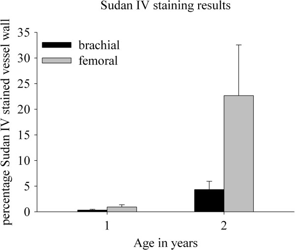
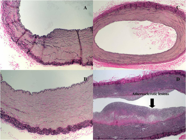
Background: The heterogeneous progression of atherosclerotic disease in the peripheral arteries is currently not well understood. In humans, artery specific disease progression is partly attributed to the local hemodynamic environments. However, despite similar hemodynamic environments, porcine brachial arteries are protected while femoral arteries are highly susceptible to advanced lesion formation. The aim of this investigation was to determine whether artery specific gene expression patterns contribute to the uneven distribution of peripheral arterial disease (PAD) in Rapacz Familial-Hypercholesterolemic (FHC) swine.
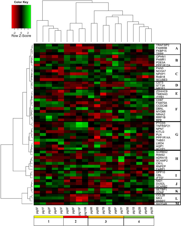
Results: Histological results confirmed rapid atherosclerotic disease progression in femoral but not brachial arteries. A total of 18,922 probe sets had sufficient signal abundance. A main effect for age and artery was observed for 1784 and 1256 probe sets, respectively. A significant age x artery interaction was found for 184 probe sets. Furthermore, comparison between arteries found a decrease from 714 to 370 differentially expressed transcripts from nine months to two years of age. Gene ontology analysis of the 56 genes with a main effect for artery and an age x artery interaction identified vascular smooth muscle contraction as enhanced biological signaling pathway.
Conclusion: This is the first investigation to report that the total number of differential genes decreases with diverging atherosclerotic disease pattern between porcine brachial and femoral arteries.
Figures

Similar articles
-
Gene expression differences in healthy brachial and femoral arteries of Rapacz familial hypercholesterolemic swine.Physiol Genomics. 2011 Jun 28;43(12):781-8. doi: 10.1152/physiolgenomics.00151.2010. Epub 2011 Apr 19. Physiol Genomics. 2011. PMID: 21505098
-
Impact of Fluoropolymer-Based Paclitaxel Delivery on Neointimal Proliferation and Vascular Healing: A Comparative Peripheral Drug-Eluting Stent Study in the Familial Hypercholesterolemic Swine Model of Femoral Restenosis.Circ Cardiovasc Interv. 2017 May;10(5):e004450. doi: 10.1161/CIRCINTERVENTIONS.116.004450. Circ Cardiovasc Interv. 2017. PMID: 28487355
-
Distinct gene expression profiles associated with Notch ligands Delta-like 4 and Jagged1 in plaque material from peripheral artery disease patients: a pilot study.J Transl Med. 2017 May 4;15(1):98. doi: 10.1186/s12967-017-1199-3. J Transl Med. 2017. PMID: 28472949 Free PMC article.
-
The Protective Role of Heme Oxygenase-1 in Atherosclerotic Diseases.Int J Mol Sci. 2019 Jul 24;20(15):3628. doi: 10.3390/ijms20153628. Int J Mol Sci. 2019. PMID: 31344980 Free PMC article. Review.
-
Structure of Atherosclerotic Plaques in Different Vascular Territories: Clinical Relevance.Curr Vasc Pharmacol. 2018 Jan 26;16(2):125-129. doi: 10.2174/1570161115666170227103125. Curr Vasc Pharmacol. 2018. PMID: 28245772 Review.
Cited by
-
ITGB2 is a central hub-gene associated with inflammation and early fibro-atheroma development in a swine model of atherosclerosis.Atheroscler Plus. 2023 Nov 15;54:30-41. doi: 10.1016/j.athplu.2023.11.001. eCollection 2023 Dec. Atheroscler Plus. 2023. PMID: 38116576 Free PMC article.
-
Development of Aortic Valve Disease in Familial Hypercholesterolemic Swine: Implications for Elucidating Disease Etiology.J Am Heart Assoc. 2015 Oct 27;4(10):e002254. doi: 10.1161/JAHA.115.002254. J Am Heart Assoc. 2015. PMID: 26508741 Free PMC article.
-
Measurements of wall shear stress and aortic pulse wave velocity in swine with familial hypercholesterolemia.J Magn Reson Imaging. 2015 May;41(5):1475-85. doi: 10.1002/jmri.24681. Epub 2014 Jun 25. J Magn Reson Imaging. 2015. PMID: 24964097 Free PMC article.
-
Dissecting the mechanism of carotid atherosclerosis from the perspective of regulation.Int J Mol Med. 2014 Dec;34(6):1458-66. doi: 10.3892/ijmm.2014.1960. Epub 2014 Oct 9. Int J Mol Med. 2014. PMID: 25318463 Free PMC article.
-
Effect of Caloric Restriction on Metabolic Dysfunction of Young Rapacz Familial Hypercholesterolemic Swine (Sus scrofa).Comp Med. 2017 Dec 1;67(6):508-517. Comp Med. 2017. PMID: 29212583 Free PMC article.
References
-
- Rosamond W, Flegal K, Friday G, Furie K, Go A, Greenlund K, Haase N, Ho M, Howard V, Kissela B, Kittner S, Lloyd-Jones D, McDermott M, Meigs J, Moy C, Nichol G, O’Donnell CJ, Roger V, Rumsfeld J, Sorlie P, Steinberger J, Thom T, Wasserthiel-Smoller S, Hong Y. Heart disease and stroke statistics–2007 update: a report from the American Heart Association Statistics Committee and Stroke Statistics Subcommittee. Circulation. 2007;115(5):e69–e171. doi: 10.1161/CIRCULATIONAHA.106.179918. - DOI - PubMed
Publication types
MeSH terms
LinkOut - more resources
Full Text Sources
Other Literature Sources
Medical
