. 2013 Oct;16(5):404-13.
doi: 10.1089/rej.2013.1433.

Inhibitory effect of hydroxysafflor yellow a on mouse skin photoaging induced by ultraviolet irradiation

Affiliations

Inhibitory effect of hydroxysafflor yellow a on mouse skin photoaging induced by ultraviolet irradiation

Song-Zhi Kong et al. Rejuvenation Res. 2013 Oct.

Abstract

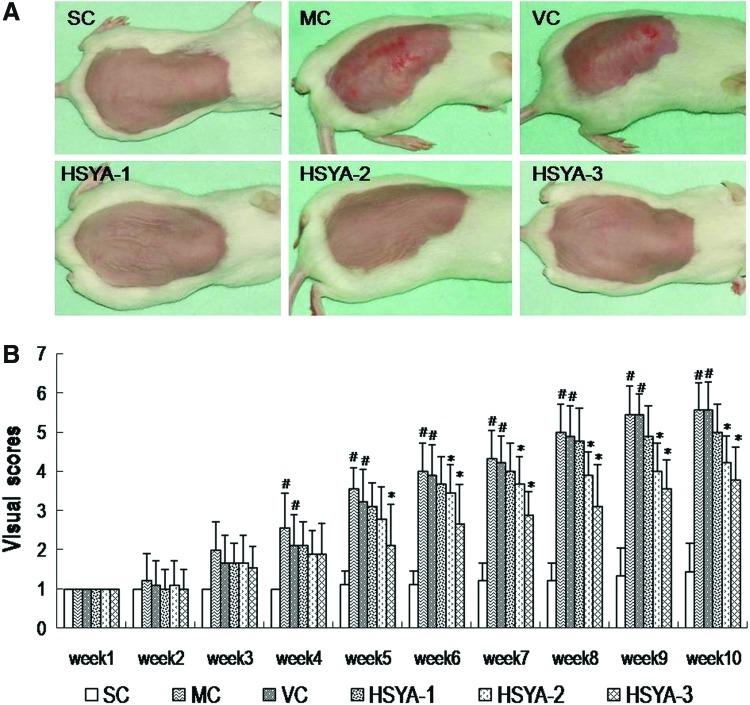
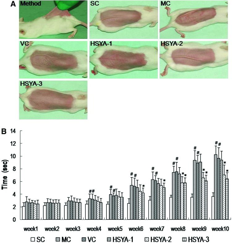
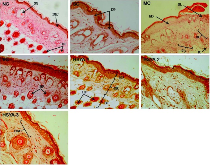
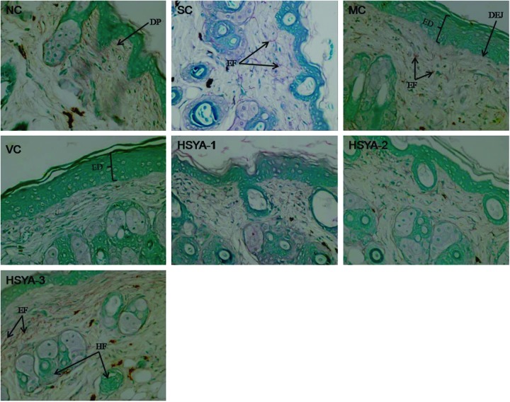
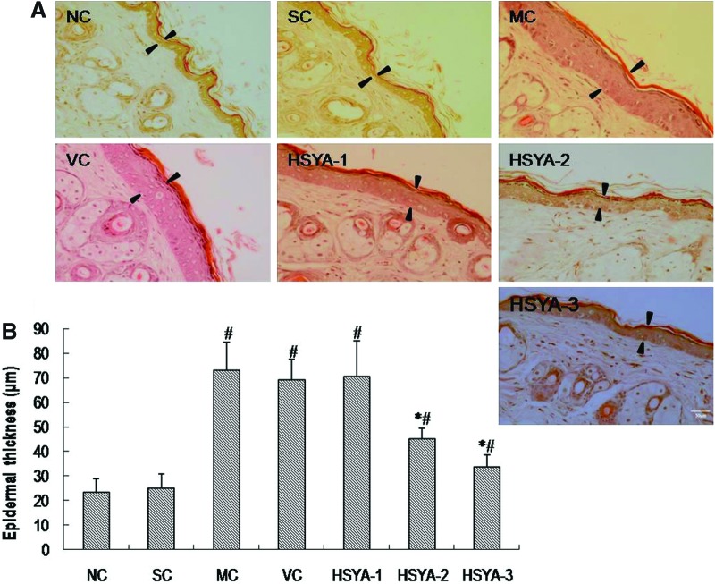
Chronic exposure to ultraviolet (UV) irradiation is believed to be the major cause of skin damage that results in premature aging of the skin, so called photoaging, characterized by increases in skin thickness, formation of wrinkles, and loss of skin elasticity. UV induces damage to skin mainly by oxidative stress and collagen degradation. In this study, we examined the photo-protective effect of hydroxysafflor yellow A (HSYA), a major active chemical component isolated from Carthamus tinctorius L., by topical application on the skin of mice. Exposure of the dorsal depilated skin of mice to UV radiation four times a week for 10 weeks induced epidermal hyperplasia, elastin accumulation, collagen degradation, etc. HSYA at the doses of 50, 100, and 200 μg/mouse was topically applied immediately following each UV exposure. The effects of HSYA were evaluated by a series of tests, including macroscopic and histopathological evaluation of skin, pinch test, and redox homeostasis of skin homogenates. Results showed that the UV-induced skin damage was significantly improved after HSYA treatment, especially at doses of 100 and 200 μg/mouse. This protective effect is possibly related to the anti-oxidative property of HSYA and mediated by promoting endogenous collagen synthesis. This is the first study providing preclinical evidence for the protective effect of HSYA against photoaging.


Figures

FIG. 1.
FIG. 1.
Chemical structure of hydroxysafflor yellow A (HSYA) isolated from Carthamus tinctorius.
FIG. 2.
FIG. 2.
Hydroxysafflor yellow A (HSYA) prevents ultraviolet (UV)-induced macroscopic skin lesions in mice in vivo. (A) Macroscopic changes in the skin of mice upon various treatments at the end of experimental period of 10 weeks. (B) Results of the visual score of different experimental groups during the 10-week study period. Data represent means±standard deviation (SD) (n=9). (#) p<0.05 compared with the SC group; (*) p<0.05 compared with the VC group. SC, shaved control; MC, model control; VC, solvent control; HSYA-1, treated with HYSA at a dose of 50 μg/mouse; HSYA-2, treated with HYSA at a dose of 100 μg/mouse; HSYA-3, treated with HYSA at a dose of 200 μg/mouse. (Color image available online at www.liebertpub.com/rej).
FIG. 3.
FIG. 3.
Evaluation of sagging by pinch testing. (A) Photographs of pinch testing carried out according to the method of Bryce and Bogdan. (B) Recovery time in the pinch test, performed at the end of week 10, in groups with or without UV exposure. Data represent means±standard deviation (SD) (n=9). (#) p<0.05 compared with the SC group; (*) p<0.05 compared with the VC group. SC, shaved control; MC, model control; VC, solvent control; HSYA-1, treated with HYSA at a dose of 50 μg/mouse; HSYA-2, treated with HYSA at a dose of 100 μg/mouse; HSYA-3, treated with HYSA at a dose of 200 μg/mouse. (Color image available online at www.liebertpub.com/rej).
FIG. 4.
FIG. 4.
Hematoxylin & Eosin (H&E) staining of mouse skin in: NC group (100×); SC group, having the same or similar characteristics to the skin in NC group (200×); MC group, showing destroyed structure and inflammation (100×; note the inflammatory cells); VC group, having the similar features to that in MC group (100×); HSYA-1 group (100×)); HSYA-2 group (200×)); HSYA-3 group (200×)). ED, epidermis; DEJ, dermal–epidermal junction; DR, dermis; SP, dermal papilla; ST, subcutaneous tissue; HF, hair follicle; SG, sebaceous glands; IC, inflammatory cells; IFZ, inflammation zone; DAC, densely arranged collagen fibers which were stacked on top of each other in an orderly arrangement; SL, skin lesion; DC, degraded collagen fibers which were messily arranged. (Color image available online at www.liebertpub.com/rej).
FIG. 5.
FIG. 5.
Gomori aldehyde fuchsin staining of mouse skin in: NC group (200×; note that the arrangement of the elastic fibers is web-like and has a random pattern. SC group, having the same or similar characteristics to the skin in NC group (200×)); MC group, showing large quantities of tangled, fractured thickened, degraded fibers (200×)); VC group, having the similar features to that in MC group (200×); HSYA-1 group (200×); HSYA-2 group (200×); HSYA-3 group (200×). DP, dermal papillae; EF, elastic fibers; ED, epidermis; DEJ, dermal–epidermal junction; HF, hair follicle. NC, untreated control; SC, shaved control; MC, model control; VC, solvent control; HSYA-1, treated with HYSA at a dose of 50 μg/mouse; HSYA-2, treated with HYSA at a dose of 100 μg/mouse; HSYA-3, treated with HYSA at a dose of 200 μg/mouse. (Color image available online at www.liebertpub.com/rej).
FIG. 6.
FIG. 6.
Induction of epidermal hyperproliferation by ultraviolet (UV) and inhibition by hydroxysafflor yellow A (HSYA). (A) Photographs (200×) of epidermal hyperproliferation in the skin of mice upon various treatments by Hematoxylin & Eosin (H&E) stain. Scale bar, 50 μm. Arrowheads indicate the width of epidermis. (B) The average thickness of the epidermis in the skin of mice upon various treatments. Data represent means±standard deviation (SD) (n=6). (#) p<0.01 compared with the SC group; (*) p<0.05 compared with the VC group. NC, untreated control; SC, shaved control; MC, model control; VC, solvent control; HSYA-1, treated with HYSA at a dose of 50 μg/mouse; HSYA-2, treated with HYSA at a dose of 100 μg/mouse; HSYA-3, treated with HYSA at a dose of 200 μg/mouse. (Color image available online at www.liebertpub.com/rej).

References

    1. Diffey BL. Solar ultraviolet radiation effects on biological system. PhysMed Biol. 1991;36:299–328. - PubMed
    1. Fisher GJ. Wang ZQ. Fatta SC. Varani J. Kang S. Voorhees JJ. Pathophysiology of premature skin aging induced by ultraviolet light. N Engl Med. 1997;337:1419–1428. - PubMed
    1. Chung JH. Photoaging in Asians. Photodermatol Photo. 2003;19:109–121. - PubMed
    1. Sams WM. Smith J. The histochemistry of chronically sun damaged skin. J Invest Dermatol. 1961;37:447–452. - PubMed
    1. Uitto J. Fazio MJ. Olsen DR. Molecular mechanisms of cutaneous aging. Age-associated connective tissue alterations in the dermis. J Am Acad Dermatol. 1989;21:614–622. - PubMed

Publication types

MeSH terms

LinkOut - more resources