. 2013 Jul 2;105(1):11-20.
doi: 10.1016/j.bpj.2013.05.032.

Nonlinear strain stiffening is not sufficient to explain how far cells can feel on fibrous protein gels

Affiliations

Nonlinear strain stiffening is not sufficient to explain how far cells can feel on fibrous protein gels

Mathilda S Rudnicki et al. Biophys J. .

Abstract

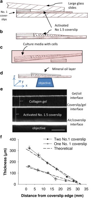
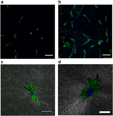
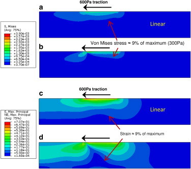
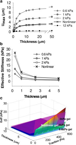
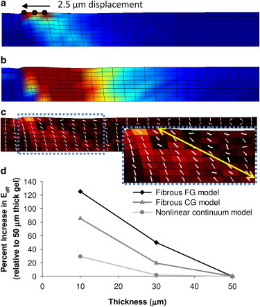
Recent observations suggest that cells on fibrous extracellular matrix materials sense mechanical signals over much larger distances than they do on linearly elastic synthetic materials. In this work, we systematically investigate the distance fibroblasts can sense a rigid boundary through fibrous gels by quantifying the spread areas of human lung fibroblasts and 3T3 fibroblasts cultured on sloped collagen and fibrin gels. The cell areas gradually decrease as gel thickness increases from 0 to 150 μm, with characteristic sensing distances of >65 μm below fibrin and collagen gels, and spreading affected on gels as thick as 150 μm. These results demonstrate that fibroblasts sense deeper into collagen and fibrin gels than they do into polyacrylamide gels, with the latter exhibiting characteristic sensing distances of <5 μm. We apply finite-element analysis to explore the role of strain stiffening, a characteristic mechanical property of collagen and fibrin that is not observed in polyacrylamide, in facilitating mechanosensing over long distances. Our analysis shows that the effective stiffness of both linear and nonlinear materials sharply increases once the thickness is reduced below 5 μm, with only a slight enhancement in sensitivity to depth for the nonlinear material at very low thickness and high applied traction. Multiscale simulations with a simplified geometry predict changes in fiber alignment deep into the gel and a large increase in effective stiffness with a decrease in substrate thickness that is not predicted by nonlinear elasticity. These results suggest that the observed cell-spreading response to gel thickness is not explained by the nonlinear strain-stiffening behavior of the material alone and is likely due to the fibrous nature of the proteins.


Figures

Figure 1
Figure 1
(a) Schematic of the experimental setup for creating the sloped-gel sample. The collagen or fibrin gel adheres to the activated No. 1.5 coverslip and is formed into the sloped shape by the upper large glass slide that is weighted with a 50 g object. (b) Schematic of the finished sample ready for cell seeding. (c) Samples are placed in a 100 mm petri dish and the right side is propped up with two No. 1 coverslips to ensure a level seeding surface. (d) Sample setup for imaging with mineral oil to minimize reflection at the edge of the gel. (e) Confocal reflectance image taken in cross-section mode shows the No. 1.5 coverslip between the bottom and middle white lines, and protein gel fibers with speckled pattern between the top two lines (10× dry objective; scale bar = 100 μm). The change in height (∼4 μm) cannot be readily seen over the field of view (∼400 μm) due to the gradual slope of the gel. (f) Validation of the slopes of fibrin gels created in the system using either one or two No. 1 coverslips.
Figure 2
Figure 2
Representative fluorescent micrographs of HLFs on thick (a) and thin (b) fibrin gels used for cell area analysis (10× dry objective; scale bars = 250 μm). Actin cytoskeleton stained with phalloidin (green in color, light grey in greyscale) and nuclei stained with Hoescht (blue in color, dark grey in greyscale) after 16 h culture period. Confocal reflectance images (in standard XYZ mode) of fibrin (c) and collagen (d) gels with fluorescent stain overlays reveal local reorganization by cells, indicating stress/strain propagation through the fibrous network. (c) 20× oil objective; scale bar = 75 μm; (d) 40× oil objective; scale bar = 25 μm.
Figure 3
Figure 3
Cell spreading as a function of gel thickness for HLFs on fibrin (red squares) and collagen (green triangles) gels, and for 3T3s on fibrin gels (blue circles). Reported as mean ± SE; HLFs on collagen, mean = 9 cells per data point, n = 4 gels; HLFs on fibrin, mean = 10 cells per data point, n = 5 gels; 3T3 on collagen, mean = 20 cells per data point, n = 2 gels. Open symbols indicate control cells on tissue culture plastic. Data from the literature for 3T3 fibroblasts (+ symbols) and hMSCs (x symbols) indicate that cells on PA gels are not affected by rigid boundaries more than ∼30 μm away. On nonlinear fibrin and collagen gels, fibroblasts sense the rigid boundary on much thicker gels (>100 μm), consistent with the spread area of hMSCs on collagen gels (star symbols), and the relationship between spread area and thickness is more gradual than on PA gels.
Figure 4
Figure 4
(a–d) Stress (a and b; in MPa) and strain (c and d; unitless) distributions for 2 kPa linear (a and c) and nonlinear (b and d) gels of 10 μm thickness with 600 Pa traction applied. Plots for the other stiffness linear gels have different magnitude but when normalized to maximum values look identical (data not shown). The stress contours are blunted and exhibit a pinched profile in the strain-stiffening material. In contrast, the strain profiles extend farther into the strain-stiffening material compared with the linear material, as can be seen by the lowest contour, representing roughly 9% of the maximum strain, touching the lower boundary in the 10-μm-thick nonlinear gel but not in the 10-μm-thick linear gel. See Supporting Material for plots for 1, 5, and 50 μm gels.
Figure 5
Figure 5
(a) For a given applied traction (600 Pa), substrate displacement decreases with decreasing thickness and increasing gel stiffness. (b) The strain-stiffening behavior causes the nonlinear material to become effectively stiffer at low thicknesses (h < 2 μm) when the applied traction is increased. (c) A surface plot of effective stiffness as a function of applied traction (100–600 Pa) and thickness (0.5–10 μm) shows that the nonlinear material (gradient colored) exhibits an effective stiffness similar to that of the 1 kPa linear material with 100 Pa applied traction, but is more stiff than the 2 kPa linear material with tractions over ∼500 Pa at low thicknesses. When viewed in greyscale, darker colors indicate lower values except immediately under the loading at the upper surface.
Figure 6
Figure 6
(a and b) Fibrous multiscale model results for the 10-μm-thick case indicate that stresses (a) and strains (b) propagate throughout the thickness of the substrate. (c) The change in degree of orientation from initially random to aligned is greatest at the surface and extends to areas near the lower boundary that are >20 μm away from the point of deformation at the surface (indicated by the arrow in c, inset). (d) The effective stiffness (proportional to peak stress/displacement and normalized to the 50-μm-thick case) is much greater for the 10- and 30-μm-thick fibrous models than for nonlinear models with identical loading, geometry, and nonlinear bulk mechanical properties, indicating a greater impact of the rigid boundary in the fibrous model. The simplified model is 75 μm long and one element in depth (4 μm), with 2.5 μm displacement applied over a small area on the top surface as indicated in panel a. Due to differences in geometry, results are not directly comparable with the radially symmetric model results in Fig. 4. Scale factors: (a) 0–100 Pa von Mises stress; (b) 0–0.1 engineering strain; (c) 0–1.0 change in degree of fiber alignment. When viewed in greyscale, darker color indicates lower stress in (a and c), and darker color indicates lower strain in outer areas but higher strain in the central region under the loading in (b). Fibrin gel (FG); collagen gel (CG).

References

    1. Saha K., Keung A.J., Healy K.E. Substrate modulus directs neural stem cell behavior. Biophys. J. 2008;95:4426–4438. - PMC - PubMed
    1. Yip C.Y., Chen J.H., Simmons C.A. Calcification by valve interstitial cells is regulated by the stiffness of the extracellular matrix. Arterioscler. Thromb. Vasc. Biol. 2009;29:936–942. - PubMed
    1. Wang H.B., Dembo M., Wang Y.L. Substrate flexibility regulates growth and apoptosis of normal but not transformed cells. Am. J. Physiol. Cell Physiol. 2000;279:C1345–C1350. - PubMed
    1. Ingber D.E. Mechanobiology and diseases of mechanotransduction. Ann. Med. 2003;35:564–577. - PubMed
    1. Vogel V., Sheetz M. Local force and geometry sensing regulate cell functions. Nat. Rev. Mol. Cell Biol. 2006;7:265–275. - PubMed

Publication types

LinkOut - more resources