. 2013 Jun 18;8(6):e66549.
doi: 10.1371/journal.pone.0066549. Print 2013.

Mitigation of Lethal Radiation Syndrome in Mice by Intramuscular Injection of 3D Cultured Adherent Human Placental Stromal Cells

Affiliations

Mitigation of Lethal Radiation Syndrome in Mice by Intramuscular Injection of 3D Cultured Adherent Human Placental Stromal Cells

Elena Gaberman et al. PLoS One. .

Abstract

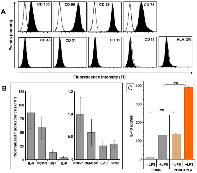
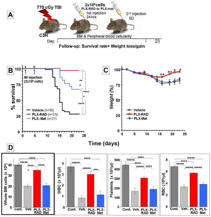
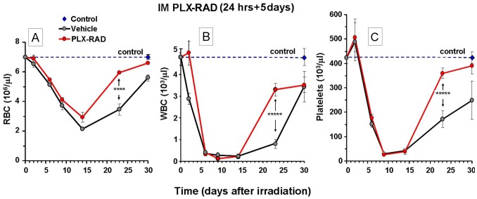
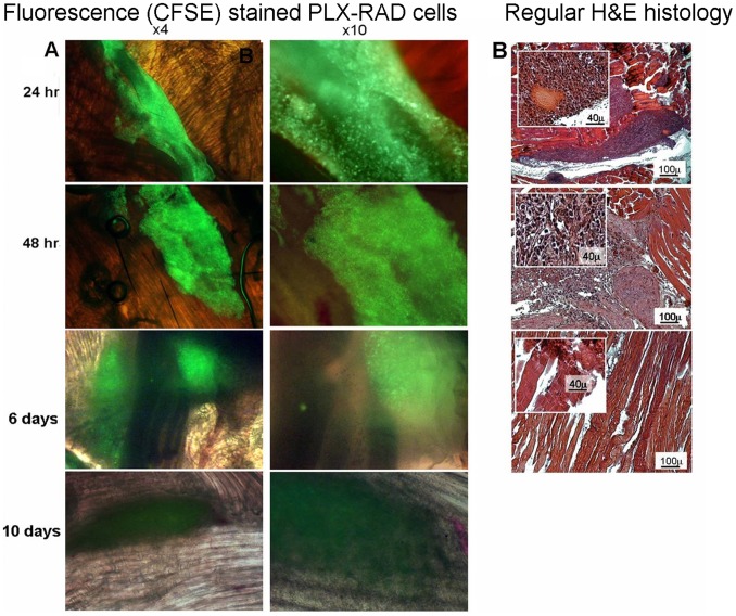
Exposure to high lethal dose of ionizing radiation results in acute radiation syndrome with deleterious systemic effects to different organs. A primary target is the highly sensitive bone marrow and the hematopoietic system. In the current study C3H/HeN mice were total body irradiated by 7.7 Gy. Twenty four hrs and 5 days after irradiation 2×10(6) cells from different preparations of human derived 3D expanded adherent placental stromal cells (PLX) were injected intramuscularly. Treatment with batches consisting of pure maternal cell preparations (PLX-Mat) increased the survival of the irradiated mice from ∼27% to 68% (P<0.001), while cell preparations with a mixture of maternal and fetal derived cells (PLX-RAD) increased the survival to ∼98% (P<0.0001). The dose modifying factor of this treatment for both 50% and 37% survival (DMF50 and DMF37) was∼1.23. Initiation of the more effective treatment with PLX-RAD injection could be delayed for up to 48 hrs after irradiation with similar effect. A delayed treatment by 72 hrs had lower, but still significantly effect (p<0.05). A faster recovery of the BM and improved reconstitution of all blood cell lineages in the PLX-RAD treated mice during the follow-up explains the increased survival of the cells treated irradiated mice. The number of CD45+/SCA1+ hematopoietic progenitor cells within the fast recovering population of nucleated BM cells in the irradiated mice was also elevated in the PLX-RAD treated mice. Our study suggests that IM treatment with PLX-RAD cells may serve as a highly effective "off the shelf" therapy to treat BM failure following total body exposure to high doses of radiation. The results suggest that similar treatments may be beneficial also for clinical conditions associated with severe BM aplasia and pancytopenia.


Conflict of interest statement

Competing Interests: Major parts of the research project were sponsored and performed in collaboration with Pluristem Therapeutics which also prepared and supplies the relevant cells on which the study is based. Authors Lena Pinzur, Nir Netzer and Zami Aberman were employed by Pluristem Therapeutics Inc. at the time of this project was performed. Pluristem Therapeutics Inc. has filed the following patent application which relate to the current study (Patent application Publication US2011/0293583 A1 and PCT/1B2012/000664)). They may have previous patents on similar other cell types which were not used in the current study. Pluristem Therapeutics Inc. does not market yet products relating to the relevant study (PLX-RAD). By the terms of the research contract. The authors and their institutes have no direct or indirect benefits from current or future patent applied by the company. The authors adhere to all the PLOS ONE policies on sharing data and materials.

Figures

Figure 1
Figure 1. In-vitro characterization of the PLX-RAD cells.
A. Representative FACS profile of cell surface markers of PLX-RAD cells batches used in the current study. The PLX-RAD cell population could be defined as having typical human mesenchymal markers (CD 105, 29, 73 and 90), with no contamination of cells expressing WBC markers (CD 45, 19, 14 and HLA-DR) or endothelial cells marker (CD31). B. Secretion profile of protein of interest by PLX-RAD cells in vitro. The protein secretion composition of PLX-RAD cell batches (n = 3) after 24 hrs incubation in EBM-2, using a protein array. Levels of IL-6, MCP-3, HGF, IL-8, FGF-7, GM-CSF, IL-10 and bFGF in the conditioned medium are shown. C. Induction of IL-10 secretion by PBMC in the presence of PLX-RAD. The PLX-RAD cells were co-cultured with PBMC with or without LPS, and IL-10 release in the media was measured by sandwich ELISA plate. PLX-RAD alone increased the IL-10 secretion significantly and induction with LPS further significantly increased the PLX-RAD effect (** = p<0.01).
Figure 2
Figure 2. The effect of IM PLX-Mat and PLX-RAD injection on the survival of 7.7 Gy total body irradiated mice.
The setup of the experiment is described in (A). 2×10⁶ PLX-RAD or PLX-Mat cells were injected IM twice, 1st injection delivered 24 hrs and 2nd injection on day 5 after irradiation. The follow-up of mice survival is shown in (B). Weight changes for the group of IM injected mice at day 1 and 5 after irradiation are presented in (C). The BM and blood counts of the surviving animal at the end of the experiment at day 23 are presented in (D). Significance: * = P<0.05, ** = P<0.01*** = P<0.005, **** = P<0.001, ***** = P<0.0001.
Figure 3
Figure 3. The effect of IM PLX-RAD injection on the survival of 7.7 Gy total body irradiated mice.
The setup of the experiment arms is described in (A). PLX-RAD cells (2×10⁶) were injected IM twice, 1st injection was done 24 or 48 or 72 hrs and 2nd injection was done on day 5 after irradiation. In another arm only a single injection on day 5 was given. The data presented are based on merged separate experimental repetitions. The total number of animals in each arm is shown in the legend. The follow-up of mice survival between the different schedules of treatment is shown in (B). The BM and blood cells profile of the surviving mice at the end of the experiments on day 23 is shown in (C). Significance: * = P<0.05, ** = P<0.01, **** = P<0.001, ***** = P<0.0001.
Figure 4
Figure 4. Time dependent alterations of the profile of the 3 peripheral blood cell lineages following irradiation and PLX-RAD treatment.
Mice were injected with PLX-RAD on day 1 and 5 following 7.7 Gy irradiation. The mice were bled and sacrificed on days 2, 6, 9, 14, 23 and 30 after irradiation. RBC (A), WBC (B) and Platelets (C) were counted in peripheral blood. The data are based on at least 8 mice which were included for each group for the different rime points. For day 23 the data are based on 31 PLX-RAD treated and 55 vehicle treated mice (from repeated experiments). The recovery on day 23 of peripheral blood cells counts of the surviving mice subjected to the different tested arms with different timing of the first IM injections is presented. The more affected animals died before the end of the experiment, mostly in the group of vehicle treated controls, leaving only the few survivors on which the relevant data are based. The profile of the WBC, platelets and RBC are presented. Significance: * = P<0.05, ** = P<0.01, **** = P<0.001, ***** = P<0.0001.
Figure 5
Figure 5. The presence of PLX-RAD cells in the injection site.
(A) 2×10⁶ CFSE labeled cells PLX-RAD cells were injected IM to 7.7 Gy irradiated mice. Direct fluorescence microscopy images with dim white light background (as described in methods and materials) at different magnifications are presented. The injected cells were observed only within the injection site in the freshly excised intact muscle. Direct fluorescence microscopy showed slow clearance of the cells within 6–10 days. (B) Regular H&E stained histological sections of samples taken 24, 48 hrs and 6 days after injection. An inset in each of the H&E histology shows a higher magnification of the area within the muscles where the injected cells resided are clearly seen.
Figure 6
Figure 6. Kinetics of whole BM cellularity in mice treated on days 1 and 5 after irradiation with IM injections of PLX-RAD or vehicle controls.
The whole BM cells were extracted from the tibias and femurs of the surviving treated animals from the 2 experimental arms on days 2, 6, 9, 14 and 23 after irradiation. Each group consisted of 8–10 mice. A clear advantage of the total cell number in the PLX-RAD injected mice was recorded along the follow up and was most significant from day 9 onward (A). In parallel samples of histological sections of decalcified bones from PLX-RAD and vehicle treated animals relative to bones from non-irradiated mice on day 9 and 23 are presented (B-G), further demonstrating the advantage of the PLX-RAD treatment in this critical time points. The histological bone sections are presented from day 9 for a non-irradiated control (B1 with magnified frames in B2 and B3), from vehicle controls (C1 with magnified frames in C2 and C3), and PLX-RAD treated mice (D1 with magnified frames in D2 and D3). The histological sections show only marginal better cellularity in the PLX-RAD treated mice on day 9 relative to vehicle controls. On day 23 after irradiation the difference was maximal, as also reflected in the relevant histological slides (F1–3 and G1–3) similar to the cellularity observed in non-irradiated control mice (E1–3). Significance: ** = p<0.01, ***** = p<0.0001.
Figure 7
Figure 7. The effect of IM injection of PLX-RAD cells on days 1 and 5 after radiation on the number of nucleated cells from whole BM and the ratio of progenitors within this cell population.
(A) Up to day 9 after irradiation, the number of the nucleated BM cells decreased sharply in both arms, followed by a faster gain in the number of total nucleated BM cells in the PLX-RAD treated animals, as compared to the vehicle controls. By day 30 a full recovery of the number of nucleated BM cells was recorded only in the group of irradiated PLX-RAD treated mice. (B) FACS analysis of the kinetics of changes in the % of CD45+/Sca-1+ of the nucleated cells (representing the progenitor cells population) was tested in both arms of mice irradiated by 7.7 Gy, PLX-RAD or vehicle control treated. Maximal increase in the proportion of these cells due to the radiation exposure was recorded on day 9 in both groups, before the onset of the critical phase of the hematopoietic syndrome. Significance: * = P<0.05, ** = P<0.01.
Figure 8
Figure 8. Evaluation of the dose modifying factor (DMF) of IM treatment with PLX-RAD.
The ratio between the total dose needed to reach 50% survival rate of animals treated with PLX-RAD cells (9.05 Gy) relative to the dose needed to yield the same effect in the controls (7.42 Gy) was calculated based on the survival of irradiated mice exposed to different doses of radiation +/− IM injection of 2×10⁶ PLX cells at days 1 and 5 after irradiation. A DMF of 1.22 was recorded. The highest supported total body irradiation dose (HSD) which still yields 100% survival in the irradiated controls can be extrapolated from this Figure to be ∼6.8 Gy.

References

    1. Williams JP, McBride WH (2011) After the bomb drops: a new look at radiation-induced multiple organ dysfunction syndrome (MODS). Int J Radiat Biol 87: 851–868. - PMC - PubMed
    1. Koenig KL, Goans RE, Hatchett RJ, Mettler FA Jr, Schumacher TA, et al. (2005) Medical treatment of radiological casualties: current concepts. Ann Emerg Med 45: 643–852. - PubMed
    1. Meineke V, Fliedner TM (2005) Radiation-induced multi-organ involvement and failure: challenges for radiation accident medical management and future research. BJR Suppl 27: 196–200.
    1. Need JT, Mothershead JL (2006) Strategic National Stockpile program: implications for military medicine. Mil Med 171: 698–702. - PubMed
    1. Andersson KG, Mikkelsen T, Astrup P, Thykier-Nielsen S, Jacobsen LH, et al. (2008) Estimation of health hazards resulting from a radiological terrorist attack in a city. Radiat Prot Dosimetry 131: 297–307. - PubMed

Publication types