. 2013 Jun 18;8(6):e66162.
doi: 10.1371/journal.pone.0066162. Print 2013.

The Observation of Highly Ordered Domains in Membranes with Cholesterol

Affiliations

The Observation of Highly Ordered Domains in Membranes with Cholesterol

Clare L Armstrong et al. PLoS One. .

Abstract

Rafts, or functional domains, are transient nano- or mesoscopic structures in the exoplasmic leaflet of the plasma membrane, and are thought to be essential for many cellular processes. Using neutron diffraction and computer modelling, we present evidence for the existence of highly ordered lipid domains in the cholesterol-rich (32.5 mol%) liquid-ordered ([Formula: see text]) phase of dipalmitoylphosphatidylcholine membranes. The liquid ordered phase in one-component lipid membranes has previously been thought to be a homogeneous phase. The presence of highly ordered lipid domains embedded in a disordered lipid matrix implies non-uniform distribution of cholesterol between the two phases. The experimental results are in excellent agreement with recent computer simulations of DPPC/cholesterol complexes [Meinhardt, Vink and Schmid (2013). Proc Natl Acad Sci USA 110(12): 4476-4481], which reported the existence of nanometer size [Formula: see text] domains in a liquid disordered lipid environment.


Conflict of interest statement

Competing Interests: The authors have declared that no competing interests exist.

Figures

Figure 1
Figure 1. Schematics of the studied systems.
a) Schematic of a lipid bilayer containing a lipid domain as studied by neutron scattering techniques using a low (top) and high (bottom) spatial resolution setup. b) In-plane representation of saturated hydrocarbon-chain lipid-cholesterol interactions in accordance with the umbrella model, whereby each lipid is associated with 2 cholesterol molecules. This structural arrangement results when the cholesterol content is 66 mol%. The blue squares represent lipid head groups, the yellow circles correspond to lipid tails, and the red circle are cholesterol molecules. c) Schematic molecular structures of DPPC and cholesterol molecules.
Figure 2
Figure 2. Phase diagram of phospholipid/cholesterol complexes, such as DMPC/cholesterol and DPPC/cholesterol, as reported by, for example,
–. Besides the well known gel and fluid phases, the so-called liquid-order phase is observed at high cholesterol concentrations. The 32.5 mol% sample, as depicted by the ⊛, was determined to be in the lo phase.
Figure 3
Figure 3. Comparison between the in-plane scans of DPPC-d62 bilayers with 32.5 mol% cholesterol using a) the conventional high energy resolution (small ΔE) setup and b) the low energy resolution (large ΔE) setup.
The data are denoted by circles with the fit shown as a solid line. A disordered structure was observed in a), while the sharp features in b) are indicative of the presence of highly ordered lipid domains. A top view of the corresponding molecular structures are shown in the insets to the Figure using the same symbols as in Figure 1 b); the quasi-Bragg reflections are indicated by vertical dashed lines and their associated Miller indices, [hkl]. Peaks resulting from the silicon substrates and the aluminum sample chamber (as described in the Materials and Methods Section) are highlighted in grey, but not accounted for in the fit.
Figure 4
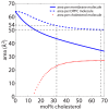
Figure 4. Area per molecule and partial areas for DPPC and cholesterol molecules as function of cholesterol concentration.
Curves were calculated using Eq. (1) from .
Figure 5
Figure 5. Results of the computer modelling.
a) The scattering function S(q||) integrated over an area of 250×250 Å2, resulting in a diffraction pattern indicative of fluid, disordered bilayers. b) S(q||) for an ordered lipid domain of size 70×70 Å2. Three sharp correlation peaks are observed. The dots represent data obtained from the computer model, while the solid lines correspond to the fits obtained from the neutron scattering experiment in Figure 3 a) and b). Areas of integration are indicated by the blue and red rectangles shown in c). c) Snapshot of a typical configuration in the computer model. The blue region corresponds to the disordered lipid matrix, and the red regions to ordered lipid domains. The total system size was 300×300 Å2 and contained 2000 lipid molecules, or 4000 lipid tails. The ordered domains were 70×70 Å2 in size and included 110 lipid molecules. d) A close up view of the computer model system, where the blue circles represent lipid tails in the fluid disordered state and the red diamonds are tails in the lo phase.
Figure 6
Figure 6. Geometry of the triple axis spectrometer.
Orientation of the sample for in-plane scans, such that the scattering vector, Q, lies in the plane of the membrane (q||). ki and kf are the incident and final neutron wave vectors (k = 2π = λ) and the c’s denote the location of collimators along the beam line.
Figure 7
Figure 7. In-plane diffraction data and peak assignments.
Data were taken with and without a PG filter, which was used to suppress higher order reflections.

References

    1. Simons K, Ikonen E (1997) Functional rafts in cell membranes. Nature 387: 569–572. - PubMed
    1. Simons K, Ikonen E (2000) How cells handle cholesterol. Science 290: 1721–1726. - PubMed
    1. Engelman DM (2005) Membranes are more mosaic than fluid. Nature 438: 578–580. - PubMed
    1. Niemelä PS, Ollila S, Hyvnen MT, Karttunen M, Vattulainen I (2007) Assessing the nature of lipid raft membranes. PLoS Comput Biol 3: e34. - PMC - PubMed
    1. Pike L (2009) The challenge of lipid rafts. J Lipid Res 50: S323. - PMC - PubMed

Publication types

MeSH terms