

Evaluation of different fiducial markers for image-guided radiotherapy and particle therapy
- PMID: 23824129
- PMCID: PMC3700523
- DOI: 10.1093/jrr/rrt071
Evaluation of different fiducial markers for image-guided radiotherapy and particle therapy
Abstract
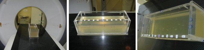
Modern radiotherapy (RT) techniques are widely used in the irradiation of moving organs. A crucial step in ensuring the correct position of a target structure directly before or during treatment is daily image guidance by computed tomography (CT) or X-ray radiography (image-guided radiotherapy, IGRT). Therefore, combinations of modern irradiation devices and imaging, such as on-board imaging (OBI) with X-rays, or in-room CT such as the tomotherapy system, have been developed. Moreover, combinations of linear accelerators and in-room CT-scanners have been designed. IGRT is of special interest in hypofractionated and radiosurgical treatments where high single doses are applied in the proximity of critical organs at risk. Radiographically visible markers in or in close proximity to the target structure may help to reproduce the position during RT and could therefore be used as external surrogates for motion monitoring. Criteria sought for fiducial markers are (i) visibility in the radiologic modalities involved in radiotherapeutic treatment planning and image guidance, such as CT and kilovoltage (kV) OBI), (ii) low production of imaging artifacts, and (iii) low perturbation of the therapeutic dose to the target volume. Photon interaction with interstitial markers has been shown to be not as important as in particle therapy, where interaction of the particle beam, especially with metal markers, can have a significant impact on treatment. This applies especially with a scanned ion beam. Recently we commenced patient recruitment at our institution within the PROMETHEUS trial, which evaluates a hypofractionation regime, starting with 4 x 10 Gy (RBE), for patients with hepatocellular carcinoma. The aim of this work is, therefore, to evaluate potential implantable fiducial markers for enabling precise patient and thus organ positioning in scanned ion beams. To transfer existing knowledge of marker application from photon to particle therapy, we used a range of commercially available markers of different forms and sizes, consisting of carbon and gold materials, and evaluated them for their potential use in the clinical setup with scanned ion beams at our institution. All markers were implanted in a standardized Alderson phantom and were examined using CT scans and orthogonal kV OBI in our clinical routine protocol. Impact on beam perturbation downstream of the markers in the plateau region of a spread-out Bragg peak (SOBP) was estimated by using radiographic films for clinical proton and carbon ion beams of high and low energies. All tested markers achieved good visibility in CT and kV OBI. Disturbances due to artifacts and dose perturbation were highest in the arbitrarily folded gold and the thickest gold marker, but especially low in the carbon marker. Dose perturbation was highest in the arbitrarily folded gold marker. In summary, the analyzed markers offer promising potential for identifying target structures in our treatment setup at HIT and will soon be used in clinical routine. However, a careful choice of marker, depending on the tumor localization and irradiation strategy, will need to be made.
Keywords: carbon ion therapy; fiducial marker; liver tumors; moving organs; raster-scanning technique.
Figures


References
-
- Combs SE, Schulz-Ertner D, Herfarth KK, et al. Advances in radio-oncology. From precision radiotherapy with photons to ion therapy with protons and carbon ions. Chirurg. 2006;77:1126–32. - PubMed
-
- Combs SE, Ellerbrock M, Haberer T, et al. Heidelberg Ion Therapy Center (HIT): Initial clinical experience in the first 80 patients. Acta Oncol. 2010;49:1132–40. - PubMed
-
- Combs SE, Jakel O, Haberer T, et al. Particle therapy at the Heidelberg Ion Therapy Center (HIT) – Integrated research-driven university-hospital-based radiation oncology service in Heidelberg, Germany. Radiother Oncol. 2010;95:41–4. - PubMed
Publication types
MeSH terms
Substances
LinkOut - more resources
Full Text Sources
Other Literature Sources
Medical
