. 2013 Jul 23;110(30):E2782-91.
doi: 10.1073/pnas.1308846110. Epub 2013 Jul 3.

Thermodynamic origins of protein folding, allostery, and capsid formation in the human hepatitis B virus core protein

Affiliations

Thermodynamic origins of protein folding, allostery, and capsid formation in the human hepatitis B virus core protein

Crispin G Alexander et al. Proc Natl Acad Sci U S A. .

Abstract

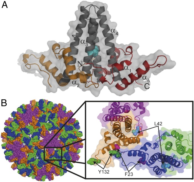
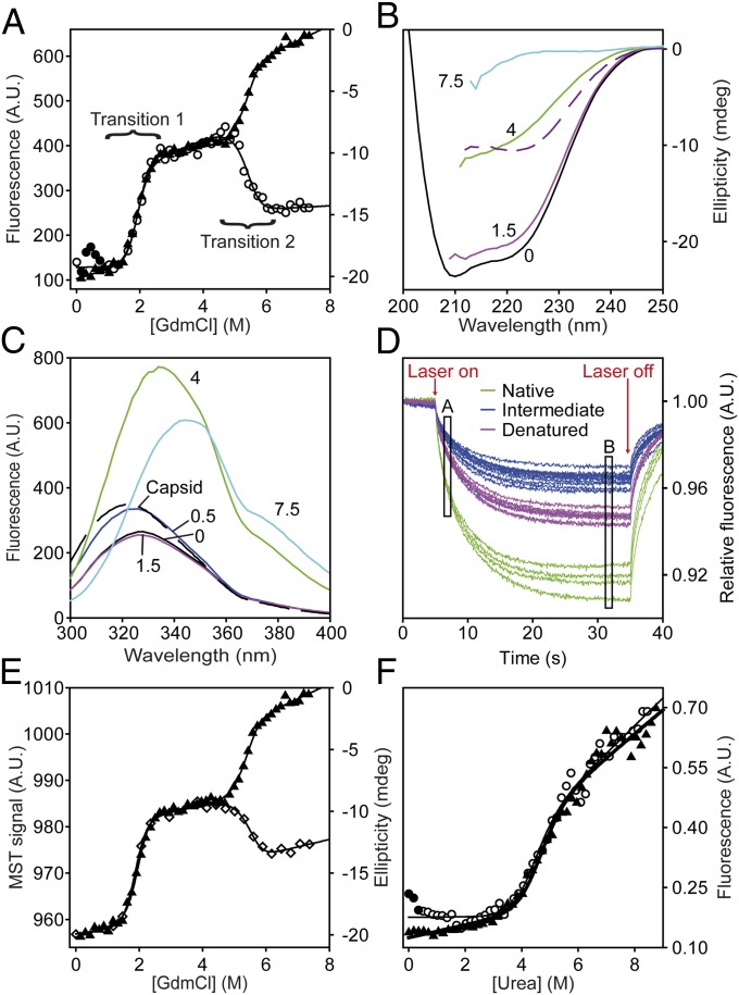
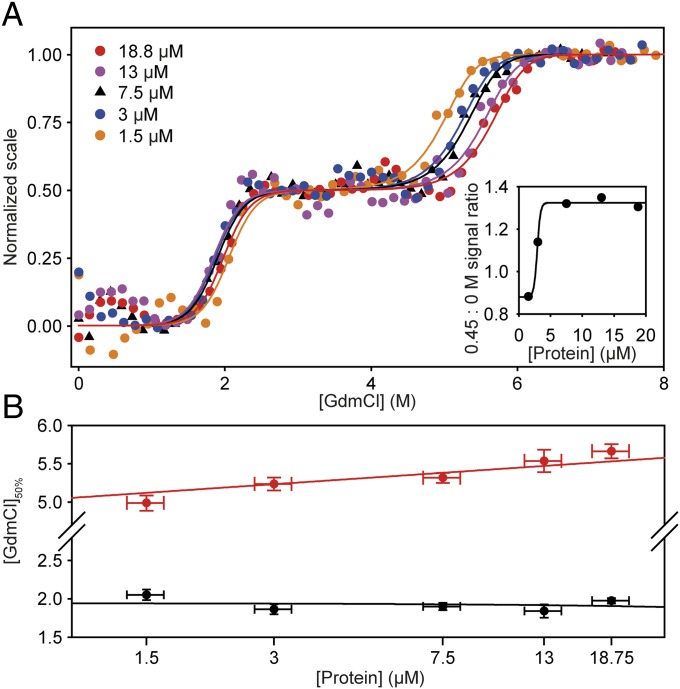
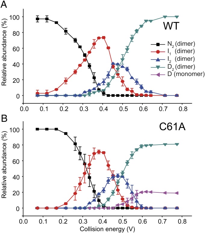
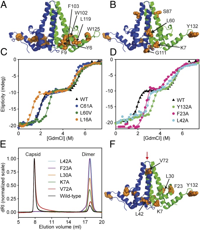
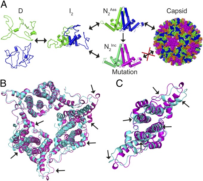
HBc, the capsid-forming "core protein" of human hepatitis B virus (HBV), is a multidomain, α-helical homodimer that aggressively forms human HBV capsids. Structural plasticity has been proposed to be important to the myriad functions HBc mediates during viral replication. Here, we report detailed thermodynamic analyses of the folding of the dimeric HBc protomer under conditions that prevented capsid formation. Central to our success was the use of ion mobility spectrometry-mass spectrometry and microscale thermophoresis, which allowed folding mechanisms to be characterized using just micrograms of protein. HBc folds in a three-state transition with a stable, dimeric, α-helical intermediate. Extensive protein engineering showed thermodynamic linkage between different structural domains. Unusual effects associated with mutating some residues suggest structural strain, arising from frustrated contacts, is present in the native dimer. We found evidence of structural gatekeepers that, when mutated, alleviated native strain and prevented (or significantly attenuated) capsid formation by tuning the population of alternative native conformations. This strain is likely an evolved feature that helps HBc access the different structures associated with its diverse essential functions. The subtle balance between native and strained contacts may provide the means to tune conformational properties of HBc by molecular interactions or mutations, thereby conferring allosteric regulation of structure and function. The ability to trap HBc conformers thermodynamically by mutation, and thereby ablate HBV capsid formation, provides proof of principle for designing antivirals that elicit similar effects.

Keywords: capsid assembly; energy landscape; protein dynamics; thermodynamic coupling.


Conflict of interest statement

The authors declare no conflict of interest.

Figures

Fig. 1.
Fig. 1.
HBc1–149 dimer structure within HBV capsids. (A) Four-helix bundle dimerization interface (black) is flanked by contact domains (orange and red). Helices are numbered, and the N and C termini of one monomer are indicated. The disulfide link between C61 of each monomer is indicated (cyan). (B) Exterior surface of a T = 4 capsid HBc1–149 (PDB ID code 1QGT) (19). Dimers around the threefold and fivefold axes are indicated in blue/green and purple/orange, respectively. (Inset) Interacting quasiequivalent HBc1–149 dimers from the fivefold (purple and orange) and threefold (blue and green) axes are shown. Hydrophobic contacts between contact domains stabilize capsids. Residues that perturb capsid formation when mutated are indicated.
Fig. 2.
Fig. 2.
Equilibrium chemical denaturation of HBc1–149. (A) GdmCl denaturation of HBc1–149 measured by far-UV CD (▲) and fluorescence emission spectroscopy (○). Solid lines are fits to an equation describing a linear three-state transition with a dimeric intermediate (SI Materials and Methods). (B) Far-UV CD spectra of HBc1–149, corresponding to the main species populated in different GdmCl solutions: 0 M (black), 1.5 M (magenta), 4 M (green), and 7.5 M (cyan). The difference spectrum (Spectrum0M − Spectrum4M) is a dashed purple line. (C) FES spectra of the species populated in GdmCl titrations (using the same colors as in B). The spectra of assembled capsids (black dashed line) and HBc1–149 in 0.5 M GdmCl (dark blue line) are similar, consistent with salt-induced capsid formation (32, 36). (D) Raw MST data for chemical denaturation of HBc1–149. Fluorescence is measured for 5 s before and after applying a 30-s heating pulse (“Laser on/Laser off”), which creates a temperature gradient of 2–3 °C (33). The MST signal is typically determined from the normalized fluorescence intensity changes between any two points (here, A and B) after the temperature gradient has been established (34). Typically, point A is selected to be a time-point shortly after the laser is switched on, whereas point B is some time after significant thermophoresis has occurred. The measured properties for N (green), I (blue), and D (purple) are shown. (E) GdmCl titrations of HBc1–149 measured by far-UV CD (▲) and MST (◇). Solid lines are best fits to a three-state transition (as in Fig. 2A). (F) Urea denaturation of HBc1–149 in the presence (◇) or absence (▲) of 1 M NaCl. Solid circles indicate species with capsid-like spectra. A.U., arbitrary units.
Fig. 3.
Fig. 3.
Effects of protein concentration on chemical denaturation of HBc1–149. (A) GdmCl-induced denaturation over a 12.5-fold concentration range of HBc1–149. For clarity, these data are shown on a normalized scale (raw data are reported in Fig. S2). Each dataset is fitted to an equation (solid lines) describing a linear three-state transition with a dimeric intermediate (Materials and Methods and SI Materials and Methods). (Inset) Ratio of native baseline signal to the signal at 0.45 M GdmCl (where GdmCl-induced capsid formation was maximal). These data define a pseudocritical concentration of ∼3 μM (monomer) (24, 36, 38). (B) Transition 1 was independent of protein concentration (black circles), but transition 2 [GdmCl]50% values (red circles) were markedly dependent on HBc1–149 concentration. These data are consistent with a change in oligomeric state of HBc1–149 during transition 2.
Fig. 4.
Fig. 4.
Gas phase unfolding profiles of HBc1–149. (A) Abundance of each state as a function of the center-of-mass collision energy (Materials and Methods and SI Materials and Methods). At low energies, N was the dominant species (black squares). As collision energy was increased, the population of N decreased and a partially unfolded state (I1) was populated (red circles). Further increases in energy induced the population of state I2 (blue triangles), which then populated the unfolded state D (green triangles) at higher energies. (B) C61A underwent a very similar unfolding process until high energies were reached; at that point, dissociation into monomeric D occurred. This illustrates gas phase dissociation of the unfolded state depends on the presence of the intermolecular disulfide link. Error bars show the SD from triplicate measurements.
Fig. 5.
Fig. 5.
Mutational analysis of HBc1–149. (A) Conserved residues that, when mutated, caused aggregation and were refractory to study colocalized to a conserved hydrophobic core between the four-helix bundle and contact domains (orange) (19). (B) Mutations that altered transition 1 stability only. (C) GdmCl titrations for representative HBc1–149 mutants: WT; C61A, the only mutant to affect transition 2 only; L16A, which destabilized both transitions; and L60V, which stabilized transition 1 only. (D) GdmCl titrations of WT HBc1–149 and capsid assembly-incompetent mutants (F23A, L42A, and Y132A). (E) Light-scattering analysis of HBc1–149 constructs that reduce (K7A, L30A, and V72A) or knock out capsid assembly (F23A and L42A) compared with WT. After dialysis into a buffer that promotes in vitro capsid formation, samples were run on an analytical Superose 6 column. HBc1–149 capsids eluted at ∼8 mL (molar mass of ∼4 × 10⁶ Da), and dimers eluted at 16 mL (molar mass of ∼3.4 × 10⁴ Da). For clarity, each capsid peak was normalized to an arbitrary value of 1 (15, 17). dRI, differential refractive index. (F) Location of residues discussed in E is shown. The binding site of an antiviral peptide is indicated with a red arrow (14, 15) (associated calorimetry data are provided in Fig. S6).
Fig. 6.
Fig. 6.
Three-state folding of HBc1–149. (A) Unfolded HBc1–149 monomers interact and form a partially structured, dimeric helical intermediate. This intermediate can thereafter fold to multiple native structures. One native conformer (HBcAss) can form capsids, whereas the other is assembly-incompetent (HBcInc). The occupancy of an alternative native structure can be tuned thermodynamically by point mutations, thereby modulating the extent of capsid formation. (B) Comparison of a previously reported crystal structure for Y132A (21) (cyan; PDB ID code 3KXS) with one determined by us under different conditions (pink; PDB ID code 4BMG). Arrows show structural variations between the dimers. These data demonstrate that native HBc1–149 dimers exhibit considerable structural variability, even within the same protein sequence. (C) Close-up of a Y132A dimer within the hexameric unit shown in B that highlights the structural differences between the same protein under different crystallization conditions.

References

    1. Onuchic JN, Wolynes PG. Theory of protein folding. Curr Opin Struct Biol. 2004;14(1):70–75. - PubMed
    1. Jackson SE. How do small single-domain proteins fold? Fold Des. 1998;3(4):R81–R91. - PubMed
    1. Fulton KF, et al. PFD: A database for the investigation of protein folding kinetics and stability. Nucleic Acids Res. 2005;33(Database issue):D279–D283. - PMC - PubMed
    1. Teague SJ. Implications of protein flexibility for drug discovery. Nat Rev Drug Discov. 2003;2(7):527–541. - PubMed
    1. World Health Organization Publication Hepatitis B vaccines: WHO position paper—Recommendations. Vaccine. 2010;28(3):589–590. - PubMed

Publication types

Substances

Associated data