. 2013 Aug;136(Pt 8):2510-26.
doi: 10.1093/brain/awt171. Epub 2013 Jul 3.

Dissecting phenotypic traits linked to human resilience to Alzheimer's pathology

Affiliations

Dissecting phenotypic traits linked to human resilience to Alzheimer's pathology

Beatriz G Perez-Nievas et al. Brain. 2013 Aug.

Abstract

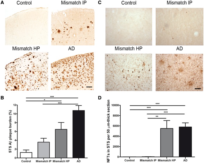
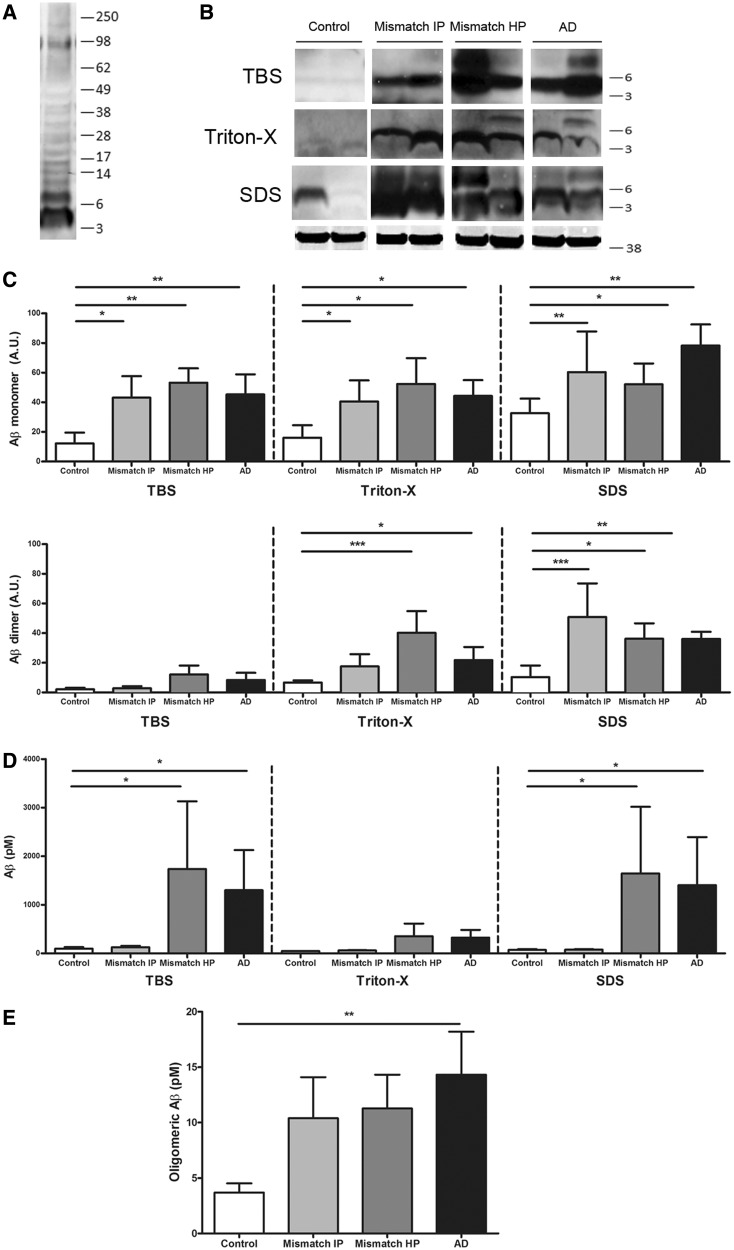
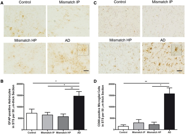
Clinico-pathological correlation studies and positron emission tomography amyloid imaging studies have shown that some individuals can tolerate substantial amounts of Alzheimer's pathology in their brains without experiencing dementia. Few details are known about the neuropathological phenotype of these unique cases that might prove relevant to understanding human resilience to Alzheimer's pathology. We conducted detailed quantitative histopathological and biochemical assessments on brains from non-demented individuals before death whose brains were free of substantial Alzheimer's pathology, non-demented individuals before death but whose post-mortem examination demonstrated significant amounts of Alzheimer's changes ('mismatches'), and demented Alzheimer's cases. Quantification of amyloid-β plaque burden, stereologically-based counts of neurofibrillary tangles, neurons and reactive glia, and morphological analyses of axons were performed in the multimodal association cortex lining the superior temporal sulcus. Levels of synaptic integrity markers, and soluble monomeric and multimeric amyloid-β and tau species were measured. Our results indicate that some individuals can accumulate equivalent loads of amyloid-β plaques and tangles to those found in demented Alzheimer's cases without experiencing dementia. Analyses revealed four main phenotypic differences among these two groups: (i) mismatches had striking preservation of neuron numbers, synaptic markers and axonal geometry compared to demented cases; (ii) demented cases had significantly higher burdens of fibrillar thioflavin-S-positive plaques and of oligomeric amyloid-β deposits reactive to conformer-specific antibody NAB61 than mismatches; (iii) strong and selective accumulation of hyperphosphorylated soluble tau multimers into the synaptic compartment was noted in demented cases compared with controls but not in mismatches; and (iv) the robust glial activation accompanying amyloid-β and tau pathologies in demented cases was remarkably reduced in mismatches. Further biochemical measurements of soluble amyloid-β species-monomers, dimers and higher molecular weight oligomers-in total brain homogenates and synaptoneurosomal preparations failed to demonstrate significant differences between mismatches and demented cases. Together, these data suggest that amyloid-β plaques and tangles do not inevitably result in neural system derangement and dementia in all individuals. We identified distinct phenotypic characteristics in the profile of brain fibrillar and soluble amyloid-β and tau accrual and in the glial response that discriminated demented and non-demented individuals with high loads of Alzheimer's pathology. Amyloid-β deposition in the form of fibrillar plaques and intimately related oligomeric amyloid-β assemblies, hyperphosphorylated soluble tau species localized in synapses, and glial activation emerged in this series as likely mediators of neurotoxicity and altered cognition, providing further insight into factors and pathways potentially involved in human susceptibility or resilience to Alzheimer's pathological changes.

Keywords: Alzheimers disease; amyloid pathology; astrocytes; microglia; resilience; tau pathology.


Figures

Figure 1
Figure 1
Amyloid-β plaque burden and number of neurofibrillary tangles in the superior temporal sulcus (STS). (A) Representative photomicrographs of 10D5 immunostained plaques in the superior temporal sulcus. (B) Demented cases with Alzheimer’s disease (AD) had a significantly higher amyloid-β plaque burden in the superior temporal sulcus when compared with intermediate probability mismatches (Mismatch IP) and controls. No significant differences were found between demented cases with Alzheimer’s disease and high probability mismatches (Mismatch HP). (C) Representative photomicrographs of PHF-1 immunostained neurofibrillary tangles in the superior temporal sulcus. (D) A significantly higher number of neurofibrillary tangles (NFTs) in the superior temporal sulcus were found in demented cases with Alzheimer’s disease when compared with intermediate probability mismatches. No significant differences were detected in the number of neurofibrillary tangles between cases with Alzheimer’s disease and high probability mismatches. Controls were essentially free of neurofibrillary tangles in this brain area. Scale bar = 150 µm; n = 8–15 per group; *P < 0.05; **P < 0.01; ***P < 0.001. One way ANOVA and post hoc Tukey test, and Kruskal-Wallis ANOVA and Dunn’s multiple comparison test, respectively.
Figure 2
Figure 2
Number of neurons, cortical thickness and synaptic markers in the superior temporal sulcus. (A) Stereologically based neuronal counts in the superior temporal sulcus (STS) showed a significant reduction by >40% in the number of neurons in demented cases with Alzheimer’s disease (AD) compared to controls. Intermediate (Mismatch IP) and high probability mismatches (Mismatch HP) showed no significant neuronal loss in this region compared with controls. (B) Superior temporal sulcus cortical thickness was significantly reduced by >20% in cases with Alzheimer’s disease but not in intermediate or high probability mismatches when compared to controls. (C) Representative image of western blot and quantification for postsynaptic marker PSD-95. Levels of PSD-95 in the superior temporal sulcus were significantly decreased in cases with Alzheimer’s disease but not in intermediate or high probability mismatches when compared with controls. (D) Representative image of western blot and quantification for presynaptic marker synaptophysin. Levels of synaptophysin in the superior temporal sulcus were significantly decreased in cases with Alzheimer’s disease but not in high probability mismatches when compared with controls. A significantly higher level of synaptophysin was detected in the superior temporal sulcus in intermediate probability mismatches compared with controls. GAPDH was used as loading control. n = 5–8 per group; *P < 0.05; **P < 0.01; ***P < 0.001. One way ANOVA and post hoc Tukey test.
Figure 3
Figure 3
Neurite trajectory analysis. (A) Representative photomicrographs of SMI-312 immunostained axons (red) and thioflavin-S labelled amyloid plaques (green) in the superior temporal sulcus. Arrowhead shows example of curved axonal segment. Arrows show examples of neuritic dystrophies. Scale bar = 50 µm. (B) Demented cases with Alzheimer’s disease (AD) had significantly curvier axons close (<50 µm) and far (>50 µm) from plaque than high and intermediate probability mismatches (Mismatch IP and HP) and controls free of Alzheimer’s disease pathology (control); ***P < 0.001. Mann Whitney test (‘close to plaque’ analysis) and Kruskal-Wallis ANOVA and Dunn’s multiple comparison test (‘far from plaque’ analysis). (C) The number of dystrophic neurites was significantly higher in cases with Alzheimer’s disease compared to high probability mismatches. n = 172–290 axonal segments ‘close to plaque’ and 238–456 axonal segments ‘far from plaque’; ***P < 0.001 Mann Whitney test.
Figure 4
Figure 4
Superior temporal sulcus thioflavin-S-positive amyloid plaque burden. (A) Demented cases with Alzheimer’s disease (AD) had a significantly higher thioflavin-S-positive plaque burden in the superior temporal sulcus when compared with intermediate and high probability mismatches (Mismatch HP and IP). n = 8–10 per group; *P < 0.05, **P < 0.01. One way ANOVA and post hoc Tukey test. (B) Representative photomicrographs of thioflavin-S stained plaques in the superior temporal sulcus. Scale bar = 100 µm.
Figure 5
Figure 5
Biochemical analyses of soluble amyloid-β species in serially extracted samples (Tris-buffered saline, 1% Triton™ X-100 and 2% SDS) containing the superior temporal sulcus. (A) Representative image of western blot showing bands recognized by 6E10 plus 82E1 antibodies. (B) Representative image of western blot for amyloid-β monomers (4 kDa) and dimers (8 kDa); GAPDH (38 kDa) is used as loading control. (C) Levels of amyloid-β monomers and dimers by western blot did not significantly differ between demented cases with Alzheimer’s disease (AD) and intermediate or high probability mismatches (Mismatch IP and HP) but were significantly higher in all three groups compared to controls (monomers in all three fractions and dimers in Triton™ X-100 and SDS fractions). (D) Two-sandwich ELISA using BNT77/BA27 antibody pair did not detect significant differences in soluble amyloid-β levels (monomers and oligomers) between cases with Alzheimer’s disease and high probability mismatches; in both groups amyloid-β levels were significantly higher in Tris-buffered saline and SDS fractions than in intermediate probability mismatches and controls. (E) Same site sandwich ELISA (82E1), specific for dimers and higher molecular weight amyloid-βspecies, did not detect significant differences between Alzheimer’s disease and high probability mismatches; Alzheimer’s disease had significantly higher levels of oligomeric amyloid-β than controls. n = 7–11 per group; *P < 0.05; **P < 0.01; ***P < 0.001; Kruskal-Wallis ANOVA and Dunn’s multiple comparison test.
Figure 6
Figure 6
Biochemical analysis of amyloid-β monomers and dimers in synaptoneurosomes and superior temporal sulcus (STS) NAB61 oligomeric amyloid-β burden. (A) Purity control of synaptoneurosome preparations. Western blot shows an enrichment in pre- and postsynaptic markers (synaptophysin and PSD-95) in synaptoneurosome compared with cytosolic fractions. (B) Representative image of western blot for amyloid-β monomers (4 kDa) and dimers (8 kDa) in synaptoneurosome fractions (6E10 plus 82E1 antibodies); actin (42 kDa) was used as loading control. (C) Levels of amyloid-β monomers and dimers in the synaptic compartment did not significantly differ between demented cases with Alzheimer’s disease (AD) and high probability mismatches (Mismatch HP); in both groups they were significantly higher than in controls. n = 6–8 per group. (D) Representative photomicrographs of NAB61 immunolabelled oligomeric amyloid-β (Aβ) deposits in the superior temporal sulcus. (E) Superior temporal sulcus NAB61 oligomeric amyloid-β burden was significantly higher in cases with Alzheimer’s disease than in high probability mismatches and controls. n = 6–14 per group; *P < 0.05; **P < 0.01; ***P < 0.001; Kruskal-Wallis ANOVA and Dunn’s multiple comparison test. Scale bar = 100 µm. C = cytosolic fraction; S = synaptoneurosome fraction.
Figure 7
Figure 7
Biochemical analysis of total tau and hyperphosphorylated monomeric and multimeric tau in synaptoneurosomes. (A) Representative images of cytosolic (C) and synaptoneurosome (S) fractions on western blot for total human tau (H7 antibody) and (C) hyperphosporylated tau (PHF-1 antibody), showing strong accumulation of tau monomers and multimers at synapses in demented Alzheimer’s disease (AD) cases. (B) Quantitative analyses for (C) cytosolic and (S) synaptic total tau and (D) hyperphosphorylated tau showed selective and significantly higher accumulation of monomeric tau (64 kDa) and multimeric tau (75-250 kDa) species in synapses in cases with Alzheimer’s disease, but not in intermediate and high probability mismatches (Mismatch IP and HP) when compared with controls. Total levels of tau monomers in the cytosol did not significantly differ between groups. Actin (42 kDa) was used as loading control. n = 6–9 per group; *P < 0.05; **P < 0.01; Kruskal-Wallis ANOVA and Dunn’s multiple comparison test.
Figure 8
Figure 8
Number of GFAP-positive astrocytes in the superior temporal sulcus and CD68-positive microglia. (A) Representative photomicrographs of GFAP-positive astrocytes. (B) Stereological counts of astrocytes in the superior temporal sulcus (STS) on sections immunostained with GFAP showed a significant increase in the amount of GFAP-positive astrocytes in demented cases with Alzheimer’s disease but not in intermediate probability (Mismatch IP) or high probability mismatches (Mismatch HP) in comparison with controls. (C) Representative photomicrographs of CD68-positive microglia cells in the superior temporal sulcus. Haematoxylin was used as a counterstain. (D) Stereological counts of microglia in the superior temporal sulcus on sections immunostained with CD68 showed a significant increase in the amount of CD68-positive microglial cells in demented cases with Alzheimer’s disease, but not in intermediate or high probability mismatches in comparison with controls. n = 8–14 per group; *P < 0.05; **P < 0.01. One way ANOVA and post hoc Tukey test, and Kruskal-Wallis ANOVA and Dunn’s multiple comparison test, respectively. Scale bar = 100 µm.

References

    1. Aizenstein HJ, Nebes RD, Saxton JA, Price JC, Mathis CA, Tsopelas ND, et al. Frequent amyloid deposition without significant cognitive impairment among the elderly. Arch Neurol. 2008;65:1509–17. - PMC - PubMed
    1. Arias E. United States life tables, 2008. Natl Vital Stat Rep. 2012;61:3. - PubMed
    1. Arnold SE, Louneva N, Cao K, Wang LS, Han LY, Wolk DA, et al. Cellular, synaptic, and biochemical features of resilient cognition in Alzheimer's disease. Neurobiol Aging. 2013;34:157–68. - PMC - PubMed
    1. Arriagada PV, Marzloff K, Hyman BT. Distribution of Alzheimer-type pathologic changes in nondemented elderly individuals matches the pattern in Alzheimer's disease. Neurology. 1992;42:1681–88. - PubMed
    1. Benilova I, Karran E, De SB. The toxic Abeta oligomer and Alzheimer's disease: an emperor in need of clothes. Nat Neurosci. 2012;15:349–57. - PubMed

Publication types

MeSH terms