. 2013 Oct;87(19):10524-38.
doi: 10.1128/JVI.00832-13. Epub 2013 Jul 3.

Mutational analysis of the binding pockets of the diketo acid inhibitor L-742,001 in the influenza virus PA endonuclease

Affiliations

Mutational analysis of the binding pockets of the diketo acid inhibitor L-742,001 in the influenza virus PA endonuclease

Annelies Stevaert et al. J Virol. 2013 Oct.

Abstract

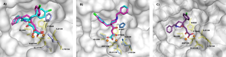
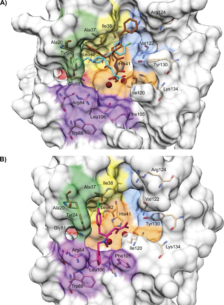
The influenza virus PA endonuclease, which cleaves capped host pre-mRNAs to initiate synthesis of viral mRNA, is a prime target for antiviral therapy. The diketo acid compound L-742,001 was previously identified as a potent inhibitor of the influenza virus endonuclease reaction, but information on its precise binding mode to PA or potential resistance profile is limited. Computer-assisted docking of L-742,001 into the crystal structure of inhibitor-free N-terminal PA (PA-Nter) indicated a binding orientation distinct from that seen in a recent crystallographic study with L-742,001-bound PA-Nter (R. M. DuBois et al., PLoS Pathog. 8:e1002830, 2012). A comprehensive mutational analysis was performed to determine which amino acid changes within the catalytic center of PA or its surrounding hydrophobic pockets alter the antiviral sensitivity to L-742,001 in cell culture. Marked (up to 20-fold) resistance to L-742,001 was observed for the H41A, I120T, and G81F/V/T mutant forms of PA. Two- to 3-fold resistance was seen for the T20A, L42T, and V122T mutants, and the R124Q and Y130A mutants were 3-fold more sensitive to L-742,001. Several mutations situated at noncatalytic sites in PA had no or only marginal impact on the enzymatic functionality of viral ribonucleoprotein complexes reconstituted in cell culture, consistent with the less conserved nature of these PA residues. Our data provide relevant insights into the binding mode of L-742,001 in the PA endonuclease active site. In addition, we predict some potential resistance sites that should be taken into account during optimization of PA endonuclease inhibitors toward tight binding in any of the hydrophobic pockets surrounding the catalytic center of the enzyme.


Figures

Fig 1
Fig 1
Chemical structure and protonation states of L-742,001.
Fig 2
Fig 2
Binding models of L-742,001 predicted by docking in inhibitor-free PA-Nter. (A and B) Clusterized binding poses obtained by docking L 742,001 into the published (14) structure of inhibitor-free PA Nter (PDB entry 2W69), using a rigid receptor (cyan) or flexible protein (magenta) approach and representing the most favorable binding energies (A) or most diffuse population of conformers (B). (C) Representative pose of L-742,001 when modeled in its zwitterionic form. Metal-binding and catalytic residues are shown in yellow. In case these residues are flexible, the alternative positions are shown in violet (A) or in pale blue (B). The two Mn2+ ions in the catalytic center are shown in orange.
Fig 3
Fig 3
View of the binding pockets for L-742,001 in PA-Nter, as predicted by docking versus X-ray crystallography. (A) Graphical representation of the hypothetical disposition of L-742,001 in PA-Nter. An analogous disposition is seen for the two conformers predicted by docking, i.e., the one representing the most favorable binding energies (in cyan) and that representing the most diffuse population of conformers (in maroon). (B) The structure in pink represents the position of L-742,001 in the crystal structure of the L-742,001-PA-Nter complex (30). The two protein structures are shown in the same orientation after structural alignment using the DALI server (74). The following colors indicate different regions in PA-Nter: orange, metal binding and catalytic residues (His41, Glu80, Asp108, Glu119, Ile120, and Lys134); yellow, pocket P1 (Ala37, Ile38, Leu42, and Lys34); blue, pocket P2 (Thr40, Val122, Arg124, Tyr130, and Phe150); purple, pocket P3 (Arg84, Trp88, Phe105, and Leu106); red, pocket P4 (Leu16 and Gly81); and green, pocket P5 (Ala20, Tyr24, and Glu26). The residues included in our mutational analysis are shown in stick presentation and are labeled. The two metal ions are colored dark red.
Fig 4
Fig 4
Dose-response curves for virus yield reduction in MDCK cells at 24 h p.i. (A) and inhibition of vRNP activity in HEK293T cells (B). Data are means ± standard errors of the means (SEM) from three experiments.
Fig 5
Fig 5
Inhibitory effect of L-742,001 on viral RNA synthesis is situated after viral entry. Light gray bars indicate that the test compound L-742,001 (50 μM), EGCG (50 μM), ribavirin (20 μM), or SA-19 (20 μM) was added to MDCK cells, and after 30 min incubation at 35°C, influenza virus A/PR/8/34 was added. Dark gray bars indicate that virus was added first and allowed to enter during 1 h of incubation, after which the compounds were added at the same concentrations as those described for the light gray bars. In both conditions, total cellular RNA was extracted at 10 h p.i. VC, untreated virus control. The number of vRNA copies was quantified by two-step real-time RT-PCR. On the y axis, the fold increase in vRNA copies is shown relative to the viral copy number added at time zero. Ribavirin and L-742,001 remain fully effective when added after virus entry. In contrast, the reported entry inhibitor SA-19 (33) as well as EGCG are inactive when added at 1 h p.i. Data shown are the means ± SEM from two independent tests.
Fig 6
Fig 6
Polymerase activity of vRNP complexes carrying mutant forms of PA. HEK293T cells were cotransfected with the four vRNP-reconstituting plasmids (including the WT or mutant form of PA) and the reporter plasmid. On the horizontal axis, the vRNP-driven firefly luciferase signal is shown relative to the WT (top axis) or in absolute values (bottom axis). Data shown are the means ± SEM from at least three independent tests.
Fig 7
Fig 7
Hypothetical interaction between L-742,001 and residue Gly81 in PA-Nter. (A to C) Predicted disposition of L-742,001 into pocket P4. In the best-scoring binding poses predicted by our docking, the benzyl group of L-742,001 is directed toward pocket P4, which contains residue Gly81 (red in all panels) inside the tunnel (A and B). Panel C shows that the benzyl group of L-742,001 may establish a favorable hydrophobic interaction with Gly81 within a distance of ∼3 Å.

References

    1. Smith GJ, Vijaykrishna D, Bahl J, Lycett SJ, Worobey M, Pybus OG, Ma SK, Cheung CL, Raghwani J, Bhatt S, Peiris JS, Guan Y, Rambaut A. 2009. Origins and evolutionary genomics of the 2009 swine-origin H1N1 influenza A epidemic. Nature 459:1122–1125 - PubMed
    1. Simonsen L, Taylor RJ, Viboud C, Miller MA, Jackson LA. 2007. Mortality benefits of influenza vaccination in elderly people: an ongoing controversy. Lancet Infect. Dis. 7:658–666 - PubMed
    1. Deyde VM, Xu X, Bright RA, Shaw M, Smith CB, Zhang Y, Shu Y, Gubareva LV, Cox NJ, Klimov AI. 2007. Surveillance of resistance to adamantanes among influenza A(H3N2) and A(H1N1) viruses isolated worldwide. J. Infect. Dis. 196:249–257 - PubMed
    1. Moscona A. 2009. Global transmission of oseltamivir-resistant influenza. N. Engl. J. Med. 360:953–956 - PubMed
    1. Hayden F. 2009. Developing new antiviral agents for influenza treatment: what does the future hold? Clin. Infect. Dis. 48(Suppl 1):S3–S13 - PubMed

Publication types

MeSH terms