. 2013 Sep;24(17):2739-52.
doi: 10.1091/mbc.E12-10-0771. Epub 2013 Jul 3.

p53 SUMOylation promotes its nuclear export by facilitating its release from the nuclear export receptor CRM1

Affiliations

p53 SUMOylation promotes its nuclear export by facilitating its release from the nuclear export receptor CRM1

Aleixo Santiago et al. Mol Biol Cell. 2013 Sep.

Abstract

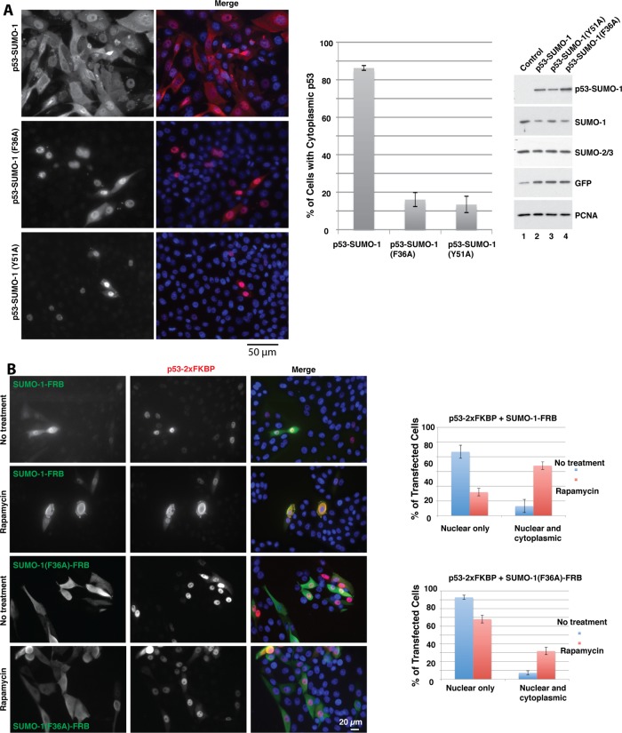
Chromosomal region maintenance 1 (CRM1) mediates p53 nuclear export. Although p53 SUMOylation promotes its nuclear export, the underlying mechanism is unclear. Here we show that tethering of a small, ubiquitin-like modifier (SUMO) moiety to p53 markedly increases its cytoplasmic localization. SUMO attachment to p53 does not affect its oligomerization, suggesting that subunit dissociation required for exposing p53's nuclear export signal (NES) is unnecessary for p53 nuclear export. Surprisingly, SUMO-mediated p53 nuclear export depends on the SUMO-interacting motif (SIM)-binding pocket of SUMO-1. The CRM1 C-terminal domain lacking the NES-binding groove interacts with tetrameric p53, and the proper folding of the p53 core domain, rather than the presence of the N- or C-terminal tails, appears to be important for p53-CRM1 interaction. The CRM1 Huntington, EF3, a subunit of PP2A, and TOR1 9 (HEAT9) loop, which regulates GTP-binding nuclear protein Ran binding and cargo release, contains a prototypical SIM. Remarkably, disruption of this SIM in conjunction with a mutated SIM-binding groove of SUMO-1 markedly enhances the binding of CRM1 to p53-SUMO-1 and their accumulation in the nuclear pore complexes (NPCs), as well as their persistent association in the cytoplasm. We propose that SUMOylation of a CRM1 cargo such as p53 at the NPCs unlocks the HEAT9 loop of CRM1 to facilitate the disassembly of the transporting complex and cargo release to the cytoplasm.


Figures

FIGURE 1:
FIGURE 1:
Rapamycin-mediated heterodimerization of p53 and SUMO inhibits p53 transactivation function. (A) Schematic diagram of p53-2xFKBP and fusion of SUMO-1 or -3 with FRB and their heterodimerization mediated by rapamycin. GFP-SUMO-3 served as a control. (B) Tethering SUMO to p53 inhibited its transactivation function. H1299 cells were transfected with a firefly luciferase reporter under the control of the p21 promoter along with a control plasmid or various combinations of the indicated DNA constructs. The transfected cells were untreated (white bars) or treated with 0.1 μM of rapamycin at 6 h after transfection (black bars). Cells were lysed for dual luciferase assay 24 h after transfection. Statistical significance of pairwise comparisons was evaluated with Student's t test; down-regulation of reporter activity by heterodimerization of p53-2xFKBP and SUMO-1-FRB is significantly different from that by the control (p < 0.05). (C) p53-SUMO-3 fusion represses endogenous p21 expression. H1299 cells were stably transduced with lentiviral vectors for tetracycline-inducible expression of wt p53 or p53-SUMO-3 fusion. The vehicle (dimethyl sulfoxide) or tetracycline was added to the parental H1299 cells and wt p53- and p53-SUMO-3–expressing cells as indicated. The cells were then exposed to vehicle or 10 μM etoposide for 18 h. The cells were lysed for Western blotting analysis with antibodies against the indicated proteins. PCNA was used as a loading control.
FIGURE 2:
FIGURE 2:
Rapamycin-mediated heterodimerization of p53 and SUMO promotes p53 nuclear export. (A) Saos-2 cells were transfected with the indicated DNA constructs. The transfected cells were untreated or treated with 0.1 μM rapamycin at 6 h after transfection. Cells were fixed for immunofluorescence microscopy 24 h after transfection. Representative images of transfected cells. (B) Quantification of p53 subcellular localization. Cells from at least 10 random microscopic fields that generally contained >200 transfected cells were examined for subcellular distribution of p53. Cells with predominant nuclear localization pattern of p53 with no visible cytoplasmic presence were counted as cells with nuclear p53, whereas those with predominant or clearly visible cytoplasmic distribution of p53 were counted as cells with cytoplasmic p53. Average percentage values of p53 subcellular distribution along with SDs.
FIGURE 3:
FIGURE 3:
The SIM-binding groove of SUMO is required for nuclear exit of SUMO-modified p53. Saos-2 cells were transfected and processed for immunofluorescence microscopy as in Figure 2. Quantification of the subcellular distribution of p53 in transfected cells was determined as in Figure 2. (A) wt SUMO-1 or mutants with a mutation in the SIM-binding groove (F36A and Y51A) of SUMO-1 were directly fused to the C-terminus of p53. These fusion constructs were expressed in Saos-2 cells, and their intracellular distributions were examined. Representative images of cells expressing these constructs. Middle, plots depicting percentage distributions of these fusions. Western blotting was done to determine the expression levels of these p53-SUMO-1 fusion constructs in the transfected Saos-2 cells using an anti-p53 antibody (DO-1) and their effects on endogenous SUMO (right). GFP expression was used as a transfection control, and the PCNA level served as a loading control. (B) Representative images of cells expressing p53-2xFKBP along with SUMO-1-FRB or SUMO-1(F36A)-FRB in the absence or presence of rapamycin. Quantifications of p53 localizations (right).
FIGURE 4:
FIGURE 4:
Tethering SUMO-3 to the C-terminus of p53 promoted its nuclear export. (A) Saos-2 cells were transfected with p53-SUMO-3 fusion or cotransfected with p53-2xFKBP or GFP-SUMO-3 along with FRB or SUMO-3-FRB constructs. One set of the transfected cells was exposed to rapamycin at 0.1 μM to induce heterodimerization 6 h after transfection. Immunofluorescence microscopy was done as in Figure 2. (B) Quantification of subcellular distributions of p53 in transfected cells as in Figure 2.
FIGURE 5:
FIGURE 5:
Effect of rapamycin-induced attachment of ubiquitin to p53 on its intracellular localization. Saos-2 cells were transfected with the indicated DNA constructs (p53-2xFKBP along with K-less-Ub(G76A)-FRB-HA or Ub(G76A)-FRB-HA). Cells were untreated or treated with rapamycin (0.1 μM) 6 h after transfection and fixed 24 h after transfection. Cells were stained with rabbit anti-HA and mouse anti-p53 (DO-1) antibodies. Goat anti-rabbit immunoglobulin G (IgG)–fluorescein and goat anti-mouse IgG-rhodamine conjugates were used as the secondary antibodies. The nuclei were stained with 4′,6-diamidino-2-phenylindole.
FIGURE 6:
FIGURE 6:
Coimmunoprecipitation of p53 and SUMO-modified p53. (A) Saos-2 cells were transfected with the indicated DNA constructs. The transfected cells were untreated or exposed to 0.1 μM rapamycin. At 24 h after transfection, cells were lysed and the extracts of the transfected cells were subjected to IP as described in Materials and Methods, and different forms of coprecipitated p53 were detected in Western blotting analysis. (B) Transfections with the specified DNA constructs, rapamycin treatment, and IP experiments were done as in A.
FIGURE 7:
FIGURE 7:
Effects of shRNA-mediated knockdown of CRM1 on SUMO-stimulated p53 nuclear export. (A) Saos-2 cells were cotransfected with the p53-SUMO-1 construct along with an indicated shRNA expression vector. Cells were fixed and subjected to immunofluorescence microscopy. Representative images of cells transfected with the indicated constructs. (B) Subcellular localization of p53 quantified as in Figure 2. Error bars are SEM. The p values of pairwise comparisons were calculated with Student's t tests. (C) Relative CRM1 mRNA levels in Saos-2 cells transfected with the indicated shRNA vectors were determined by quantitative real-time PCR. The results are average values of biological triplicate samples along with SEM. The p values were assessed with Student's t tests.
FIGURE 8:
FIGURE 8:
Tethering SUMO-1 to monomeric p53 failed to promote its nuclear export. Saos-2 cells were transfected with the indicated p53 constructs along with the SUMO-1-FRB-HA construct. p53 del11-27 contains a deletion of amino acid residues 11–27. The L348A/L350P double mutation within the tetramerization domain abolishes p53 oligomerization. The p53 del11-27/L348A/L350P construct carries both del11-27 deletion and the L348A/L350P double mutation. The transfected cells were untreated or exposed to rapamycin and then processed for immunostaining as in Figures 2 and 3.
FIGURE 9:
FIGURE 9:
Interaction between p53 and CRM1 as determined in yeast two-hybrid assays. (A) The indicated CRM1 constructs fused to Gal4-BD were introduced to yeast cells along with various p53 constructs fused to Gal4-AD. The transformants of each hybrid combination were restreaked in duplicate on plates with SD medium lacking lysine and histidine. Yeast two-hybrid assays of the Gal4-BD-Mdm2 construct with Gal4-AD-p53 hybrid were done as a positive control (sector 1). (B) Yeast cells were transformed with the indicated combinations of Gal4-AD-p53 and Gal4-BD-CRM1 (aa 571–1071) hybrids. The resulting transformants were grown overnight in the permissive medium (SD lacking lysine, leucine, and uracil) at 30°C. The overnight cultures were serially diluted and plated on the permissive medium (left) and nonpermissive medium (SD lacking histidine and lysine; right). The transformation with Gal4-BD-p300 and Gal4-AD-p53 was used as a positive control (first row). Representative results of at least two distinct colonies with similar growth phenotype for each combination. (C) Sequence alignments of the HEAT9 loop of CRM1 with known SIMs from the indicated human proteins (left) and among the corresponding HEAT9 loop sequences of various CRM1 orthologues from the indicated species (right). The hydrophobic core of the SIMs is boxed. The numbers in each sequence refer to beginning and ending residues in each protein. (D) Full-length CRM1 or a fragment encompassing aa 375–463 (HEAT9 loop) fused to Gal4-BD was tested for interaction with indicated SUMO constructs fused to Gal4-AD. Yeast two-hybrid assays were done as in A.
FIGURE 10:
FIGURE 10:
Effects of p53 SUMO modification on its interaction with CRM1. (A) Saos-2 cells were transfected with the indicated DNA constructs. The extracts of the transfected cells were subjected to IP as in Figure 6. Coprecipitated GFP-CRM1 was detected with an anti-GFP antibody. (B) The indicated DNA constructs were transfected into Saos-2 cells. IP assays and Western blotting were done as in A. (C) Saos-2 cells were transfected with RFP-p53 alone or together with GFP-CRM1 or GFP-CRM1 V430K as indicated. Cells were fixed for fluorescence microscopy 24 h after transfection as in Figure 2. Representative images are shown. Quanti­fications of subcellular distribution of p53 were done as in Figure 2 (bottom).
FIGURE 11:
FIGURE 11:
Effect of mutations in the SIM-binding groove of the p53-SUMO-1 fusion and the HEAT9 loop of CRM1 on their localization at NPCs. (A) The GFP-CRM1 (full-length) construct was cotransfected with an indicated p53-SUMO-1 fusion construct to Saos-2 cells. The transfected cells were fixed for immunofluorescence microscopy as in Figure 2. (B) The GFP-CRM1 (full-length) V430K mutant was cotransfected with an indicated p53-SUMO-1 fusion construct into Saos-2 cells. The transfected cells were fixed for immunofluorescence microscopy as described. (C) The p53-SUMO-1 fusion construct was coexpressed with the GFP-CRM1 V430K mutant in Saos-2 cells. The transfected cells were fixed 24 h after transfection and stained with antibodies to p53 and Nup153. p53, CRM1, and Nup153 were detected in the blue, green and red channels, respectively. Colocalization of CRM1, p53-SUMO-1, and Nup153 at the NPCs, as well as that of CRM1 and p53-SUMO-1 in the cytoplasm, is denoted with white arrows. Bottom, images of NPC colocalization of p53-SUMO-1, CRM1 V430K mutant, and Nup153 shown at a higher magnification, corresponding to the boxed area at the top. Lack of colocalization of CRM1 with the bright spots of p53-SUMO-1 in the nucleoplasm is indicated with a yellow arrow. (D) A model explaining a potential regulatory role for the cargo SUMOylation in cargo release and the disassembly of a CRM1 export complex. p53 (tetramer) is shown as the cargo (yellow oval). The HEAT9 loop is depicted as a hairpin in red. N, N-terminus; C, C-terminus; S1, SUMO-1. See the text for details.

Similar articles

Cited by

References

    1. Askjaer P, Jensen TH, Nilsson J, Englmeier L, Kjems J. The specificity of the CRM1-Rev nuclear export signal interaction is mediated by RanGTP. J Biol Chem. 1998;273:33414–33422. - PubMed
    1. Bischoff FR, Klebe C, Kretschmer J, Wittinghofer A, Ponstingl H. RanGAP1 induces GTPase activity of nuclear Ras-related Ran. Proc Natl Acad Sci USA. 1994;91:2587–2591. - PMC - PubMed
    1. Carter S, Bischof O, Dejean A, Vousden KH. C-terminal modifications regulate MDM2 dissociation and nuclear export of p53. Nat Cell Biol. 2007;9:428–435. - PubMed
    1. Carter S, Vousden KH. p53-Ubl fusions as models of ubiquitination, sumoylation and neddylation of p53. Cell Cycle. 2008;7:2519–2528. - PubMed
    1. Chang CC, et al. Structural and functional roles of Daxx SIM phosphorylation in SUMO paralog-selective binding and apoptosis modulation. Mol Cell. 2011;42:62–74. - PubMed

Publication types

MeSH terms

LinkOut - more resources