. 2013 Jul 3;5(192):192ra85.
doi: 10.1126/scitranslmed.3006055.

Bactericidal antibiotics induce mitochondrial dysfunction and oxidative damage in Mammalian cells

Affiliations

Bactericidal antibiotics induce mitochondrial dysfunction and oxidative damage in Mammalian cells

Sameer Kalghatgi et al. Sci Transl Med. .

Abstract

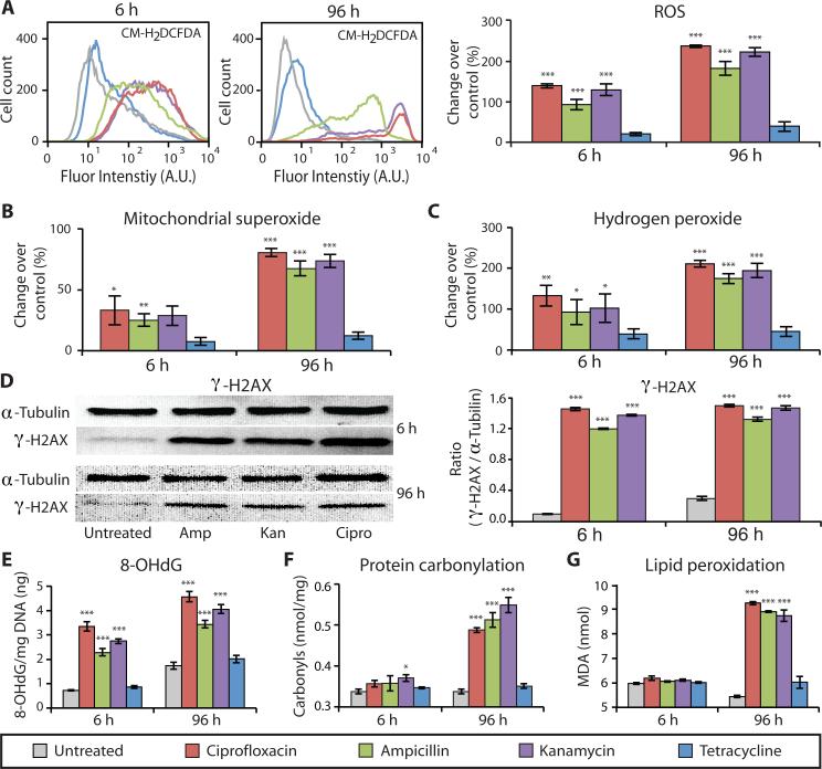
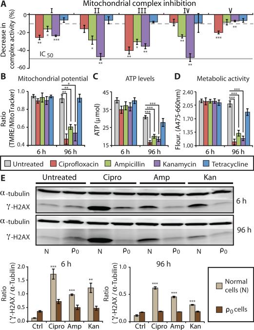
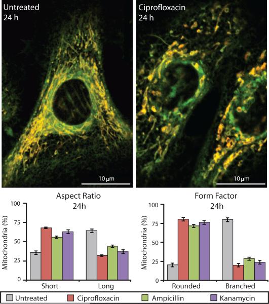
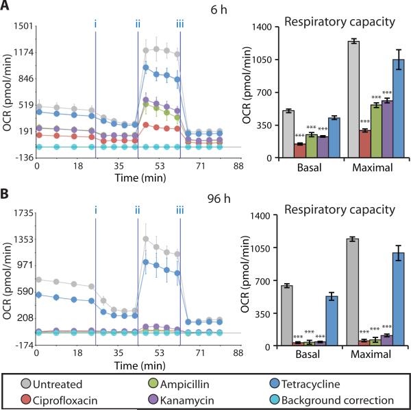
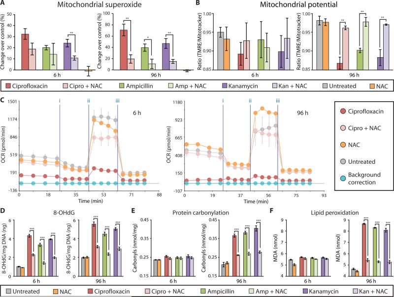
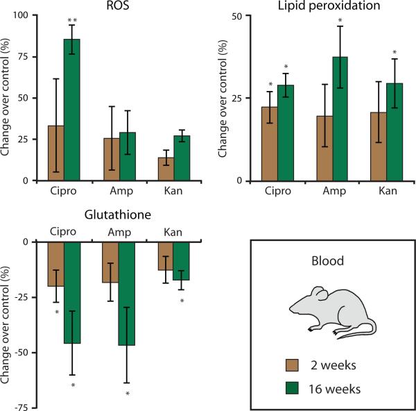
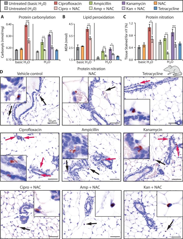
Prolonged antibiotic treatment can lead to detrimental side effects in patients, including ototoxicity, nephrotoxicity, and tendinopathy, yet the mechanisms underlying the effects of antibiotics in mammalian systems remain unclear. It has been suggested that bactericidal antibiotics induce the formation of toxic reactive oxygen species (ROS) in bacteria. We show that clinically relevant doses of bactericidal antibiotics-quinolones, aminoglycosides, and β-lactams-cause mitochondrial dysfunction and ROS overproduction in mammalian cells. We demonstrate that these bactericidal antibiotic-induced effects lead to oxidative damage to DNA, proteins, and membrane lipids. Mice treated with bactericidal antibiotics exhibited elevated oxidative stress markers in the blood, oxidative tissue damage, and up-regulated expression of key genes involved in antioxidant defense mechanisms, which points to the potential physiological relevance of these antibiotic effects. The deleterious effects of bactericidal antibiotics were alleviated in cell culture and in mice by the administration of the antioxidant N-acetyl-l-cysteine or prevented by preferential use of bacteriostatic antibiotics. This work highlights the role of antibiotics in the production of oxidative tissue damage in mammalian cells and presents strategies to mitigate or prevent the resulting damage, with the goal of improving the safety of antibiotic treatment in people.


Figures

Figure 1
Figure 1. Bactericidal antibiotics cause oxidative damage to mammalian cells
ROS and oxidative damage were measured in human mammary epithelial cells (MCF10A) after 6 and 96 h of treatment with bactericidal [ciprofloxacin (10 μg/ml), ampicillin (20 μg/ml), kanamycin (25 μg/ml)] or bacteriostatic [tetracycline (10 μg/ml)] antibiotics, compared to untreated cells. (A) ROS was quantified using CM-H2DCFDA by flow cytometry (histograms on left) and a microplate spectrophotometer (bar graphs on right). (B) Mitochondrial superoxide was measured using MitoSox Red. (C) Hydrogen peroxide release was quantified by measuring Amplex Red fluorescence. (D) Antibiotic-induced DNA damage was evaluated by Western blot analysis to measure the abundance of the phosphorylated histone protein, γ-H2AX, compared to α-tubulin serving as the loading control (quantification in the bar graph). (E) Additionally, oxidative DNA damage was assessed by measuring the abundance of 8-hydroxy-2‘-deoxyguanosine (8-OHdG). (F) Oxidative protein damage, protein carbonylation, was detected using an enzyme-linked immunosorbent assay (ELISA). (G) Lipid peroxidation was evaluated by measuring malondialdehyde (MDA). All bar graphs display means ± S.E.M. (n ≥ 3). Comparisons between treatments and untreated controls were made using a Student's t test (*p < 0.5, **p < 0.01, ***p < 0.001).
Figure 2
Figure 2. Bactericidal antibiotics induce mitochondrial dysfunction
(A) The effects of bactericidal [ciprofloxacin (10 μg/ml), ampicillin (20 μg/ml), kanamycin (25 μg/ml)] or bacteriostatic [tetracycline, (10 μg/ml)] antibiotics on the function of individual, isolated ETC protein complexes was measured. Bars represent the change in activity of individual antibiotic-treated complexes compared to untreated complexes. The dashed grey line represents the maximum inhibition (10%) seen across several, independent negative controls. The solid grey line represents the IC50 of positive control drugs shown to inhibit specific target complexes. (See fig. S7 for all positive and negative control results.) (B) Mitochondrial membrane potential was quantified using TMRE and MitoTracker Green. (C) ATP levels were measured using a luciferin/luciferase assay. (D) Metabolic activity was measured using a colorimetric tetrazolium dye (XTT). (E) DNA damage in normal (N) and mtDNA-depleted (ρ0) MCF10A cells was measured by Western blot to evaluate the ratio of γ-H2AX compared to the loading control, α-tubulin. Quantification is shown below the blots. All bar graphs display means ± S.E.M. (n ≥ 3). Comparisons between treatments and untreated controls were made using a Student's t test (*p < 0.5, **p < 0.01, ***p < 0.001).
Figure 3
Figure 3. Mitochondria in bactericidal antibiotic–treated cells show an abnormal, profission state
Mitochondrial morphology was measured in primary human mammary epithelial cells using TMRE and MitoTracker Green. Untreated samples (left image) show mitochondria with normal morphology, which are long and highly branched. Ciprofloxacin-treated cells (10 μg/ml) (right image) had abnormally short and truncated mitochondria. The percentages of mitochondria with a short versus long aspect ratio and rounded versus branched form factor were measured for untreated and bactericidal antibiotic–treated cells. Data are means ± S.E.M. (n ≥ 5).
Figure 4
Figure 4. Bactericidal antibiotics decrease mitochondrial basal respiration and maximal respiratory capacity
(A and B) Oxygen consumption rate (OCR) was measured in MCF10A cells after 6 h (A) and 96 h (B) of bactericidal [ciprofloxacin (10 μg/ml), ampicillin (20 μg/ml), kanamycin (25 μg/ml)] or bacteriostatic [tetracycline, (10 μg/ml)] antibiotic treatment. Cells were treated with antibiotics followed by the Seahorse OCR protocol including treatment with three mitochondrial ETC complex inhibitors: (i) oligomycin, (ii) FCCP, and (iii) antimycin A. OCR measured before (i) represents basal respiration, while OCR measured between (ii) and (iii) represents the maximal respiratory capacity. Representative Seahorse OCR plots are shown and the bar graphs are means ± S.D. for n = 3. Comparisons between treatments and untreated controls were made using a Student's t test (***p < 0.001).
Figure 5
Figure 5. NAC rescues bactericidal antibiotic–induced oxidative damage in vitro
MCF10A cells were incubated with and without NAC (10 mM) for 2 h, followed by treatment with ciprofloxacin (10 μg/ml), ampicillin (20 μg/ml), kanamycin (25 μg/ml), or tetracycline (10 μg/ml) for 6 and 96 h. (A) Mitochondrial superoxide was measured using MitoSox Red. (B) Mitochondrial membrane potential was quantified using TMRE and MitoTracker Green. (C) OCR was measured using the Seahorse XF24 flux analyzer, and a representative diagram testing the ciprofloxacin + NAC is shown. (See fig. S10 and S11 for ampicillin and kanamycin). (D) Oxidative DNA damage was assessed by quantification of 8-OHdG. (E) Protein carbonyls were quantified to evaluate oxidative protein damage. (F) Lipid peroxidation was measured by quantifying malondialdehyde (MDA). Data are means ± S.E.M. (n ≥ 3). Comparisons between treatment + NAC and treatments were made using a Student's t test (*p < 0.5, **p < 0.01, ***p < 0.001).
Figure 6
Figure 6. Bactericidal antibiotics induce oxidative damage in mice
Oxidative stress markers were measured in blood drawn from wild-type mice treated with ciprofloxacin (12.5 mg/kg/day), ampicillin (28.5 mg/kg/day), or kanamycin (15 mg/kg/day) for 2 or for 16 weeks. Data are means ± S.E.M. (n ≥ 3 animals per treatment group). Comparisons between treatments and untreated controls were made using a Student's t test (*p < 0.5, **p < 0.01).
Figure 7
Figure 7. Oxidative damage induced in mouse mammary gland tissue by bactericidal antibiotics is rescued by an antioxidant
Mouse mammary glands were harvested 16 weeks after being treated with bactericidal antibiotics [ciprofloxacin (12.5 mg/kg/day), ampicillin (28.5 mg/kg/day), kanamycin (15 mg/kg/day)], with and without NAC (1.5 g/kg/day), or a bacteriostatic antibiotic [tetracycline (27 mg/kg/day)]. Ciprofloxacin requires basic water (pH 8.0) to dissolve, thus the antibiotic treatments in each plot have been grouped according to their control treatments: basic H2O (pH = 8) for ciprofloxacin, and H2O for ampicillin, kanamycin, and tetracycline. (A and B) Protein carbonylation and lipid peroxidation were measured in mouse mammary gland tissue collected from treated mice. (C) Mammary tissue was stained with an anti-nitrotyrosine antibody to measure oxidative protein damage (nitration). Quantification of the protein nitration was defined as the percent of total tissue area that was stained with the anti-nitrotyrosine antibody. Data are means ± S.E.M. (n ≥ 3 animals per treatment group). Comparisons between treatment + NAC and treatments were made using a Student's t test (*p < 0.5, **p < 0.01, ***p < 0.001). (D) Representative IHC images that were quantified in (C). Red arrows indicate protein damage foci (nitration) to ductal epithelial cells, black arrows point to protein damage to connective tissue cells (adipocytes and stromal cells).

References

    1. Brummett RE, Fox KE. Aminoglycoside-induced hearing loss in humans. Antimicrob Agents Chemother. 1989;33:797. - PMC - PubMed
    1. Mingeot-Leclercq MP, Tulkens PM. Aminoglycosides: nephrotoxicity. Antimicrob Agents Chemother. 1999;43:1003. - PMC - PubMed
    1. Khaliq Y, Zhanel G. Fluoroquinolone-associated tendinopathy: a critical review of the literature. Clin Infect Dis. 2003;36:1404. - PubMed
    1. Kohanski MA, Dwyer DJ, Hayete B, Lawrence CA, Collins JJ. A common mechanism of cellular death induced by bactericidal antibiotics. Cell. 2007;130:797. - PubMed
    1. Dwyer DJ, Kohanski MA, Hayete B, Collins JJ. Gyrase inhibitors induce an oxidative damage cellular death pathway in Escherichia coli. Mol Syst Biol. 2007;3:91. - PMC - PubMed

MeSH terms