. 2013 Jun 18;8(6):e66985.
doi: 10.1371/journal.pone.0066985. Print 2013.

Autophagic Killing Effects against Mycobacterium tuberculosis by Alveolar Macrophages from Young and Aged Rhesus Macaques

Affiliations

Autophagic Killing Effects against Mycobacterium tuberculosis by Alveolar Macrophages from Young and Aged Rhesus Macaques

Sophia A Pacheco et al. PLoS One. .

Abstract

Non-human primates, notably rhesus macaques (Macaca mulatta, RM), provide a robust experimental model to investigate the immune response to and effective control of Mycobacterium tuberculosis infections. Changes in the function of immune cells and immunosenescence may contribute to the increased susceptibility of the elderly to tuberculosis. The goal of this study was to examine the impact of age on M. tuberculosis host-pathogen interactions following infection of primary alveolar macrophages derived from young and aged rhesus macaques. Of specific interest to us was whether the mycobactericidal capacity of autophagic macrophages was reduced in older animals since decreased autophagosome formation and autophagolysosomal fusion has been observed in other cells types of aged animals. Our data demonstrate that alveolar macrophages from old RM are as competent as those from young animals for autophagic clearance of M. tuberculosis infection and controlling mycobacterial replication. While our data do not reveal significant differences between alveolar macrophage responses to M. tuberculosis by young and old animals, these studies are the first to functionally characterize autophagic clearance of M. tuberculosis by alveolar macrophages from RM.


Conflict of interest statement

Competing Interests: The authors have declared that no competing interests exist.

Figures

Figure 1
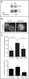
Figure 1. Assessment of autophagy in alveolar macrophages from adult RM.
A. Western analysis was performed on cell lysates from resting control (C) or autophagic (Rap) alveolar macrophages using antibody against LC-3 and actin. The LC-3 antibody reacts with both free cytosolic LC3-I (18 kDa) and membrane associated LC3-II (16 kDa). Levels of LC3-II were normalized to actin by densitometry and the ratio of LC3-II/actin given below the blot. A representative image from a single rhesus macaque (RM) sample is shown. B. Immunofluorescence microscopy was performed on control and rapamycin-treated alveolar macrophages using primary antibody against LC-3. C. The number of cells possessing LC-3+ puncta (top) and the average number of LC3+ vacuoles in LC-3+ cells (bottom) were quantified (n = 6 NHP samples, n>50 macrophages in each condition). The difference between the number of cells possessing LC-3+ puncta was significantly different between control and autophagic cells (***, p<0.001; **, p<0.01; ANOVA).
Figure 2
Figure 2. Bactericidal capacity of autophagic macrophages from adult RM.
A. Alveolar macrophages were infected at an MOI of 5∶1 with M. tuberculosis CDC1551. Bacterial colony forming units (cfu) were determined following control treatment, 4 h treatment with 50 µg/mL rapamycin (rap) to induce autophagy, and 4 h treatment with 50 µg/mL rapamycin and 10 mM 3-methyladenine (3-MA) to block autophagy. Viability is expressed as % survival relative to the number of viable bacteria in untreated resting control macrophages. Each symbol represents the average of three triplicate infections for each condition using RM sample. The average and standard deviation of all samples are shown. The difference between bacterial survival in control and autophagic macrophages was significant (**, p<0.01; ANOVA). B. Immunofluorescence microscopy was performed on control and autophagic alveolar macrophages infected with fluorescently labeled M. tuberculosis CDC1551 (green). Primary antibodies against either the autophagosomal marker LC3 (top) or the lysosomal marker LAMP (bottom) and DyeLight 594-conjugated secondary antibody were used. Arrows indicate a representative mycobacterium co-localized with LAMP-1. C. The number of M. tuberculosis that co-localize with LAMP-1 were quantified. The difference between control and autophagic cells was not significantly different (p = 0.0586; ANOVA; n = 6 NHP samples).
Figure 3
Figure 3. Comparison of innate immune functions of alveolar macrophages of adult and aged RM.
A. Phagocytic capacity of alveolar macrophages from adult and aged RM was measured. Alveolar macrophages were incubated with fluorescently labeled M. tuberculosis CDC1551 for 45 minutes, and then extracellular bacteria removed by washing. Cells were fixed, and infected macrophages were observed by fluorescence microscopy. A macrophage was considered infected if it had internalized at least one bacterium. Each symbol represents the % infection of each RM sample. The average and standard deviation of all samples for each group is shown. n>100 macrophages for each RM sample. B. Alveolar macrophages from aged RM were infected at an MOI of 5∶1 with M. tuberculosis CDC1551. Bacterial cfu were determined following control treatment, 4 h treatment with 50 µg/mL rapamycin (rap) to induce autophagy, and 4 h treatment with 50 µg/mL rapamycin and 10 mM 3-methyladenine (3-MA) to block autophagy. Viability is expressed as % survival relative to the number of viable bacteria in untreated resting control macrophages. Each symbol represents the average of three triplicate infections for each condition using RM sample. The average and standard deviation of all samples are shown. The difference between bacterial survival in control and autophagic macrophages was significant (***, p<0.001; *, p<0.05; ANOVA).
Figure 4
Figure 4. Bactericidal capacity of resting alveolar macrophages.
Alveolar macrophages were infected at an MOI of 5∶1 with M. tuberculosis CDC1551. Bacterial survival was followed over seven days by harvesting the infected monolayers at the indicated timepoints and plating serial dilutions. The assay was performed in triplicate and the average and standard deviation for each time point are shown for each RM sample. The reference number for each RM is noted in the legend. Bacterial survival in alveolar macrophages from adult (left) and aged (right) RM is shown. There was no significant difference in mycobacterial survival between groups at t = 7d (Student's t-test, p = 0.0693).

References

    1. Clemens D, Lee B, Horwitz M (2000) Deviant expression of Rab5 on phagosomes containing the intracellular pathogens Mycobacterium tuberculosis and Legionella pneumophila is associated with altered phagosomal fate. Infect Immun 68: 2671–2684. - PMC - PubMed
    1. Clemens D, Horwitz M (1996) The Mycobacterium tuberculosis phagosome interacts with early endosomes and is accessible to exogenously administered transferrin. J Exp Med 184: 1349–1355. - PMC - PubMed
    1. Sturgill-Koszycki S, Schlesinger P, Chakraborty P, Haddix P, Collins H, et al. (1994) Lack of acidification in Mycobacterium phagosomes produced by exclusion of the vesicular proton-ATPase. Science 263: 678–681. - PubMed
    1. Via L, Deretic D, Ulmer R, Hibler N, Huber L, et al. (1997) Arrest of mycobacterial phagosome maturation is caused by a block in vesicle fusion between stages controlled by rab5 and rab7. J Biol Chem 272: 13326–13331. - PubMed
    1. Via L, Fratti R, McFalone M, Pagan-Ramos E, Deretic D, et al. (1998) Effects of cytokines on mycobacterial phagosome maturation. J Cell Sci 111: 897–905. - PubMed

Publication types

LinkOut - more resources