. 2013 Jun 25;8(6):e67256.
doi: 10.1371/journal.pone.0067256. Print 2013.

Interleukin-21 enhances rituximab activity in a cynomolgus monkey model of B cell depletion and in mouse B cell lymphoma models

Affiliations

Interleukin-21 enhances rituximab activity in a cynomolgus monkey model of B cell depletion and in mouse B cell lymphoma models

Cecile M Krejsa et al. PLoS One. .

Abstract

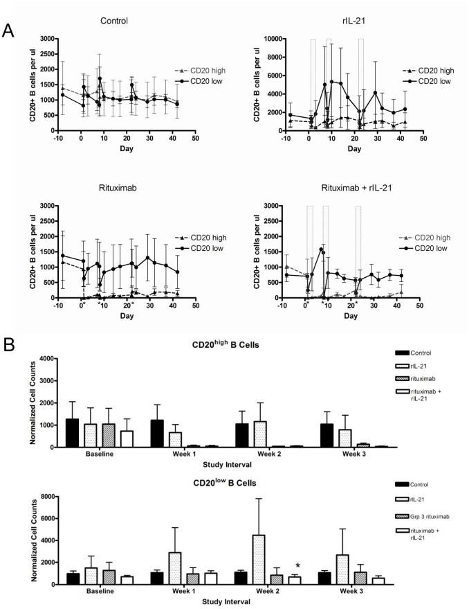
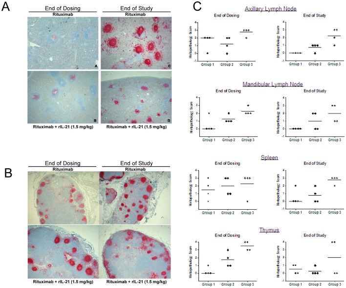
Rituximab, a monoclonal antibody targeting CD20 on B cells, is currently used to treat many subtypes of B cell lymphomas. However, treatment is not curative and response rates are variable. Recombinant interleukin-21 (rIL-21) is a cytokine that enhances immune effector function and affects both primary and transformed B cell differentiation. We hypothesized that the combination of rIL-21 plus rituximab would be a more efficacious treatment for B cell malignancies than rituximab alone. We cultured human and cynomolgus monkey NK cells with rIL-21 and found that their activity was increased and proteins associated with antibody dependent cytotoxicity were up-regulated. Studies in cynomolgus monkeys modeled the effects of rIL-21 on rituximab activity against CD20 B cells. In these studies, rIL-21 activated innate immune effectors, increased ADCC and mobilized B cells into peripheral blood. When rIL-21 was combined with rituximab, deeper and more durable B cell depletion was observed. In another series of experiments, IL-21 was shown to have direct antiproliferative activity against a subset of human lymphoma cell lines, and combination of murine IL-21 with rituximab yielded significant survival benefits over either agent alone in xenogeneic mouse tumor models of disseminated lymphoma. Therefore, our results do suggest that the therapeutic efficacy of rituximab may be improved when used in combination with rIL-21.


Conflict of interest statement

Competing Interests: The authors have read the journal’s policy and have the following conflicts: at the time this research was conducted all the authors were employees of ZymoGenetics, Inc., the commercial funder of the research. However, this does not alter the authors’ adherence to all PLOS ONE policies on sharing data and materials.

Figures

Figure 1
Figure 1. Treatment with rIL-21 enhances ADCC activity in human NK cells.
(A) Induction of granzyme B in NK cells cultured with rIL-21, IgG or both. (B, C) Purified human NK cells were cultured with rIL-21 (25 ng/ml) or media for 3 days and then placed in ADCC assays with control IgG or rituximab (2 µg/ml). Percentage lysis in representative assays against DOHH2 targets (B), and lysis of Ramos cells compared to Namalwa cells (C) are shown. Insets show CD20 expression on target cell lines. Data shown are representative results from 2 separate co-culture experiments (A) and from separate experiments using 6 NK cell donors and 5 lymphoma lines (B, C). E:T = effector to target cell ratio.
Figure 2
Figure 2. Effects of rIL-21 on normal human B cells.
(A) IL-21R expression on B cells, T cells and monocytes. Median and individual mean fluorescence intensity (MFI) data from 5–6 donors are shown. Right panel shows higher expression of IL-21R on CD21+ B cell subset. (B, C) Effect of rIL-21 culture on B cell phenotype and expression of cell surface markers. Median percentage change from 48 hr control cultures, and significance levels of t tests are shown. (B) rIL-21 treatment shifts CD20+ B cells to class-switched phenotype. (C) Expression of surface markers following rituximab and rIL-21 cultures.
Figure 3
Figure 3. Activation of innate effector cells in cynomolgus monkeys treated with rIL-21.
(A) ADCC activity in PBMC preparations in animals treated with vehicle control, weekly rituximab (0.05 mg/kg), rIL-21 (0.5 mg/kg for 3 days), or a combination of rituximab (0.05 mg/kg) and rIL-21 (0.5 mg/kg for 3 days). Gray bars indicate rIL-21 dosing days; rituximab was co-administered before the first rIL-21 dose in each cycle (days 1, 8, and 20). (B) Percentage of peripheral blood NK cells and cytotoxic T lymphocytes (CTL) staining positive for perforin. Gray bars in C indicate dosing days, error bars show standard error of the mean (SEM). (A, B) N = 3 animals per group.
Figure 4
Figure 4. Changes in immune cell markers.
Graphs show changes in serum soluble CD25 (sCD25) and CD14+, CD16+, CD11b/cbright, and CD64+ cells in peripheral blood of vehicle control group and group treated with rIL-21 alone (0.5 mg/kg, 3 days). E:T = effector to target cell ratio. Dosing with rIL-21 (0.5 mg/kg) was by slow i.v. injection on days 1–3, 8–10 and 22–24. Gray bars indicate rIL-21 dosing days, error bars show SEM. N = 3 animals per group.
Figure 5
Figure 5. Effects on B cells in cynomolgus monkeys treated with combination rIL-21 and rituximab.
The trapezoidal method was used to calculate peripheral cell counts over time (AUC) for the first 7–8 days of each dosing cycle and normalized to a daily average cell count. Graphs show CD20high and CD20low cells in animals treated with vehicle, weekly rituximab (0.05 mg/kg), rIL-21 (0.5 mg/kg for 3 days), or rituximab co-administered before the first rIL-21 dose in each cycle (days 1, 8, and 20). (B) Rituximab in combination with rIL-21 decreased the counts of CD20low B cells, compared to rIL-21 alone, after the second dosing cycle. Open bars indicate rIL-21 dosing days; rituximab was co-administered before the first rIL-21 dose in each cycle (days 1, 8, and 20, indicated by asterix). Note: scales are expanded for control and rituximab-treated groups compared to rIL-21-treated group. (B) Percentage change in normalized CD20low B cell counts after each dosing cycle. (*p<0.05, repeated measures ANOVA comparing combination groups to 0.5 mg/kg rIL-21 group only). Error bars show SEM. (A, B) N = 3 animals/group.
Figure 6
Figure 6. rIL-21 enhances rituximab-mediated B cell depletion in cynomolgus monkeys.
B cell depletion in animals treated with 10 mg/kg rituximab (Group 1) and in animals given 10 mg/kg rituximab plus rIL-21 at 0.3 mg/kg (Group 2) or 1.5 mg/kg (Group 3) for four weekly doses, followed by a dose-free period of 30 days. Immunohistochemistry of CD20+ cells in spleen (A) and mandibular lymph node (B) at end of dosing (terminal sacrifice) and end of the study. Images were collected using an Olympus BX51 microscope equipped with a PlanApo 2×/0.08 lens and U-CMAD3 camera, and captured with Colorview and analySIS Soft Imaging System software (Olympus, Tokyo Japan). (C) Lymphoid atrophy scores in lymphoid tissues collected at the end of dosing and at the end of the study. Histopathology scores for severity of atrophy were subjectively assigned as 0, none; 1, minimal; 2, slight; 3, moderate; 4, marked. Bar indicates median group score.
Figure 7
Figure 7. rIL21 induced STAT1 signaling and growth inhibition for IM-9 but not Raji lymphoma cells.
Growth curves for (A) IM-9 and (B) Raji cells when cultured with 50 ng/ml of rIL-21 after 3 days pre-treatment under the same conditions. Insets show phospho-STAT1 signaling IM-9 and Raji cells after 15 min culture with 10 ng/ml IL-21. Lower panels show survival curves for SCID mice (6–10/group) injected i.v. with 106 cells of the (C) IM-9, (D) Raji, or (E) H-S Sultan lymphoma lines. Human rIL-21 or control treatment (PBS) was administered by 28-day mini-osmotic pump implanted one day after tumor cell injection.
Figure 8
Figure 8. Murine IL-21 plus rituximab prolongs the survival of SCID mice bearing disseminated lymphoma tumor cells.
(A, B) SCID mice (n = 10/group) were injected i.v. with 106 lymphoma cells and then treated with rituximab alone (20 µg on days 3, 7, 11, 15, 19), mIL-21 alone (100 µg days 1–5), or mIL-21 and rituximab. Significant prolongation of survival was observed in the rituximab plus mIL-21 group when compared to rituximab alone (p = 0.0006) in the HS-Sultan model (A). Mice in the Raji model (B) were treated as above, except that mIL-21 was given on days 3–7 and rituximab was given on days 5, 9, 13, 17, and 21. Mice in the combination group survived longer than those treated with rituximab alone (p = 0.0079). (C, D, E) Test of effector cell function with Raji lymphoma models established in SCID or NOD/SCID mice as described above. (C) NOD/SCID mice with impaired NK cells compared to SCID mice. Equivalent survival times were observed for NOD/SCID and SCID mice given rituximab plus rIL-21 (p = 0.012) or rituximab alone. (D) SCID mice injected i.p. with 50 µg of anti-Gr-1 antibody on day -1, 4, 9 and 14 to deplete granulocytes or with PBS (control). Survival was significantly decreased for depleted mice given rituximab plus rIL-21 (p = 0.012) or rituximab alone (p = 0.001) compared with non-depleted mice. (E) SCID mice injected i.v. with liposomes containing clodronate to deplete macrophages or PBS (control). Survival was significantly decreased for depleted mice given rituximab plus rIL-21 treatment (p = 0.0115) or rituximab alone (p = 0.0011) compared with non-depleted mice.

References

    1. Asao H, Okuyama C, Kumaki S, Ishii N, Tsuchiya S, et al. (2001) Cutting edge: the common gamma-chain is an indispensable subunit of the IL-21 receptor complex. J Immunol 167: 1–5. - PubMed
    1. Habib T, Senadheera S, Weinberg K, Kaushansky K (2002) The common gamma chain (gamma c) is a required signaling component of the IL-21 receptor and supports IL-21-induced cell proliferation via JAK3. Biochemistry 41: 8725–8731. - PubMed
    1. Parrish-Novak J, Dillon SR, Nelson A, Hammond A, Sprecher C, et al. (2000) Interleukin 21 and its receptor are involved in NK cell expansion and regulation of lymphocyte function. Nature 408: 57–63. - PubMed
    1. Ettinger R, Sims GP, Fairhurst AM, Robbins R, da Silva YS, et al. (2005) IL-21 induces differentiation of human naive and memory B cells into antibody-secreting plasma cells. J Immunol 175: 7867–7879. - PubMed
    1. Jin H, Carrio R, Yu A, Malek TR (2004) Distinct activation signals determine whether IL-21 induces B cell costimulation, growth arrest, or Bim-dependent apoptosis. J Immunol 173: 657–665. - PubMed

Publication types

MeSH terms

LinkOut - more resources