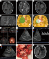
Intraoperative ultrasound assistance in resection of intracranial meningiomas
- PMID: 23825911
- PMCID: PMC3696709
- DOI: 10.3978/j.issn.1000-9604.2013.06.13
Intraoperative ultrasound assistance in resection of intracranial meningiomas
Abstract
Objective: Intracranial meningiomas, especially those located at anterior and middle skull base, are difficult to be completely resected due to their complicated anatomy structures and adjacent vessels. It's essential to locate the tumor and its vessels precisely during operation to reduce the risk of neurological deficits. The purpose of this study was to evaluate intraoperative ultrasonography in displaying intracranial meningioma and its surrounding arteries, and evaluate its potential to improve surgical precision and minimize surgical trauma.
Methods: Between December 2011 and January 2013, 20 patients with anterior and middle skull base meningioma underwent surgery with the assistance of intraoperative ultrasonography in the Neurosurgery Department of Shanghai Huashan Hospital. There were 7 male and 13 female patients, aged from 31 to 66 years old. Their sonographic features were analyzed and the advantages of intraoperative ultrasonography were discussed.
Results: The border of the meningioma and its adjacent vessels could be exhibited on intraoperative ultrasonography. The sonographic visualization allowed the neurosurgeon to choose an appropriate approach before the operation. In addition, intraoperative ultrasonography could inform neurosurgeons about the location of the tumor, its relation to the surrounding arteries during the operation, thus these essential arteries could be protected carefully.
Conclusions: Intraoperative ultrasonography is a useful intraoperative technique. When appropriately applied to assist surgical procedures for intracranial meningioma, it could offer very important intraoperative information (such as the tumor supplying vessels) that helps to improve surgical resection and therefore might reduce the postoperative morbidity.
Keywords: Intraoperative ultrasonography; intracranial meningioma.
Figures


References
-
- Riemenschneider MJ, Perry A, Reifenberger G. Histological classification and molecular genetics of meningiomas. Lancet Neurol 2006;5:1045-54 - PubMed
-
- Mawrin C, Perry A.Pathological classification and molecular genetics of meningiomas. J Neurooncol 2010;99:379-91 - PubMed
-
- Perry A, Louis DN, Scheithauer BW, et al. Meningiomas. In: Louis DN, Ohgaki H, Wiestler OD, et al. eds. WHO Classification of Tumours of the Nervous System. Lyon: IARC, 2007:163-72.
-
- Alexiou GA, Gogou P, Markoula S, et al. Management of meningiomas. Clin Neurol Neurosurg 2010;112:177-82 - PubMed
LinkOut - more resources
Full Text Sources
