. 2013 Jun 27:19:1387-96.
Print 2013.

Evaluation of the percentage of ganglion cells in the ganglion cell layer of the rodent retina

Affiliations

Evaluation of the percentage of ganglion cells in the ganglion cell layer of the rodent retina

Cassandra L Schlamp et al. Mol Vis. .

Abstract

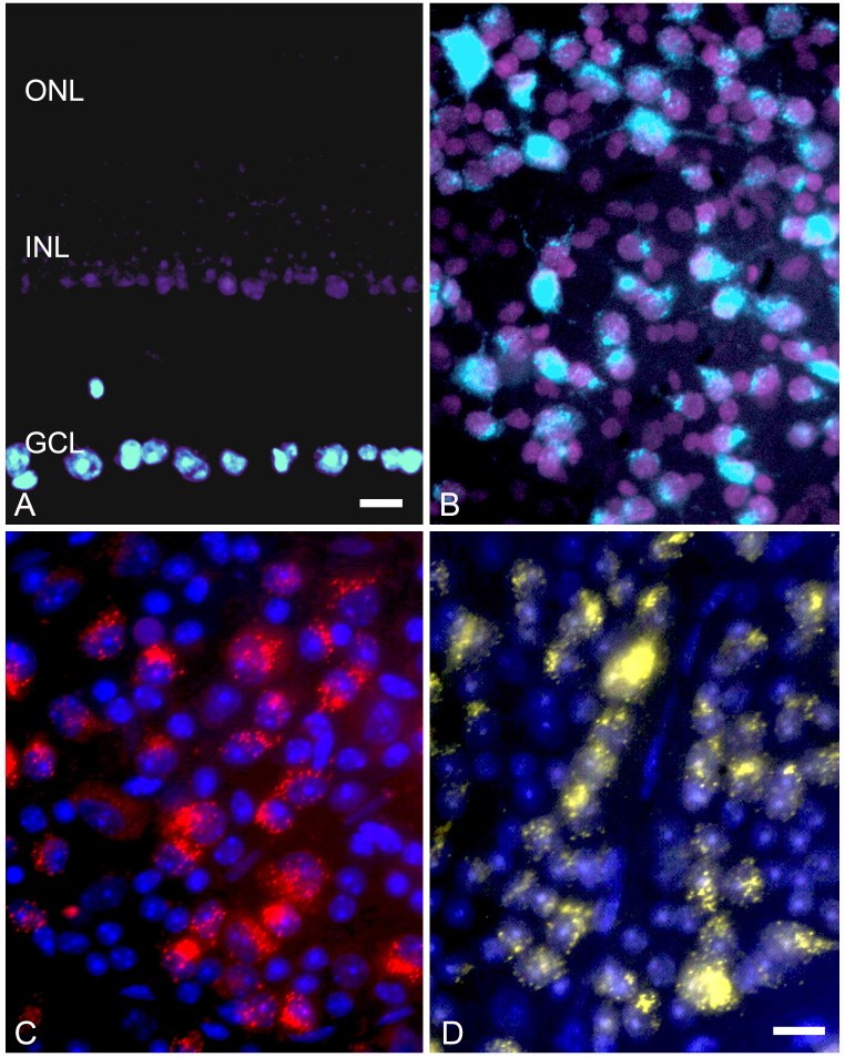
Purpose: Retinal ganglion cells comprise a percentage of the neurons actually residing in the ganglion cell layer (GCL) of the rodent retina. This estimate is useful to extrapolate ganglion cell loss in models of optic nerve disease, but the values reported in the literature are highly variable depending on the methods used to obtain them.

Methods: We tested three retrograde labeling methods and two immunostaining methods to calculate ganglion cell number in the mouse retina (C57BL/6). Additionally, a double-stain retrograde staining method was used to label rats (Long-Evans). The number of total neurons was estimated using a nuclear stain and selecting for nuclei that met specific criteria. Cholinergic amacrine cells were identified using transgenic mice expressing Tomato fluorescent protein. Total neurons and total ganglion cell numbers were measured in microscopic fields of 10(4) µm(2) to determine the percentage of neurons comprising ganglion cells in each field.

Results: Historical estimates of the percentage of ganglion cells in the mouse GCL range from 36.1% to 67.5% depending on the method used. Experimentally, retrograde labeling methods yielded a combined estimate of 50.3% in mice. A retrograde method also yielded a value of 50.21% for rat retinas. Immunolabeling estimates were higher at 64.8%. Immunolabeling may introduce overestimates, however, with non-specific labeling effects, or ectopic expression of antigens in neurons other than ganglion cells.

Conclusions: Since immunolabeling methods may overestimate ganglion cell numbers, we conclude that 50%, which is consistently derived from retrograde labeling methods, is a reliable estimate of the ganglion cells in the neuronal population of the GCL.


Figures

Figure 1
Figure 1
Histograph showing the percentage of ganglion cells that comprise the neurons in the ganglion cell layer. Ganglion cells were identified from retinas retrogradely labeled with FluoroGold and counterstained with TO-PRO-3. Data were collected from a total of 11 retinas (C57BL/6 mice). The percentage of ganglion cells did not differ among different retinal quadrants (ANOVA, p=0.798). Similarly, the percentage of neurons as ganglion cells did not vary significantly from the central to peripheral retina of each quadrant (p=0.09, data not shown).
Figure 2
Figure 2
Images of mouse retinas with ganglion cells identified with retrograde labeling. A and B: A plastic section and whole mount of mouse (A) and rat (B) retinas, respectively, are labeled with a mixture of FluoroGold and 4',6-diamidino-2-phenylindole (DAPI, see Methods) stereotactically injected into the superior colliculus. The section in (A) shows that DAPI (purple) leaks from ganglion cells in the ganglion cell layer (GCL) and penetrates as far as the innermost layer of the inner nuclear layer (INL), while the outer nuclear layer (ONL) is unstained. FluoroGold (light blue) remains in the ganglion cells of the GCL. The whole mounted retina shows the distribution of FluoroGold/DAPI positive cells, relative to the cells stained with DAPI only. These images are electronically enhanced from digitized 35 mm color slide film. C: The whole mount of a retina is stained with retrograde 1,1'-dioctadecyl-3,3,3 3′-tetramethylindocarbocyanine perchlorate (DiI) label (red, DAPI counter stain). D: The whole mount of a retina is stained with retrograde hydroxystilbamidine (FluoroGold; yellow, TO-PRO-3 counterstain). In the latter two examples, the retrograde dyes were applied using gel-foam soaked pledgets placed over the exposed superior colliculus. Size bar in A=20 µm. Size bar in B, C, D=10 µm.
Figure 3
Figure 3
Immunofluorescent image of NeuN labeling of presumptive ganglion cells and cholinergic amacrine cells. A: The merged image of a region of a whole mounted retina from mice expressing tomato fluorescent protein in cholinergic amacrine cells (red, panel C), immunolabeled for neuronal-specific nuclear protein (NeuN; green, panel D), and counterstained with 4',6-diamidino-2-phenylindole (DAPI, blue, panel B) is shown. Some neuron-like cells do not label with NeuN (exemplar indicated by arrowhead). NeuN colocalizes with the label for cholinergic amacrines (exemplar indicated with an arrow), in addition to other cell types (asterisk). Size bar=10 µm.
Figure 4
Figure 4
Immunofluorescent image of BRN3 positive ganglion cells. A: The merged image of BRN3A (red, panel C), BRN3B (green, panel D) and counterstained with 4',6-diamidino-2-phenylindole (DAPI, blue, panel B) is shown. This double labeling reveals four distinct classes of cell types. A cluster of cells that do not label with either BRN antibody are indicated with an asterisk. Most cells expressing BRN3 are positive for both proteins (example indicated with a straight arrow), while a minority express either BRN3A alone (example indicated with a bent arrow) or BRN3B alone (example indicated with an arrowhead). Size bar=10 µm.

References

    1. Jeon C-J, Strettoi E, Masland RH. The major cell populations of the mouse retina. J Neurosci. 1998;18:8936–46. - PMC - PubMed
    1. Fernandes KA, Harder JM, Fornarola LB, Freeman RS, Clark AF, Pang IH, John SWM, Libby RT. JNK2 and JNK3 are major regulators of axonal injury-induced retinal ganglion cell death. Neurobiol Dis. 2012 epub ahead of print. - PMC - PubMed
    1. Quigley HA, Cone FE, Gelman SE, Yang Z, Son JL, Oglesby EN, Pease ME, Zack DJ. Lack of neuroprotection against experimental glaucoma in c-Jun N-terminal kinase 3 knockout mice. Exp Eye Res. 2011;92:299–305. - PMC - PubMed
    1. Zhang Y, Kim IJ, Sanes JR, Meister M. The most numerous ganglion cell type of the mouse retina is a selective feature detector. Proc Natl Acad Sci USA. 2012;109:E2391–8. - PMC - PubMed
    1. Chindasub P, Lindsey JD, Duong-Polk K, Leung CK, Weinreb RN. Inhibition of histone deacetylases 1 and 3 protects injured retinal ganglion cells. Invest Ophthalmol Vis Sci. 2013;54:96–102. - PMC - PubMed

Publication types

LinkOut - more resources