Robot-assisted right ureteral polypectomy: A case report
- PMID: 23826056
- PMCID: PMC3699091
- DOI: 10.5489/cuaj.1392
Robot-assisted right ureteral polypectomy: A case report
Abstract
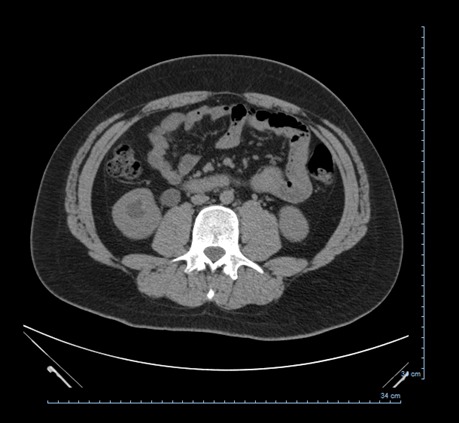
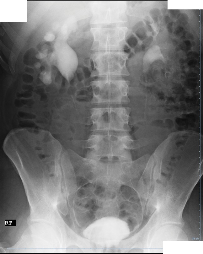
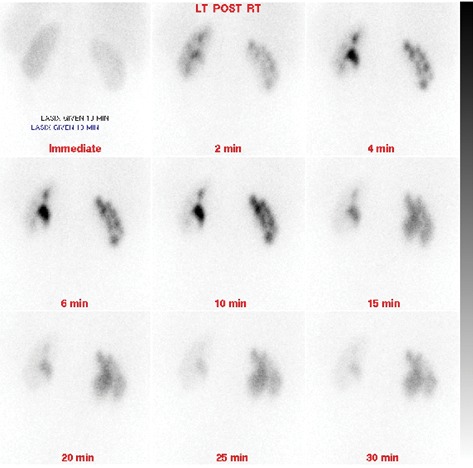
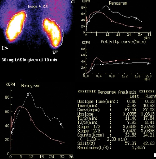
Ureteral polyps are a rare cause of ureteral obstruction in the adult and pediatric populations. Fibroepitheial polyps (FEP) are the most common type of ureteral polyps. This clinical entity is very rare, warranting periodic clinical review by practitioners, and new advancements in laparoscopy allow new surgical approaches to its cure. We present the case of a 20-year-old male with right-sided flank pain. He was found to have right ureteropelvic junction (UPJ) obstruction and subsequently underwent laparoscopic robotic-assisted right collecting system exploration, excision of polyps and right ureteropyeloplasty. Ureteral polyps were excised and determined to be fibroepithelial in origin based on the pathological report. Our case highlights the importance of having FEP in the differential diagnosis of ureteral obstruction. We also found that laparoscopic robot-assisted polypectomy is a safe and acceptable surgical option for the excision of ureteral polyps.
Figures
References
-
- Melicow MM, Findlay HV. Primary benign tumors of ureter: review of literature and report of case. Surg Gynecol Obstet. 1932;54:680–9.
-
- Harvin HJ. Ureteral Fibroepithelial Polyp on MDCT Urography. AJR Am J Roentgenol. 2006;187:W434–5. - PubMed
LinkOut - more resources
Full Text Sources
Other Literature Sources