. 2013 Jun 24;8(6):e66814.
doi: 10.1371/journal.pone.0066814. Print 2013.

MicroRNA-21 knockout improve the survival rate in DSS induced fatal colitis through protecting against inflammation and tissue injury

Affiliations

MicroRNA-21 knockout improve the survival rate in DSS induced fatal colitis through protecting against inflammation and tissue injury

Chenzhang Shi et al. PLoS One. .

Abstract

Background: MicroRNA-21 (miR-21) is overexpressed in most inflammatory diseases, but its physiological role in gut inflammation and tissue injury is poorly understood. The goal of this work is to understand the role of miR-21 in colitis and damage progression of intestine in a genetically modified murine model.

Methods: Experimental colitis was induced in miR-21 KO and wild-type (WT) mice by 3.5% dextran sulphate sodium (DSS) administration for 7 days. Disease activity index(DAI), blood parameters, intestinal permeability, histopathologic injury, cytokine and chemokine production, and epithelial cells apoptosis were examined in colons of miR-21 KO and WT mice.

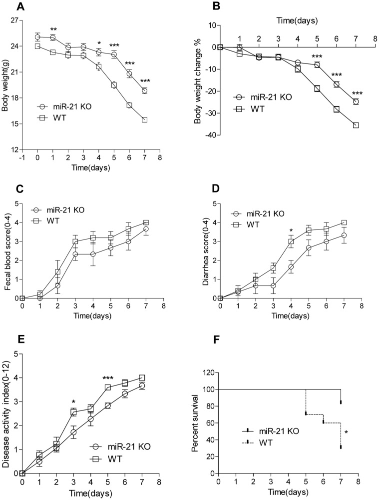
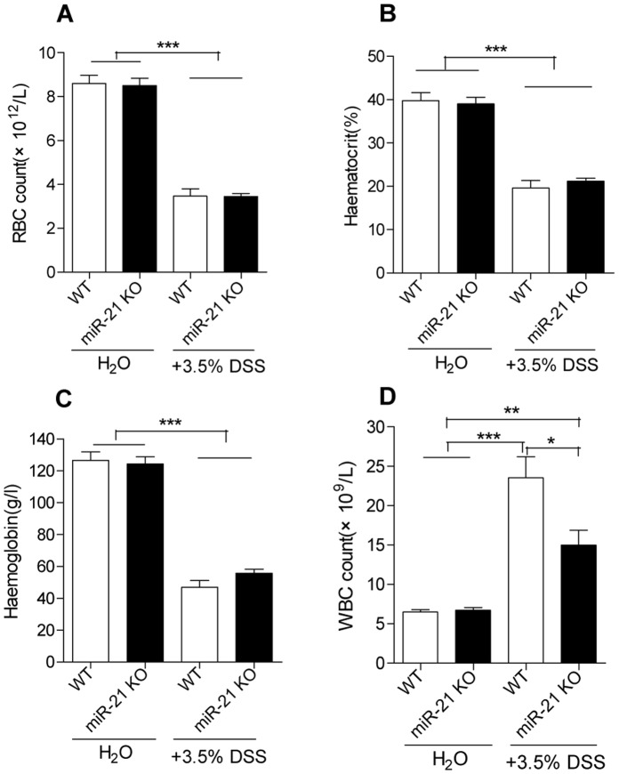
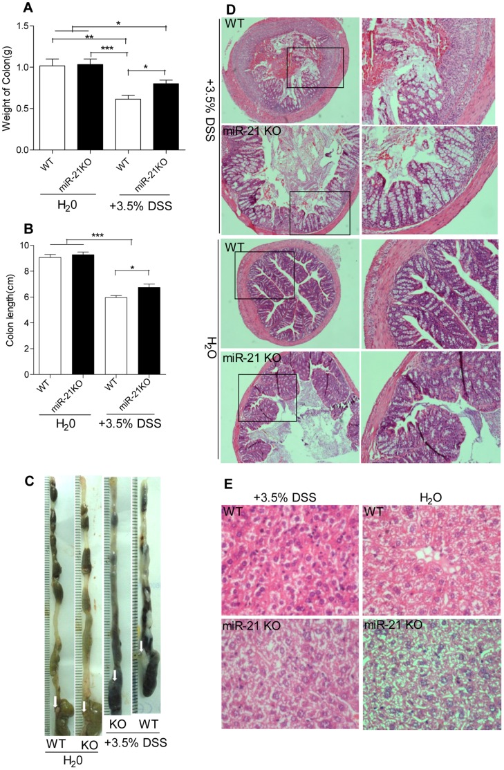
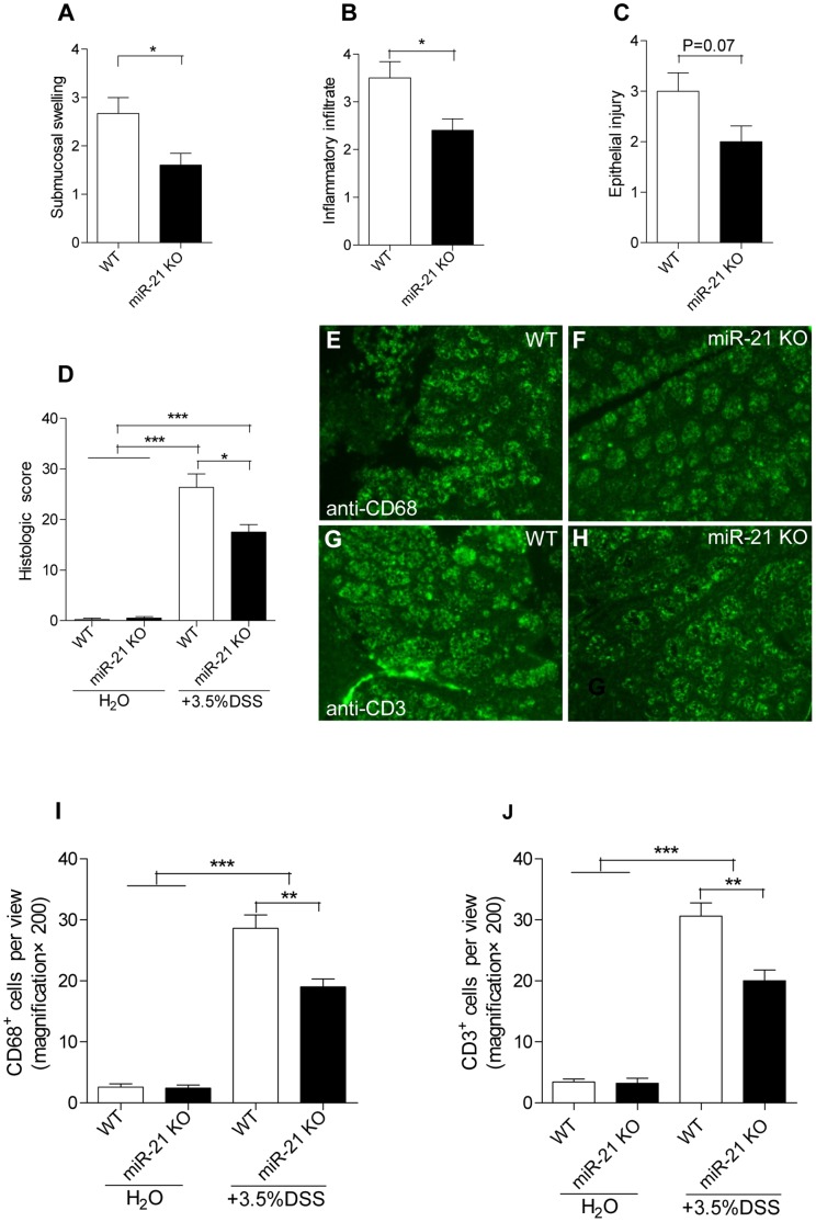
Results: miR-21 was overexpressed in intestine of inflammatory bowel diseases (IBD) and acute intestinal obstruction (AIO) patients when compared with normal intestinal tissues. Likewise, miR-21 was up-regulated in colon of IL-10 KO mice when compared with control mice. WT mice rapidly lost weight and were moribund 5 days after treatment with 3.5% DSS, while miR-21 KO mice survived for at least 6 days. Elevated leukocytes and more severe histopathology were observed in WT mice when compared with miR-21 KO mice. Elevated levels of TNF-α and macrophage inflammatory protein-2(MIP-2) in colon culture supernatants from WT mice exhibited significant higher than miR-21 KO mice. Furthermore, CD3 and CD68 positive cells, intestinal permeability and apoptosis of epithelial cells were significantly increased in WT mice when compared with miR-21 KO mice. Finally, we found that miR-21 regulated the intestinal barrier function through modulating the expression of RhoB and CDC42.

Conclusion: Our results suggest that miR-21 is overexpressed in intestinal inflammation and tissue injury, while knockout of miR-21 in mice improve the survival rate in DSS-induced fatal colitis through protecting against inflammation and tissue injury. Therefore, attenuated expression of miR-21 in gut may prevent the onset or progression of inflammatory bowel disease in patients.


Conflict of interest statement

Competing Interests: The authors have declared that no competing interests exist.

Figures

Figure 1
Figure 1. Expression of miR-21 in inflammation- and injury-involved intestine and miR-21 KO mice.
(A) Representative photomicrographs in situ detection (200×magnification, n = 3 per group) and (B) QRT-PCR (n = 5 per group) shown miR-21 is overexpressed in intestinal tissue of IBD and AIO patients when compared with normal intestinal tissues (NIT) of human. (C) miR-21 is overexpressed in inflammatory colon tissue of IL-10 KO mice (n = 8) compared with WT mice (n = 8). (D) The expression of miR-21 in colon is significantly increased in DSS-treated WT mice compare to control mice (n = 5 per group). (E) Loss of expression of miR-21 in small intestine and colon of miR-21 KO mice (n = 5) compared with WT mice (n = 5). (F) PCR genotyping of miR-21 KO mice DNAs. (mean±SEM, *p<0.05, **p<0.01,***p<0.001; Student’s t test).
Figure 2
Figure 2. Severe experimentally induced colitis in miR-21 KO mice.
MiR-21 KO and WT mice in C57BL/6J background were continuously administered 3.5% dextran sodium sulphate (DSS) freely available in drinking water. (A) Changes in body weights in WT (n = 10) and miR-21 KO (n = 6) mice were measured. (*p<0.05, **p<0.01, ***p<0.001; Student’s t test). Disease severity was measured daily and expressed in terms of (B) body weight loss,(C) fecal blood, (D) diarrhea, and (E) disease activity index (n = 6 per time point for miR-21 KO mice compared with WT control n = 10, *p<0.05, ***p<0.001, Student’s t test). (F) Percentage survival (n = 6 for miR-21 KO mice and n = 10 for WT mice, *p<0.05, log-rank test).
Figure 3
Figure 3. Blood parameters in miR-21 KO and WT mice following experimentally induced colitis.
(A) Red blood cell (RBC) count, (B) Haematocrit, (C) Haemoglobin, and (D) White blood cell (WBC) counts in blood of miR-21 KO and WT mice were measured on 4th day of DSS administration (mean±SEM, n = 6 per group, *p<0.05, **p<0.01,***p<0.001, Student’s t test).
Figure 4
Figure 4. miR-21 KO mice have a attenuation in the severity of DSS-colitis.
(A) Colon weight and (B) Colon length of miR-21 KO and WT mice were measured on day 7 or time of dying following DSS treatment. (mean±SEM, n≥6 per group, *p<0.05, ***p<0.001 by Student’s t test.) (C) Representative images of miR-21 KO and WT colon following of DSS or water treatment. (D) Representative photomicrographs showing epithelial damage and colonic inflammation (H&E stain, 40×magnification, higher magnification photomicrographs on the right×100) in miR-21 KO and WT mice following of DSS or water administration. (E) Representative photomicrographs showing liver damage and inflammation (H&E stain, 400×magnification) in miR-21 KO and WT mice following of DSS or water administration.
Figure 5
Figure 5. miR-21 KO mice have attenuation in intestinal epithelial injury and inflammatory infiltration.
(A) Submucosal swelling, (B) Inflammatory infiltration, and (C) Epithelial injury were assessed for (D) histological colitis. (mean±SEM, n = 5 for miR-21 KO mice and n = 6 for WT mice, *p<0.05, **p<0.01,***p<0.001, Student’s t test). Colon tissue sections from miR-21 KO and WT mice were immunostained with antibodies for (E, F) CD68 and (G, H) CD3 (200×magnification). (I, J) Numbers of positive stained cells were counted per view. (mean±SEM, n = 5 per group, **p<0.01, ***p<0.001, Student’s t test).
Figure 6
Figure 6. Effects of miR-21 KO on mucosal cytokine production, intestinal permeability and epithelial cells apoptosis before and after DSS treatment.
Expression of macrophage inflammatory protein-2 (MIP-2) (A,C)and tumor necrosis factor α (TNF-α) (B,D) in WT and miR-21 KO mice were measured in serum (C,D) and colon culture supernatants (A,B) (mean±SEM, n = 6 per group of DSS treatment, n = 4 per group of water treatment, *p<0.05, **p<0.01,***p<0.001, Student’s t test). (E) In vivo intestinal permeability of WT and miR-21 KO mice before and after DSS treatment were measured by gavage with FITC-Dextran (mean±SEM, n = 5 per group, **p<0.01, ***p<0.001, Student’s t test). Fluorescence micrographs of colons in WT (F, H) and miR-21 KO (G, I) mice stained for terminal deoxynucleotidyl transferase–mediated deoxyuridine triphosphate nick-end labeling (TUNEL) (green). The panels are representative of 5 WT and 5 miR-21 KO mice (200×magnification). (J) Apoptotic cells were quantified by counting the apoptotic cells/field. (mean±SEM, n = 5 per group, *p<0.05, ***p<0.001, Student’s t test).
Figure 7
Figure 7. Effects of miR-21 KO on expression of relevant gene.
QRT-PCR were performed to measure the expression of miR-21(A), PDCD4(B), Cdc25A(C), NF-κB(D), Cyclin D1(E), Cdc42 (F), and RhoB (G) in colon of miR-21 KO and WT mice after administration of 3.5% DSS for 7 days.(mean±SEM, n = 6 per group, *p<0.05,***p<0.001, Student’s t test). Meanwhile, we measured the expression of CDC42(H) and RhoB (I) in colon of IL-10 KO and WT mice. (mean±SEM, n = 8 per group, *p<0.05, ***p<0.001, Student’s t test).

References

    1. Hatley ME, Patrick DM, Garcia MR, Richardson JA, Bassel-Duby R, et al. (2010) Modulation of K-Ras-dependent lung tumorigenesis by MicroRNA-21. Cancer Cell 18: 282–293. - PMC - PubMed
    1. Medina PP, Nolde M, Slack FJ (2010) OncomiR addiction in an in vivo model of microRNA-21-induced pre-B-cell lymphoma. Nature 467: 86–90. - PubMed
    1. Thum T, Gross C, Fiedler J, Fischer T, Kissler S, et al. (2008) MicroRNA-21 contributes to myocardial disease by stimulating MAP kinase signalling in fibroblasts. Nature 456: 980–984. - PubMed
    1. Asangani IA, Rasheed SA, Nikolova DA, Leupold JH, Colburn NH, et al. (2008) MicroRNA-21 (miR-21) post-transcriptionally downregulates tumor suppressor Pdcd4 and stimulates invasion, intravasation and metastasis in colorectal cancer. Oncogene 27: 2128–2136. - PubMed
    1. Zhu S, Wu H, Wu F, Nie D, Sheng S, et al. (2008) MicroRNA-21 targets tumor suppressor genes in invasion and metastasis. Cell Res 18: 350–359. - PubMed

Publication types

MeSH terms