. 2013 Jun 24;8(6):e66840.
doi: 10.1371/journal.pone.0066840. Print 2013.

'Cand. Actinochlamydia clariae' gen. nov., sp. nov., a unique intracellular bacterium causing epitheliocystis in catfish (Clarias gariepinus) in Uganda

Affiliations

'Cand. Actinochlamydia clariae' gen. nov., sp. nov., a unique intracellular bacterium causing epitheliocystis in catfish (Clarias gariepinus) in Uganda

Andreas Steigen et al. PLoS One. .

Abstract

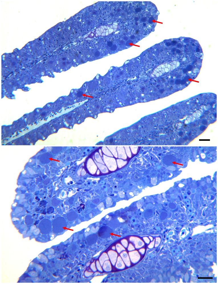
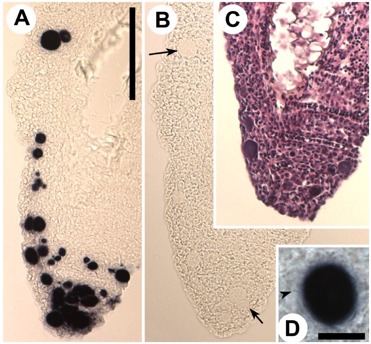
Background and objectives: Epitheliocystis, caused by bacteria infecting gill epithelial cells in fish, is common among a large range of fish species in both fresh- and seawater. The aquaculture industry considers epitheliocystis an important problem. It affects the welfare of the fish and the resulting gill disease may lead to mortalities. In a culture facility in Kampala, Uganda, juveniles of the African sharptooth catfish (Clarias gariepinus) was observed swimming in the surface, sometimes belly up, showing signs of respiratory problems. Histological examination of gill tissues from this fish revealed large amounts of epitheliocysts, and also presence of a few Ichthyobodo sp. and Trichodina sp.

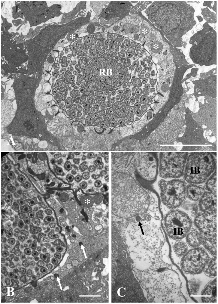
Methods and results: Sequencing of the epitheliocystis bacterium 16S rRNA gene shows 86.3% similarity with Candidatus Piscichlamydia salmonis causing epitheliocystis in Atlantic salmon (Salmo salar). Transmission electron microscopy showed that the morphology of the developmental stages of the bacterium is similar to that of members of the family Chlamydiaceae. The similarity of the bacterium rRNA gene sequences compared with other chlamydia-like bacteria ranged between 80.5% and 86.3%. Inclusions containing this new bacterium have tubules/channels (termed actinae) that are radiating from the inclusion membrane and opening on the cell surface or in neighbouring cells.

Conclusions: Radiation of tubules/channels (actinae) from the inclusion membrane has never been described in any of the other members of Chlamydiales. It seems to be a completely new character and an apomorphy. We propose the name Candidatus Actinochlamydia clariae gen. nov., sp. nov. (Actinochlamydiaceae fam. nov., order Chlamydiales, phylum Chlamydiae) for this new agent causing epitheliocystis in African sharptooth catfish.


Conflict of interest statement

Competing Interests: The authors have declared that no competing interests exist.

Figures

Figure 1
Figure 1. Semi-thin sections of infected primary lamellas from African sharptooth catfish.
Pictures of semi-thin sections of the primary gill lamellas from African sharptooth catfish infected with Candidatus Actinochlamydia clariae. Arrows point to cysts of variable sizes. The majority of the cysts are located towards the apical part of the primary lamellas. A) Bar = 60.0 µm. B) Bar = 30.0 µm.
Figure 2
Figure 2. Cysts of Candidatus Actinochlamydia clariae.
High magnification pictures of cysts containing Ca. A. clariae. Figures A) and B) show medium size cysts with tubules/channels radiating from the inclusion giving the cysts its distinct morphology. A) and B) Bars = 30.0 µm. B) A tangential section of the cyst clearly shows that the projections are tubules/channels (ring). C) This figure shows a large cyst, about 30 µm in diameter, containing mostly EBs (asterisk). C) Bar = 30.0 µm.
Figure 3
Figure 3. Infected cell with inclusion membrane and RBs.
A) Picture of a newly infected cell that contains a few RBs that is surrounded by a thin inclusion membrane. The tubular extensions (arrows) from the inclusion membrane are thin and short. Abundant amounts of mitochondria and small vesicles are present in the cytosol of the host cell close to the inclusion membrane. Bar = 1.0 µm. B) A later stage in the early development of Ca. A. clariae. The inclusion contains RBs only, and the tubular extensions (arrows) from the inclusion membrane are longer and more distinct. The inclusion is surrounded by large vesicles (asterisks) and mitochondria. The host cell nucleus (Nu) seems to be slightly distorted. Strands of DNA can be seen in the RBs. Bar = 2.0 µm.
Figure 4
Figure 4. Morphology of tubular extensions.
These two figures show the tubules from the inclusion. Picture A) shows transverse sections of the tubules close to the inclusion membrane. The tubules are irregular, star-shaped, with electron lucent material close to the inclusion membrane. Bar = 1.0 µm. B) shows a section through a neighbouring cells showing that the tubules, containing electron dense material, are extending into this cell. Cell nucleus (Nu). Bar = 1.0 µm.
Figure 5
Figure 5. Sections through inclusion membrane.
Sections through the membrane of an inclusion containing IBs and EBs. A) Tubular extensions from the inclusion are penetrating into the cytosol of a neighbouring cell. Note that there is no cell membrane separating the tubular opening from the cytosol of the neighbouring cell. Bar = 0.5 µm. B) This is a magnification of the area where two tubules enter the cytosol in figure 5A. Bar = 0.2 µm. C) Tubule from a smaller inclusion penetrating into the cytosol of a neighbouring cell. There seem to be a slight accumulation of fibrils (actin?) in the cytosol where the tubule enters. Note the large vesicles close to the inclusions in infected cells. Cell membrane (arrows). Bar = 0.2 µm. D) This is a section through the inclusion membrane of a cyst containing mainly late IBs and EBs. The inclusion membrane is very thick, probably due to insertion of bacterial proteins (arrow). Bar = 0.5 µm.
Figure 6
Figure 6. Section through inclusion with IBs.
Sections through inclusions containing mainly IBs. The IBs have condensed nucleoids, but the rest of the content of the bacteria is not condensed. A) Bar = 5.0 µm. B) Bar = 0.5 µm. C) Bar = 0.5 µm.
Figure 7
Figure 7. Section through inclusion with EBs.
Section through a large inclusion from the gills of Clarias gariepinus containing mainly EBs. A) The cyst is beginning to open to the gill surface (arrow), and the host cell cytosol shows signs of degeneration. A neighbouring cell contains an inclusion with RBs only (asterisk). Bar = 5.0 µm. B) Section through EB showing the cap area with associated protein structures (arrow). Tangential section through the cap area (ring) showing the hexagonal arrangement of the proteins. Bar = 0.2 µm. C) Section through free EBs showing the smooth cap areas (arrows) with proteins, an electron dense core (nucleoid), and condensed cytoplasm consisting mainly of ribosomes. Bar = 0.2 µm.
Figure 8
Figure 8. Degenerating epitheliocysts.
Pictures of degenerating epitheliocysts from Clarias gariepinus that contains inclusion with RBs (RB) and IBs (IB). The host cell cytoplasm loose the normal electron density and the mitochondria increase in size and become spherical (asterisks). The morphology of the tubular extensions from the inclusion changes from the normal irregular, star-shape (in transverse section), to round tubules (arrows). A) Bar = 5.0 µm. B) Bar = 1.0 µm. C) Bar = 0.5 µm.
Figure 9
Figure 9. Paraffin sections from gill tissues.
The sections of gill tissue from Clarias gariepinus have been processed for in situ hybridization or stained with HES. A) Primary filament showing dark-blue stained Candidatus Actinochlamydia clariae inclusions, stained with antisense DIG-labelled RNA-probe against Ca. A. clariae 16S rRNA. Cells with inclusions are particularly frequent at the filament tip. Bar = 100.0 µm. B) Same primary filament stained with a sense probe, demonstrating absence of staining in the inclusions (examples indicated by arrows). Bar = 100.0 µm. C) HES stained section of the same filament tip. Bar = 100.0 µm. D) Magnification of an IHC stained inclusion, where the actiniae are discernible (arrowhead). Bar 10.0 = µm.
Figure 10
Figure 10. Phylogenetic tree.
The phylogenetic tree shows the relationship between Candidatus Actinochlamydia clariae from African sharptooth catfish and selected members of other families within the order Chlamydiales. The best-fitting nucleotide substitution model was used during maximum likelihood analysis and the tree was bootstrapped (50 000 quartet puzzling steps) in TREE_PUZZLE. The scale bar shows the number of nucleotide substitutions as a proportion of branch lengths.

References

    1. Plehn M (1920) Neue Parasiten in Haut und Kiemen von Fishen. Ichthyochytrium und Mucophilus. Zentbl Bakteriolog P 85: 275–281.
    1. Hoffman GL, Dunbar CE, Wolf K, Zwillenberg LO (1969) Epitheliocystis, a new infectious disease of the bluegill (Lepomis macrochirus). Antonie van Leeuwenhoek 35: 146–158. - PubMed
    1. Moulder JW (1964) The psittacosis group as bacteria. Wiley, NY.
    1. Storz J, Page LA (1971) Taxonomy of the Chlamydiae: reasons for classifying Organisms of the Genus Chlamydia, Family Chlamydiaceae, in a Separate Order, Chlamydiales ord. nov. Int J Syst Evol Microbiol 21(4): 332–334.
    1. Garrity GM, Holt JG (2001) The Road Map to the Manual. In: Bergey’s Manual of Systematic Bacteriology, 2nd edn, vol 1, The Archaea and the Deeply Branching and Phototrophic Bacteria (ed Boone, Castenholz and Garrity). Springer, NY: 119–166.

Publication types

MeSH terms

Associated data

LinkOut - more resources