. 2013 Jun 24;8(6):e67003.
doi: 10.1371/journal.pone.0067003. Print 2013.

SLE peripheral blood B cell, T cell and myeloid cell transcriptomes display unique profiles and each subset contributes to the interferon signature

Affiliations

SLE peripheral blood B cell, T cell and myeloid cell transcriptomes display unique profiles and each subset contributes to the interferon signature

Amy M Becker et al. PLoS One. .

Abstract

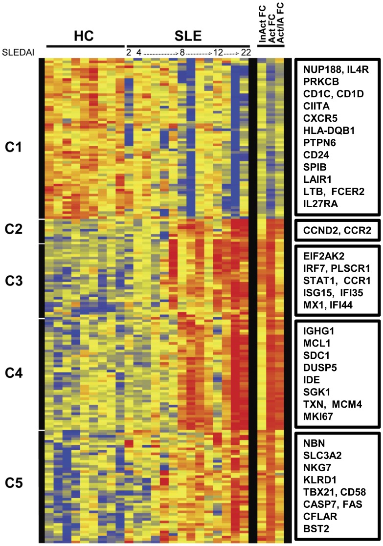
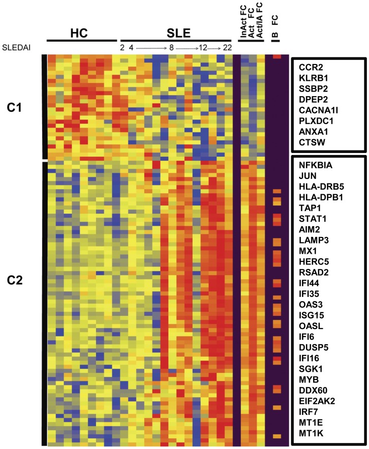
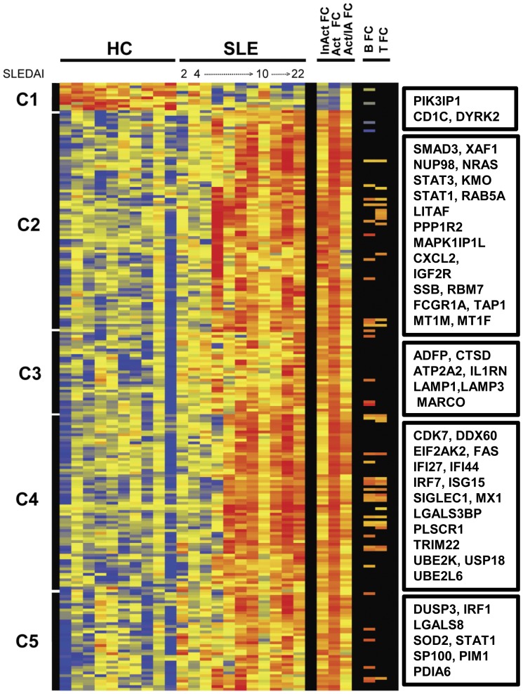
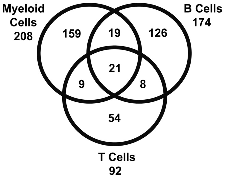
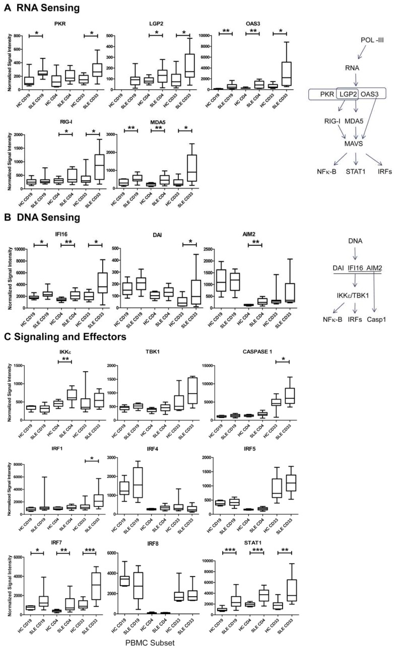
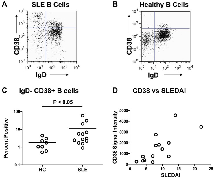
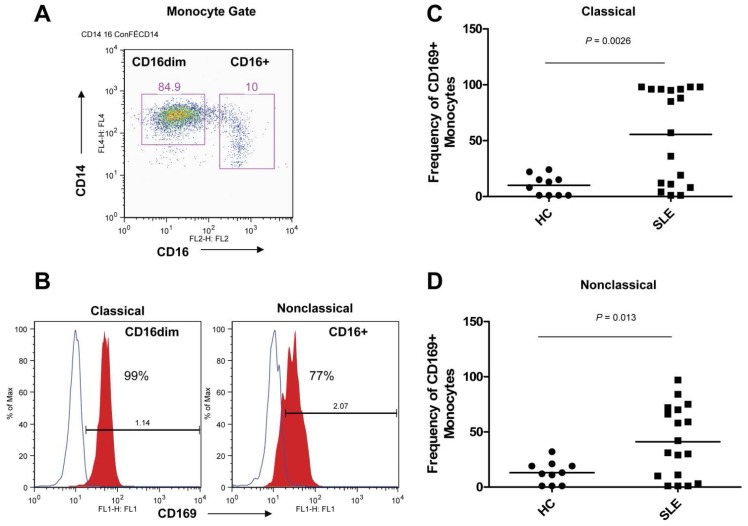
Systemic lupus erythematosus (SLE) is a chronic autoimmune disease that is characterized by defective immune tolerance combined with immune cell hyperactivity resulting in the production of pathogenic autoantibodies. Previous gene expression studies employing whole blood or peripheral blood mononuclear cells (PBMC) have demonstrated that a majority of patients with active disease have increased expression of type I interferon (IFN) inducible transcripts known as the IFN signature. The goal of the current study was to assess the gene expression profiles of isolated leukocyte subsets obtained from SLE patients. Subsets including CD19(+) B lymphocytes, CD3(+)CD4(+) T lymphocytes and CD33(+) myeloid cells were simultaneously sorted from PBMC. The SLE transcriptomes were assessed for differentially expressed genes as compared to healthy controls. SLE CD33(+) myeloid cells exhibited the greatest number of differentially expressed genes at 208 transcripts, SLE B cells expressed 174 transcripts and SLE CD3(+)CD4(+) T cells expressed 92 transcripts. Only 4.4% (21) of the 474 total transcripts, many associated with the IFN signature, were shared by all three subsets. Transcriptional profiles translated into increased protein expression for CD38, CD63, CD107a and CD169. Moreover, these studies demonstrated that both SLE lymphoid and myeloid subsets expressed elevated transcripts for cytosolic RNA and DNA sensors and downstream effectors mediating IFN and cytokine production. Prolonged upregulation of nucleic acid sensing pathways could modulate immune effector functions and initiate or contribute to the systemic inflammation observed in SLE.


Conflict of interest statement

Competing Interests: Dr. Nancy Olsen has equity interest in a company, ArthroChip LLC, which develops assays for autoimmune diseases and she has also obtained funding from MedImmune, Novo Nordisk, Genentech/Roche and Human Genome Sciences. Dr. David Karp has received research funding from Human Genome Sciences. None of these funds were used for the current study. This does not alter the authors' adherence to all the PLOS ONE policies on sharing data and materials.

Figures

Figure 1
Figure 1. SLE B cell transcriptomes.
Hierarchical cluster dendrogram of expressed genes that differed significantly between peripheral blood CD19+ B cells isolated from SLE patients and healthy controls (HC). The SLE samples are ordered by increasing SLEDAI score. The bars to the left indicate expressed gene clusters. Color changes indicate the expression level relative to the average (log2 scale). Red is increased expression, yellow is unchanged and blue is decreased expression. Indicated on the right are fold change in inactive SLE compared to HC (InAct FC), active SLE compared to HC (Act FC) and active SLE compared to inactive SLE (Act/IA FC). Select transcripts are identified for each cluster in boxes (right). For the entire list of transcripts in order as they appear on the dendrogram see supplementary Table S1.
Figure 2
Figure 2. SLE CD4+ T cell transcriptomes.
Hierarchical cluster dendrogram of expressed genes that differed significantly between sorted peripheral blood CD4+ T cells isolated from SLE patients and healthy controls (HC) as described in figure 1. Also indicated in the columns to the right are differentially expressed genes found in both B cell and T cell compartments of SLE (B FC). Select transcripts are identified for each cluster in boxes (right). For the entire list of transcripts ordered by clusters from the dendrogram see supplementary Table S2.
Figure 3
Figure 3. SLE Myeloid cell transcriptomes.
Hierarchical cluster dendrogram of expressed genes that differed significantly between sorted peripheral blood CD33+ myeloid cells isolated from SLE patients and healthy controls (HC) as described in figure 1. Also indicated on the right are differentially expressed genes from the B cell compartment of SLE (B FC) or the T cell compartment of SLE (T FC) that were shared with myeloid cells. Select transcripts are identified for each cluster in boxes (right). For the entire list of transcripts ordered by clusters from the dendrogram see supplementary Table S3.
Figure 4
Figure 4. SLE Subsets Up-regulate Unique Transcriptional Profiles.
A Venn diagram demonstrating shared and unique differentially expressed transcripts of SLE myeloid cells, B cells and T cells. Of the 474 combined transcripts only 4.4% (21) were shared by all three subsets, whereas 69% (329) of the transcripts were unique to a particular subset at the threshold set for the described primary analysis.
Figure 5
Figure 5. DNA and RNA Sensors are Transcriptionally Active in SLE Subsets.
A secondary analysis was performed with all normalized data in order to compare transcripts for specific pathways (19). (A) RNA sensing molecules (B) DNA sensing molecules and (C) downstream signaling and effector molecules were up-regulated in the three subsets as shown (Welch’s t-test, p<0.05*, p<0.01**, p<0.001***).
Figure 6
Figure 6. Validation of Array Results by CD38 Protein Expression.
(A) Increased CD38+ IgD expression by CD19+ gated B cells from an SLE patient and (B) CD38+ IgD expression by CD19+ gated B cells from a HC. A total of 5×10⁴ PBMC were assessed from blood studied in the gene expression arrays by incubation with anti-CD38-APC, anti-IgD-FITC and anti-CD19-PE, followed by flow cytometric analysis. (C) Frequency of CD38+ IgD CD19+ B cells was assessed on available PBMC analyzed for gene expression. There was a statistically significant difference (P<0.05) in CD38+ IgD+ CD19+ B cells between SLE patients (n = 13) and healthy controls (n = 8). (D) CD38 signal intensity values for transcripts obtained from microarrays correlated (rP = 0.7542 and P≤0.005) with SLEDAI scores for SLE B cells (n = 14).
Figure 7
Figure 7. Confirmation of Myeloid Array Results by CD169 Protein Expression
. The high expression of CD169 (SIGLEC1) by myeloid cells in the arrays was confirmed by analysis of CD33+CD14+ myeloid cells in a second cohort. (A) CD14+ myeloid cells were gated for CD16dim and CD16bright myeloid cells. (B) Histograms of CD169 expression on classical (CD14+CD16dim) and nonclassical (CD14+CD16+) myeloid cells. (C) Frequency of CD169+ cells in the classical (CD14+CD16dim) myeloid subset comparing HC to SLE patients. (D) Frequency of CD169+ cells in the nonclassical (CD14+CD16+) myeloid subset comparing HC to SLE patients.

References

    1. Crow MK (2008) Collaboration, genetic associations, and lupus erythematosus. N Engl J Med 358: 956–961. - PubMed
    1. Petri M (1995) Clinical features of systemic lupus erythematosus. Curr Opin Rheumatol 7: 395–401. - PubMed
    1. Arbuckle MR, McClain MT, Rubertone MV, Scofield RH, Dennis GJ, et al. (2003) Development of autoantibodies before the clinical onset of systemic lupus erythematosus. N Engl J Med 349: 1526–1533. - PubMed
    1. Heinlen LD, Ritterhouse LL, McClain MT, Keith MP, Neas BR, et al. (2010) Ribosomal P autoantibodies are present before SLE onset and are directed against non-C-terminal peptides. J Mol Med (Berl) 88: 719–727. - PMC - PubMed
    1. Mathian A, Gallegos M, Pascual V, Banchereau J, Koutouzov S (2011) Interferon-alpha induces unabated production of short-lived plasma cells in pre-autoimmune lupus-prone (NZBxNZW)F1 mice but not in BALB/c mice. Eur J Immunol 41: 863–872. - PMC - PubMed

Publication types

MeSH terms