. 2013 Jun 27;8(6):e67294.
doi: 10.1371/journal.pone.0067294. Print 2013.

Down-Regulated miR-30a in Clear Cell Renal Cell Carcinoma Correlated with Tumor Hematogenous Metastasis by Targeting Angiogenesis-Specific DLL4

Affiliations

Down-Regulated miR-30a in Clear Cell Renal Cell Carcinoma Correlated with Tumor Hematogenous Metastasis by Targeting Angiogenesis-Specific DLL4

Qing Bo Huang et al. PLoS One. .

Abstract

Background: Endothelial DLL4 plays an important role in controlling of tumor angiogenesis, which is required for tumor invasive growth and metastasis. However, the regulation of DLL4 in clear cell renal cell carcinoma (ccRCC) has not yet been systematically elucidated.

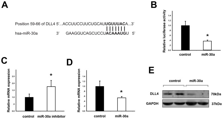
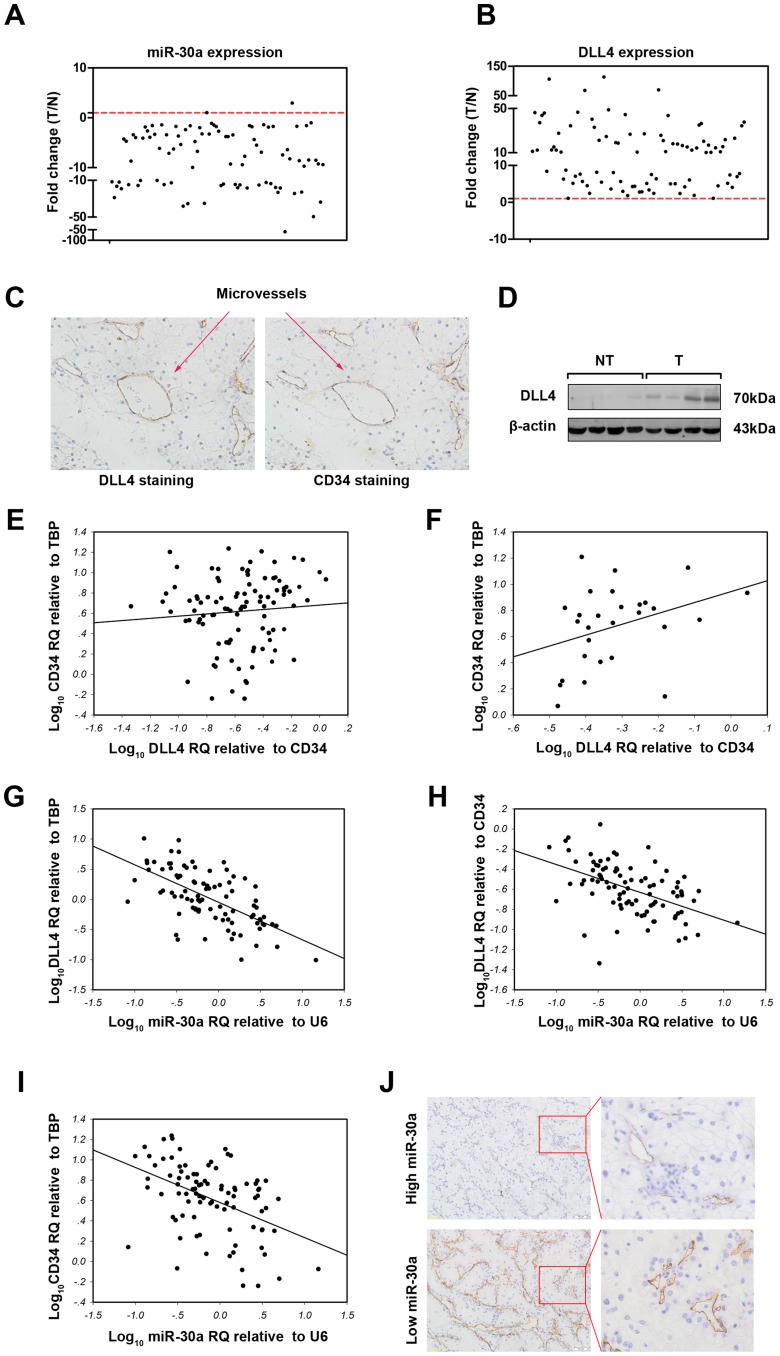
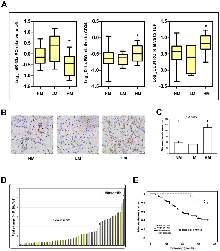
Methodology: We performed bioinformatical analysis to explore miRNAs targeting DLL4. miR-30a was selected as a representative to validate its functional association in endothelial cell. Then, the expressions of DLL4 and mature miR-30a from 90 cases of ccRCC and 28 cases of nonmatched adjacent non-tumor tissues were measured by quantitative real-time PCR. Finally, the expression of miR-30a was correlated with DLL4 expression, tumor features (metastatic condition and microvessel density), and patient metastasis-free survival. The univariate and multivariate analyses were performed to select the risk factors associated with hematogenous metastasis, respectively.

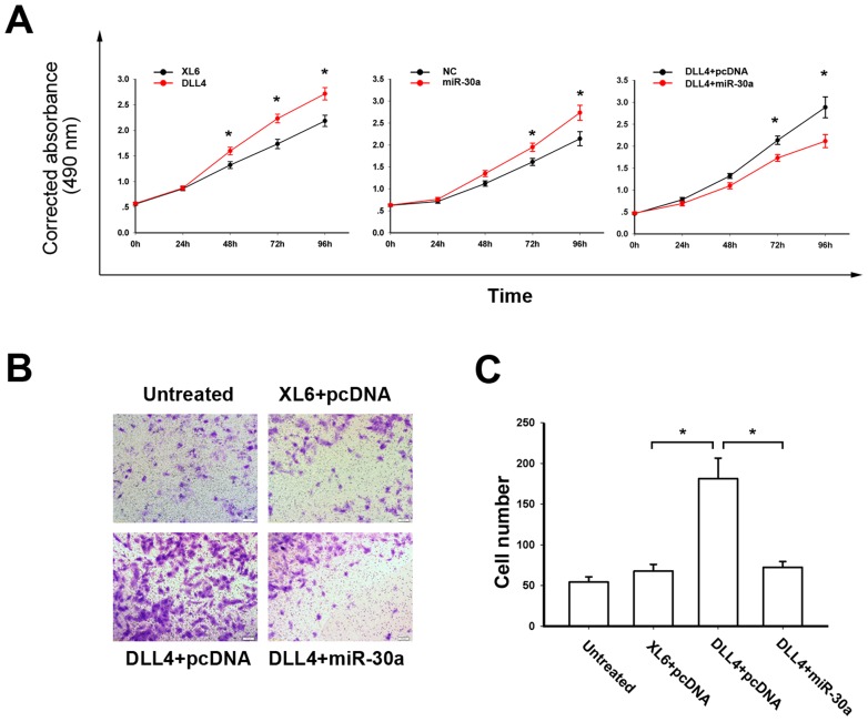
Principal findings: miR-30a negatively regulated DLL4 and inhibited the proliferation and migration of endothelial cells. DLL4 was up-regulated in ccRCC and further increased in hematogenous metastatic cases, while miR-30a was down-regulated in tumor tissues and further decreased in hematogenous metastatic ccRCC (student t test, all p<0.05). Additionally, expression of miR-30a was inversely correlated with expression of DLL4 and microvessel density (linear correlation analysis, both p<0.05). Low-level miR-30a also indicated a higher probability of developing metastasis (log-rank test, p = 0.010). Most importantly, miR-30a expression was an independent predictor of ccRCC hematogenous metastasis by the univariate analysis and binary logistic regression model (both p<0.05).

Conclusions: Down-regulated miR-30a in ccRCC was associated with tumor hematogenous metastasis through increasing microvessel density by targeting angiogenesis-specific DLL4.


Conflict of interest statement

Competing Interests: The authors have declared that no competing interests exist.

Figures

Figure 1
Figure 1. DLL4 is a direct target of miR-30a.
(A) Putative complementary site of DLL4 mRNA 3′-UTR for the seed region of miR-30a identified by prediction programs. (B) Endothelial HUVEC-C cells were transfected with DLL4 3′-UTR vector and miR-30a vector or negative control vector (pcDNA3.0) for 48 hours. Relative luciferase activities of DLL4 3′-UTR vector was reduced by miR-30a. (C) HUVEC-C cells were transfected with miR-30a inhibitor or negative control (scramble miRNA) for 24 hours. Relative mRNA expression of DLL4 was increased by miR-30a inhibitor. (D) HUVEC-C cells were transfected with miR-30a or negative control (pcDNA3.0) for 24 hours. Relative mRNA expression of DLL4 was decreased by miR-30a. (E) Western blotting showed that DLL4 was decreased by miR-30a after 48 hours of transfection. Data are represented as the means ± standard errors of the means (SEM). *statistics significant.
Figure 2
Figure 2. miR-30a and DLL4 played opposite roles in the proliferation and migration of HUVEC-C cells.
(A) DLL4-transfected HUVEC-C cells gained more proliferation capacity than control cells detected by MTS assay (left panel), while miR-30a suppressed HUVEC-C cell proliferation relative to control cells (middle panel). HUVEC-C cells were co-transfecting with either miR-30a and DLL4 or DLL4 and pcDNA3.0 and showed that miR-30a partially inhibited the DLL4-stimulated proliferation (right panel). (B) HUVEC-C cells transfected with DLL4 vector and miRNAs control vector (pcDNA) gained more migration capacity than cells transfected with double negative control vectors (XL6+pcDNA). However, HUVEC-C cells transfected with DLL4 vector and miR-30a gained less migration capacity than cells transfected with DLL4 and pcDNA vectors. (C) The numbers of migrated cells in the four groups are shown in bars. Data are presented as the mean ± SEM. *statistics significant.
Figure 3
Figure 3. Associations of DLL4 and miR-30a expression with MVD in ccRCC.
The expressions of miR-30a (A) and DLL4 (B) in 90 cases of ccRCC and 28 cases of adjacent non-tumor tissues were examined by real-time PCR using U6 and TBP as the internal controls, respectively. Each case of ccRCC was further normalized to the mean levels of 28 cases of adjacent non-tumor tissues. (C) Immunohistochemical staining showed that DLL4 was mainly expressed in the endothelium of ccRCC angiogenesis. (D) Western blotting showed that DLL4 expression was significantly higher in RCC (T) than in adjacent non-tumor tissues (NT). (E) MVD represented by CD34 showed no association with DLL4 density (normalized to CD34, r = 0.452, p = 0.080). The gene expression levels in each ccRCC sample were log10 transformed before linear correlation analysis was performed. (F) MVD was positively correlated with DLL4 density in about 30% of ccRCC samples (n = 28) with the highest DLL4 density (Spearman correlation analysis, coefficient = 0.388, p = 0.042). (G) DLL4 expression was inversely correlated with miR-30a expression in ccRCC (linear correlation analysis, r = -0.632, p<0.001). (H) DLL4 density (normalized to CD34) was also inversely correlated with miR-30a expression in ccRCC (linear correlation analysis, r = -0.493, p<0.001). (I) CD34 expression (MVD) was inversely correlated with miR-30a expression in ccRCC (linear correlation analysis, r = -0.454, p<0.001). (J) miR-30a expression was divided into low or high levels at percentile of 70%. CD34-staining immunohistochemistry showed that less MVD in the high-level miR-30a group than in the low-level miR-30a group.
Figure 4
Figure 4. Association of miR-30a with ccRCC hematogenous metastasis and metastasis-free survival.
(A) Real-time PCR analyses of miR-30a expression (left panel), DLL4 density (middle panel), and MVD (right panel) in non–metastatic (NM; n = 65), lymphatic metastatic (n = 6) and hematogenous metastatic (HM; n = 19) ccRCC. *p<0.05 based on a comparison between the HM and NM groups. Each box represents the median and 75th and 25th percentile values, and the bars represent minimums and maximums. (B) CD34 staining MVD in ccRCC including NM, LM, HM. (C) Statistical result of MVD in the three groups. (D) A total of 65 cases of non-metastatic RCC were grouped into two, “low miR-30a” (n = 50) and “high miR-30a” (n = 15) according to miR-30a expression at the threshold giving the lowest p value of log-rank test comparing metastasis-free survival between the two groups. (E) Kaplan-Meier graph representing the probability of metastasis-free survival in ccRCC from the two groups. The status was defined as “occurred metastasis or not”, “High” referred to “High-level of miR-30a expression”, while “Low” referred to “Low-level of miR-30a expression”. Log-rank test was used to estimate and compare the probability of metastasis-free survival of the two groups. The p value was from a log-rank test.

References

    1. Jemal A, Bray F (2011) Center MM, Ferlay J, Ward E, et al (2011) Global cancer statistics. CA Cancer J Clin 61: 69–90. - PubMed
    1. Bukowski RM (1997) Natural history and therapy of metastatic renal cell carcinoma: the role of interleukin-2. Cancer 80: 1198–1220. - PubMed
    1. Gupta K, Miller JD, Li JZ, Russell MW, Charbonneau C (2008) Epidemiologic and socioeconomic burden of metastatic renal cell carcinoma (mRCC): a literature review. Cancer Treat Rev 34: 193–205. - PubMed
    1. Motzer RJ, Bacik J, Schwartz LH, Reuter V, Russo P, et al. (2004) Prognostic factors for survival in previously treated patients with metastatic renal cell carcinoma. J Clin Oncol 22: 454–463. - PubMed
    1. Folkman J (2002) Role of angiogenesis in tumor growth and metastasis. Semin Oncol 29: 15–18. - PubMed

MeSH terms