

Metal binding is critical for the folding and function of laminin binding protein, Lmb of Streptococcus agalactiae
- PMID: 23826314
- PMCID: PMC3691195
- DOI: 10.1371/journal.pone.0067517
Metal binding is critical for the folding and function of laminin binding protein, Lmb of Streptococcus agalactiae
Abstract
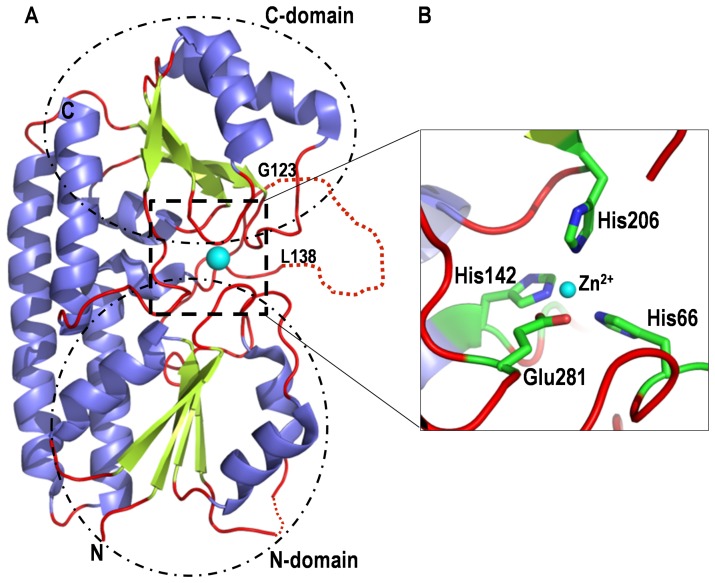
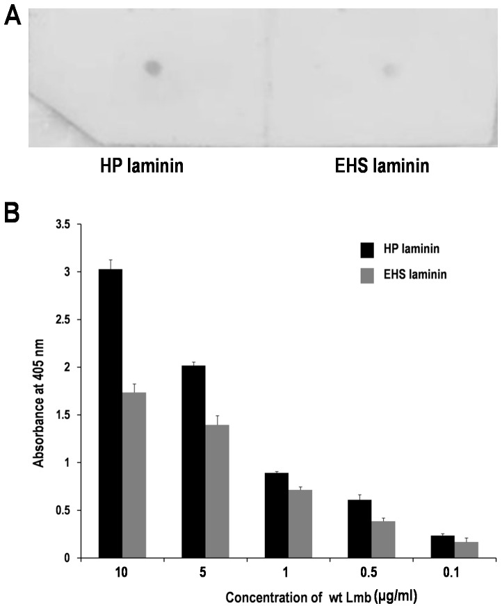
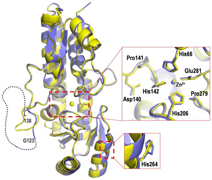
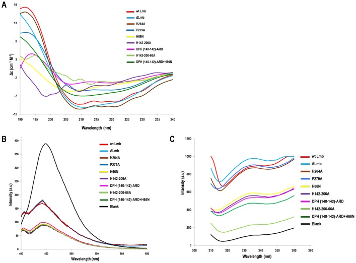
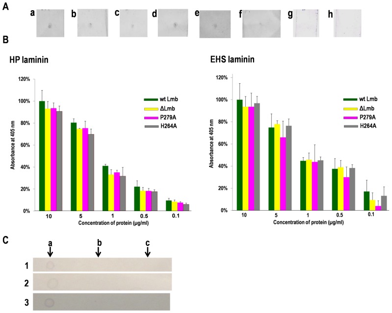
Lmb is a 34 kDa laminin binding surface adhesin of Streptococcus agalactiae. The structure of Lmb reported by us recently has shown that it consists of a metal binding crevice, in which a zinc ion is coordinated to three highly conserved histidines. To elucidate the structural and functional significance of the metal ion in Lmb, these histidines have been mutated to alanine and single, double and triple mutants were generated. These mutations resulted in insolubility of the protein and revealed altered secondary and tertiary structures, as evidenced by circular dichroism and fluorescence spectroscopy studies. The mutations also significantly decreased the binding affinity of Lmb to laminin, implicating the role played by the metal binding residues in maintaining the correct conformation of the protein for its binding to laminin. A highly disordered loop, proposed to be crucial for metal acquisition in homologous structures, was deleted in Lmb by mutation (ΔLmb) and its crystal structure was solved at 2.6 Å. The ΔLmb structure was identical to the native Lmb structure with a bound zinc ion and exhibited laminin binding activity similar to wild type protein, suggesting that the loop might not have an important role in metal acquisition or adhesion in Lmb. Targeted mutations of histidine residues confirmed the importance of the zinc binding crevice for the structure and function of the Lmb adhesin.
Conflict of interest statement
Figures
Similar articles
-
Molecular dynamics simulation of metal free structure of Lmb, a laminin-binding adhesin of Streptococcus agalactiae: metal removal and its structural implications.J Biomol Struct Dyn. 2019 Feb;37(3):714-725. doi: 10.1080/07391102.2018.1438923. Epub 2018 Feb 23. J Biomol Struct Dyn. 2019. PMID: 29421962
-
Structure of laminin-binding adhesin (Lmb) from Streptococcus agalactiae.Acta Crystallogr D Biol Crystallogr. 2009 Dec;65(Pt 12):1262-9. doi: 10.1107/S0907444909038359. Epub 2009 Nov 17. Acta Crystallogr D Biol Crystallogr. 2009. PMID: 19966412
-
The Adc/Lmb System Mediates Zinc Acquisition in Streptococcus agalactiae and Contributes to Bacterial Growth and Survival.J Bacteriol. 2016 Nov 18;198(24):3265-3277. doi: 10.1128/JB.00614-16. Print 2016 Dec 15. J Bacteriol. 2016. PMID: 27672194 Free PMC article.
-
Lmb, a protein with similarities to the LraI adhesin family, mediates attachment of Streptococcus agalactiae to human laminin.Infect Immun. 1999 Feb;67(2):871-8. doi: 10.1128/IAI.67.2.871-878.1999. Infect Immun. 1999. PMID: 9916102 Free PMC article.
-
Structural and genetic analysis of laminin-nidogen interaction.Ann N Y Acad Sci. 1998 Oct 23;857:130-42. doi: 10.1111/j.1749-6632.1998.tb10113.x. Ann N Y Acad Sci. 1998. PMID: 9917838 Review.
Cited by
-
Bacillus anthracis S-layer protein BslA binds to extracellular matrix by interacting with laminin.BMC Microbiol. 2016 Aug 11;16(1):183. doi: 10.1186/s12866-016-0802-8. BMC Microbiol. 2016. PMID: 27514510 Free PMC article.
-
Prevalence, serotypes and virulence genes of Streptococcus agalactiae isolated from pregnant women with 35-37 weeks of gestation.BMC Infect Dis. 2021 Jan 14;21(1):73. doi: 10.1186/s12879-020-05603-5. BMC Infect Dis. 2021. PMID: 33446117 Free PMC article.
-
Antimicrobial Resistance and Virulence Genes of Streptococcus Agalactiae Isolated from Mastitis Milk Samples in China.J Vet Res. 2022 Dec 16;66(4):581-590. doi: 10.2478/jvetres-2022-0069. eCollection 2022 Dec. J Vet Res. 2022. PMID: 36846045 Free PMC article.
-
Current research update on group B streptococcal infection related to obstetrics and gynecology.Front Pharmacol. 2024 Jun 17;15:1395673. doi: 10.3389/fphar.2024.1395673. eCollection 2024. Front Pharmacol. 2024. PMID: 38953105 Free PMC article. Review.
-
Novel Nonsense Mutation in SLC39A13 Initially Presenting as Myopathy: Case Report and Review of the Literature.Mol Syndromol. 2018 Feb;9(2):100-109. doi: 10.1159/000485881. Epub 2018 Jan 24. Mol Syndromol. 2018. PMID: 29593477 Free PMC article.
References
-
- Schuchat A (1999) Group B streptococcus. Lancet 353: 51–56. - PubMed
-
- Tenenbaum T, Spellerberg B, Adam R, Vogel M, Kim KS, et al. (2007) Streptococcus agalactiae invasion of human brain microvascular endothelial cells is promoted by the laminin-binding protein Lmb. . Microbes Infect. 9: 714–720. - PubMed
-
- Ragunathan P, Spellerberg B, Ponnuraj K (2009) Structure of laminin-binding adhesin (Lmb) from Streptococcus agalactiae. Acta Crystallogr D 65: 1262–1269. - PubMed
Publication types
MeSH terms
Substances
LinkOut - more resources
Full Text Sources
Other Literature Sources
