. 2013 Jun 24;8(6):e67522.
doi: 10.1371/journal.pone.0067522. Print 2013.

edn1 and hand2 Interact in early regulation of pharyngeal arch outgrowth during zebrafish development

Affiliations

edn1 and hand2 Interact in early regulation of pharyngeal arch outgrowth during zebrafish development

Mark M Sasaki et al. PLoS One. .

Abstract

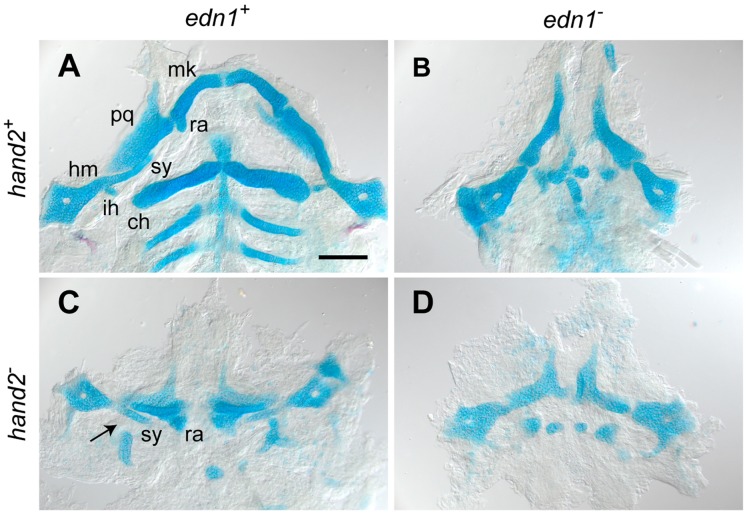
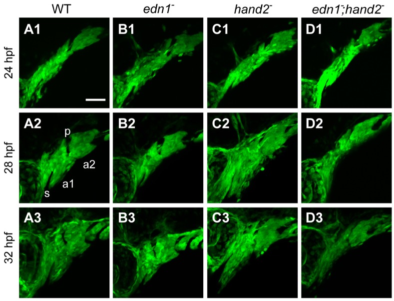
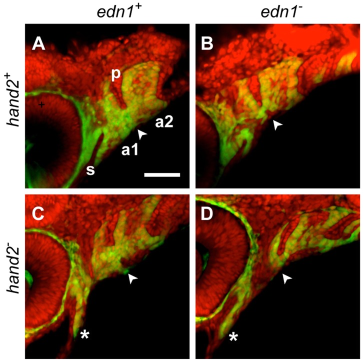
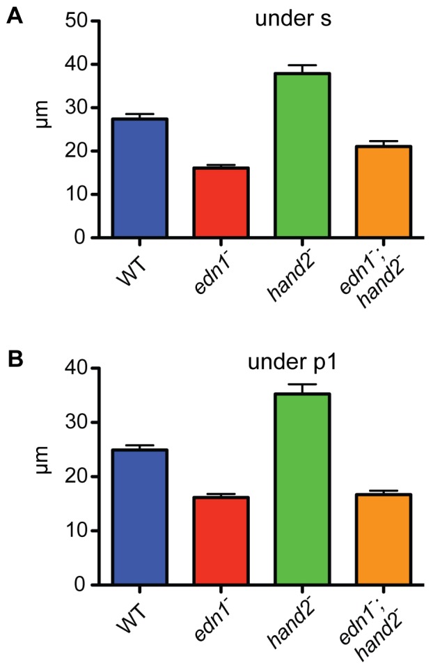
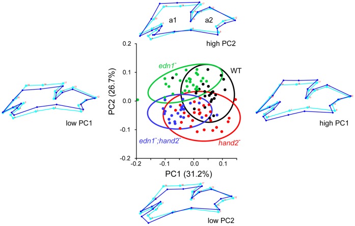
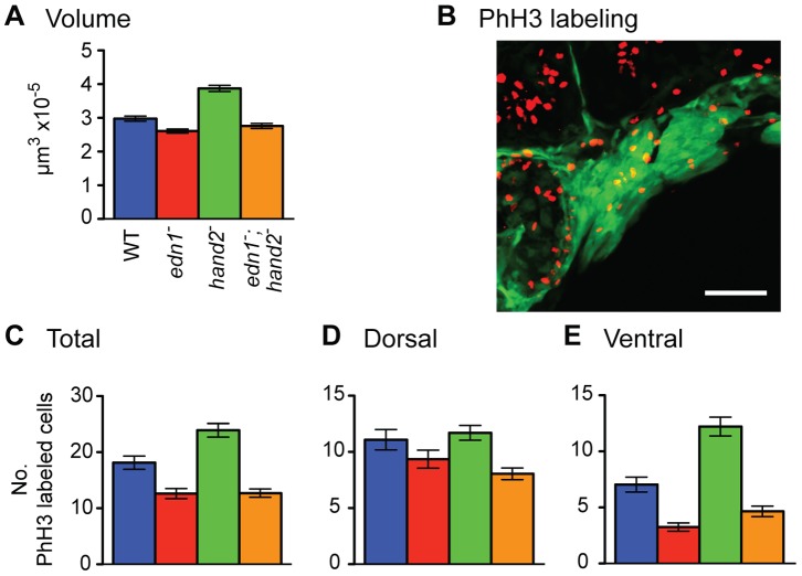
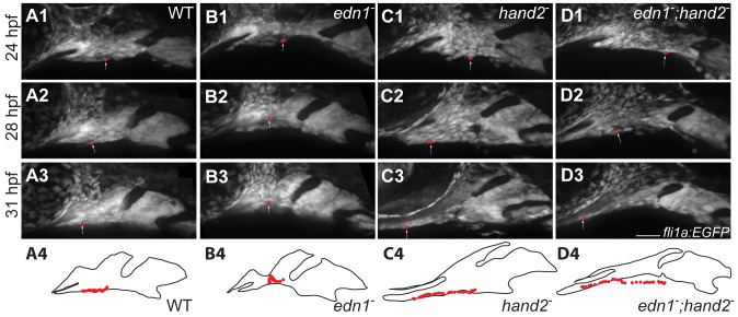
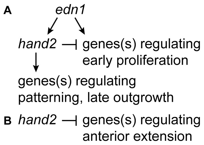
Endothelin-1 (Edn1) signaling provides a critical input to development of the embryonic pharygneal arches and their skeletal derivatives, particularly the articulating joints and the ventral skeleton including the lower jaw. Previous work in zebrafish has mostly focused on the role of Edn1 in dorsal-ventral (DV) patterning, but Edn1 signaling must also regulate tissue size, for with severe loss of the pathway the ventral skeleton is not only mispatterned, but is also prominently hypoplastic--reduced in size. Here we use mutational analyses to show that in the early pharyngeal arches, ventral-specific edn1-mediated proliferation of neural crest derived cells is required for DV expansion and outgrowth, and that this positive regulation is counterbalanced by a negative one exerted through a pivotal, ventrally expressed Edn1-target gene, hand2. We also describe a new morphogenetic cell movement in the ventral first arch, sweeping cells anterior in the arch to the region where the lower jaw forms. This movement is negatively regulated by hand2 in an apparently edn1-independent fashion. These findings point to complexity of regulation by edn1 and hand2 at the earliest stages of pharyngeal arch development, in which control of growth and morphogenesis can be genetically separated.


Conflict of interest statement

Competing Interests: The authors have declared that no competing interests exist.

Figures

Figure 1
Figure 1. edn1 and hand2 are required for ventral jaw cartilage development.
4 dpf zebrafish skeletons were cartilage and bone stained with Alcian Blue and Alizarin Red, dissected and flat-mounted. (A) WT, with cartilages of the first two arches indicated: In the first arch the dorsal palatoquadrate (pq) and ventral Meckel’s cartilage (mk) articulate, Meckel’s cartilage includes a distinctive retroarticular process (ra) adjacent to the joint. In the second arch the more dorsal hyosymplectic cartilage is subdivided into the hyomandibular (hm) and symplectic (sy) regions, and the prominent ventral cartilage is the ceratohyal (ch). The interhyal cartilage (ih) forms a small hinge within the joint region. (B) In the edn1 larva, ventral cartilages and elements of the joint regions are missing or prominently disrupted. Unidentifiable elements are scattered near the ventral midline. (C) The hand2 larva exhibits ventral reductions similar to edn1, but the arch 2 symplectic cartilage and joint (arrow) are present and the arch 1 retroarticular process is expanded rather than missing. (D) The edn1;hand2 larva exhibits defects similar to edn1; in particular, the symplectic cartilage and retroarticular process cannot be identified. Anterior is upward and right is towards the left. Scale bar: 100 µm.
Figure 2
Figure 2. edn1 and hand2 are required for proper growth and morphogenesis of the early pharyngeal arches.
Projections of lateral confocal images of embryos expressing fli1a:EGFP taken at 24, 28, and 32 hpf. This figure provides an overview of changes in the first two arches (a1, a2, separated by the first pharyngeal pouch, p) that are quantified and more fully documented in subsequent figures. Dorsal is to the top in each panel, and anterior is to the left. No differences in arch morphology are observed among the four genotypes at 24 hpf (upper row of panels). Subsequently the arches prominently lengthen along the DV axis; this DV extension is evidently reduced in the edn1 mutant (B2,B3) and edn1;hand2 double mutant (D2,D3) compared to WT and the hand2 mutant. The arches (particularly the first), also shorten along the AP axis but there appears to be no differences in AP shortening among the genotypes. The hand2 mutant also shows marked expansion of mesenchyme ventral to the stomodeum (s) in the anterior first arch (C2,C3). Scale bar: 50 µm.
Figure 3
Figure 3. Counterstaining the fli1a:EGFP-expressing pharyngeal arches (a1, a2) with the nucleic acid stain SYTO 59 (red) clarifies the ectomesenchymal phenotypes (double-labeled).
The DV ventral extension differences (arrowheads) are apparent ventral to the red-stained first pharyngeal pouch (p). Compared to the WT (A) this ventral region of mesenchyme is reduced in the edn1 mutant (B) and double mutant (D), and expanded in the hand2 mutant (C). Anterior extension of mesenchyme ventral to the stomodeum (s) is prominent in the hand2 mutant and double mutant (*, C, D).
Figure 4
Figure 4. Quantification of the DV extension phenotypes supports edn1 being epistatic to hand2 in a single regulatory pathway.
DV-length measurements were made for each group, beneath the end of the invaginated stomodeum (A) and beneath the first pharyngeal pouch (B, location indicated by arrowheads in Figure 3). In both locations, and compared to the WT, the edn1 mutant shows reduced DV extension, the hand2 mutant shows increased DV extension and the double mutant most closely resembles the edn1 mutant. Tukey-Kramer analysis supports these three groupings as being significantly different (P<0.05), whereas the differences between edn1 and the double mutant are insignificant.
Figure 5
Figure 5. Quantification of arch morphology by PCA reveals that edn1 and hand2 regulate two prominent features of arch shape, DV extension (PC1) and ventral arch 1 anterior extension (PC2).
Together these two deformations account for 58% of the total shape variation within the dataset, which uses landmarks (numbered in the accompanying wireframe diagrams) to outline the fli1a:EGFP-expressing tissue of the first two pharyngeal arches at 28 hpf. Shape change along the AP axis is minimal. We interpret the nature of the shape changes from the wireframes. For each, the light blue wireframe shows the consensus configuration and the dark blue wireframe shows the deformation associated with change (+ or -) along a PC axis. In the plot itself the individual measurements are grouped by genotype with a different color and 95% ellipses for each (sampling 23 or more individuals in each group). The data show overlap among the groups, yet Procrustes distance measurements reveal all the groups are significantly different from one another (P<0.0001 by permutation; data not shown). WT and the hand2 single mutant score high on PC1, whereas the edn1 single mutant and the double mutant score low on PC1. WT and the edn1 mutant score high on PC2, whereas the hand2 mutant and the double mutant score low. Hence the double mutant phenotype is additive in this analysis, combining the shape features of both single mutants.
Figure 6
Figure 6. edn1 positively and hand2 negatively regulate the volume of pharyngeal arch fli1a:EGFP expressing mesenchyme (A) and ventral domain proliferation of neural crest derived cells (B–E).
Data are for 28 hpf, a minimum of 23 individuals of each genotype were sampled. (B) shows an example 2-color image of phospho-histone H3 labeling for mitosing cells (red) and fli1a:EGFP expression (green). (C–E) show mean counts of double-labeled cells ±SEM. Tukey-Kramer comparison of the mean volumes (in A) reveals that the edn1 mutant volume is significantly lower than WT, and that the hand2 mutant volume is significantly higher (P<0.05). Statistically the double mutant phenotype is neither different from the edn1 mutant nor WT, suggesting some degree of phenotypic rescue of arch volume when hand2 does not function. Tukey-Kramer analysis of the levels of proliferation in both the total mesenchyme (C) and ventral sector of mesenchyme (E) reveals three statistically distinctive classes, WT, the single hand2 mutant, and the single edn1 plus edn1;hand2 mutant class (P<0.05). Genotypic differences for the dorsal mesenchyme (D) are insignificant.
Figure 7
Figure 7. hand2 negatively regulates anterior flow of ventral arch 1 cells.
fli1a:EGFP animals were imaged by time lapse microscopy from 24–32 hpf. Panels are excerpts from the movie S1 in which maximum confocal projections are presented. Red balls represent the location of an individual cell that was manually tracked in each frame. The positions of tracked cells are overlaid in the outlines. Cells travel from near the midpoint of arch1 to a location posterior to the stomodeum and the eye in wild types and edn1 mutants (A, B). Cells originating at a similar location in hand2 and hand2;edn1 double mutants travel much further, to a position well under the stomodeum and the eye (C, D). Scale bar: 50 µm.
Figure 8
Figure 8. Regulatory genetic network for control of growth and morphogenesis in the pharyngeal arches by edn1 and hand2.
(A) The model assumes that regulation of early proliferation in the ventral arches, in which edn1 is epistatic to hand2, can account for the DV extension phenotype explored in this study. The positive pathway regulating patterning and late outgrowth accounts for the general reduction of elements in the ventral skeleton, as well skeletal disruption because of downregulation of downsteam patterning genes studied elsewhere , , , . This model predicts a temporally dynamic switching in hand2 function, from negative to positive, which can be explored in future studies. (B) We propose hand2 negatively regulates the anterior extension movement in the ventral first arch independent of edn1 function.

Similar articles

Cited by

References

    1. Schilling TF, Piotrowski T, Grandel H, Brand M, Heisenberg CP, et al. (1996) Jaw and branchial arch mutants in zebrafish I: branchial arches. Development 123: 329–344. - PubMed
    1. Piotrowski T, Schilling TF, Brand M, Jiang YJ, Heisenberg CP, et al. (1996) Jaw and branchial arch mutants in zebrafish II: anterior arches and cartilage differentiation. Development 123: 345–356. - PubMed
    1. Clouthier DE, Schilling TF (2004) Understanding endothelin-1 function during craniofacial development in the mouse and zebrafish. Birth Defects Res C Embryo Today 72: 190–199. - PubMed
    1. Kimmel CB, Walker MB, Miller CT (2007) Morphing the hyomandibular skeleton in development and evolution. J Exp Zool B Mol Dev Evol 308: 609–624. - PubMed
    1. Thomas T, Kurihara H, Yamagishi H, Kurihara Y, Yazaki Y, et al. (1998) A signaling cascade involving endothelin-1, dHAND and Msx1 regulates development of neural-crest-derived branchial arch mesenchyme. Development 125: 3005–3014. - PubMed

Publication types

MeSH terms

Substances