. 2013 Jun 24;8(6):e67679.
doi: 10.1371/journal.pone.0067679. Print 2013.

Dapper antagonist of catenin-1 cooperates with Dishevelled-1 during postsynaptic development in mouse forebrain GABAergic interneurons

Affiliations

Dapper antagonist of catenin-1 cooperates with Dishevelled-1 during postsynaptic development in mouse forebrain GABAergic interneurons

Annie Arguello et al. PLoS One. .

Abstract

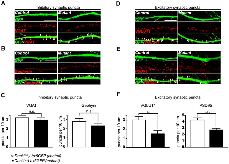
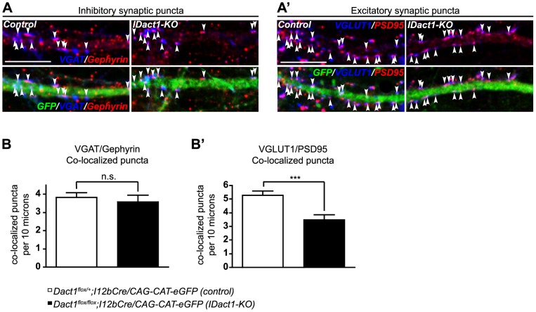
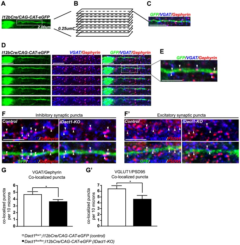
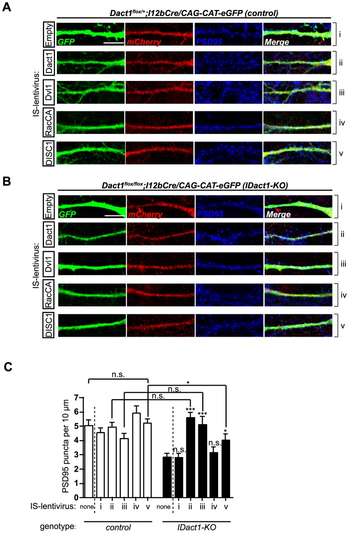
Synaptogenesis has been extensively studied along with dendritic spine development in glutamatergic pyramidal neurons, however synapse development in cortical interneurons, which are largely aspiny, is comparatively less well understood. Dact1, one of 3 paralogous Dact (Dapper/Frodo) family members in mammals, is a scaffold protein implicated in both the Wnt/β-catenin and the Wnt/Planar Cell Polarity pathways. We show here that Dact1 is expressed in immature cortical interneurons. Although Dact1 is first expressed in interneuron precursors during proliferative and migratory stages, constitutive Dact1 mutant mice have no major defects in numbers or migration of these neurons. However, cultured cortical interneurons derived from these mice have reduced numbers of excitatory synapses on their dendrites. We selectively eliminated Dact1 from mouse cortical interneurons using a conditional knock-out strategy with a Dlx-I12b enhancer-Cre allele, and thereby demonstrate a cell-autonomous role for Dact1 during postsynaptic development. Confirming this cell-autonomous role, we show that synapse numbers in Dact1 deficient cortical interneurons are rescued by virally-mediated re-expression of Dact1 specifically targeted to these cells. Synapse numbers in these neurons are also rescued by similarly targeted expression of the Dact1 binding partner Dishevelled-1, and partially rescued by expression of Disrupted in Schizophrenia-1, a synaptic protein genetically implicated in susceptibility to several major mental illnesses. In sum, our results support a novel cell-autonomous postsynaptic role for Dact1, in cooperation with Dishevelled-1 and possibly Disrupted in Schizophrenia-1, in the formation of synapses on cortical interneuron dendrites.


Conflict of interest statement

Competing Interests: The authors have declared that no competing interests exist.

Figures

Figure 1
Figure 1. Dact1 expression in the developing forebrain.
In situ hybridization (Dact1) in rostral to caudal coronal sections through wild type E14.5 (A) and E18.5 brains (A'). Arrowheads in A indicate Dact1 expression in the deep migratory stream (subventricular zone/intermediate zone) of ganglionic eminence-derived interneurons within the developing cortex. In situ hybridization (Dact1) at E14.5. B Dact1 expression in Dlx1−/−;Dlx2−/− mice (bottom) compared to wild type (top) and higher magnification (B'), shows the absence (closed arrowheads) of Dact1 expression in Dlx1−/−;Dlx2−/− mutants in the usual position of the deep migratory stream, indicating that Dact1 is normally expressed in the immature interneurons that fail to migrate in this Dlx double mutant. Asterisks in B' indicate the shift in Dact1 mRNA expression from the subventricular zone to the ventricular zone of the ganglionic eminences in Dlx1−/−;Dlx2−/− mice. Dact1 remains expressed in the marginal zone of the cortex in the double mutants (open arrowheads), reflecting Dlx-independent expression in developing excitatory neurons. cpu, caudate-putamen; ctx, cortex; fr ctx, frontal cortex; gp, globus pallidus; hip, hippocampus; sep, septum; svz, subventricular zone. Scale bars = 500 µm.
Figure 2
Figure 2. Dact1−/− mice have no major defects in the migratory paths of ganglionic eminence-derived GABAergic interneurons.
In situ hybridization (Lhx6) in rostral to caudal coronal sections through E12.5 (A), E14.5 (A'), and E18.5 (A″) Dact1−/− brains (bottom) compared to wild type (top). Arrowheads indicate Lhx6 expression in the subventricular zone (closed) and marginal zone (open) of the cortex. In situ hybridization (Dlx1) in E12.5 (B), E14.5 (B'), and E18.5 (B″) Dact1−/− mice (bottom) compared to wild type (top). cpu, caudate-putamen; cp, cortical plate; hip, hippocampus; lge, lateral ganglionic eminence; mge, medial ganglionic eminence; NAcc, nucleus accumbens; poa, preoptic area; se, septum; st, striatum; vp, ventral pallidum. Scale bars = 500 µm.
Figure 3
Figure 3. Cortical interneurons from constitutive Dact1 mutant mice have fewer excitatory synapses on primary dendrites.
Primary cortical cultures were prepared from postnatal day 0 Dact1−/−;Lhx6GFP (right) and control (left) brains, fixed at day in vitro 15, and synaptic puncta counted along GFP labeled primary dendrites from the cell soma to the first major branch point (arrowheads). Inhibitory synaptic puncta were visualized using antibodies against VGAT (presynaptic, A) and Gephyrin (postsynaptic, B) with each marker counted irrespective of co-localization with the other; C Quantification per 10 µm of primary dendrite length in control (open bars) and constitutive Dact1 mutant neurons (closed bars). Excitatory synaptic puncta were visualized using antibodies against VGLUT1 (presynaptic, D) and PSD95 (postsynaptic, E) with each marker counted irrespective of co-localization with the other; F Quantification per 10 µm of primary dendrite length. Data shown are mean ± sem of at least 3 independent experiments, collected from at least 3 mice per genotype, 10–15 neurons per animal. **p<0.01; ***p<0.001; n.s., not significant. Scale bars = 10 µm.
Figure 4
Figure 4. Reduction of excitatory synapses in Dact1 mutant cortical interneurons is cell-autonomous.
Primary cortical cultures were prepared from postnatal day 0 Interneuron-specific Dact1 mutant (IDact1-KO) (right) and control (left) brains, fixed at day in vitro 15, and pre- and post-synaptic co-localized puncta counted along GFP labeled primary dendrites from the cell soma to the first major branch point (arrowheads). VGAT/Gephyrin (inhibitory, A) and Vglut1/PSD95 (excitatory, A') co-localized puncta (arrowheads) along the GFP+ dendrite in control (left) and IDact1-KO (right) mice. Quantification of co-localized inhibitory (B) and excitatory (B') pre- and postsynaptic puncta in control (open bars) and IDact1-KO mutants (closed bars). Data shown are mean ± sem of at least 2 independent experiments, collected from at least 2 mice per genotype, 10–15 neurons per animal. ***p<0.001; n.s., not significant. Scale bars = 10 µm.
Figure 5
Figure 5. Interneuron-specific Dact1 mutant mice (IDact1-KO) have fewer excitatory and inhibitory synapses along cortical interneuron dendrites.
A-E Experimental design. A Composite z-stack image of a GFP+ interneuron located in upper layers of the primary somatosensory cortex, selected for synaptic puncta quantification from postnatal day 30 mouse brain sections. B This representative composite z-stack image is composed of 9 optical sections, 0.25 microns apart, encompassing the proximal dendrite. C Example of optical section used to confirm pre- and post-synaptic co-localization along the GFP+ dendrite. D Representative serial z-stack images used to confirm synaptic puncta co-localization in each z-plane. Co-localized puncta visible in adjacent serial sections are scored only once to avoid duplicate counting of individual synapses. E Boxed region is shown at the higher magnification, as employed to identify: VGAT/Gephyrin (inhibitory, F) and Vglut1/PSD95 (excitatory, F') co-localized puncta (arrowheads) along the GFP+ dendrite in control (left) and IDact1-KO (right) mice. Quantification of co-localized inhibitory (G) and excitatory (G') pre- and post-synaptic puncta in control (open bars) and IDact1-KO mutants (closed bars). Data shown are mean ± sem of at least 3 independent experiments, collected from at least 3 mice per genotype, 5-10 neurons per animal. *p<0.05. Scale bars = 5 µm.
Figure 6
Figure 6. Interneuron-specific expression of Dact1, Dvl1, or DISC1 rescues excitatory synapse numbers in IDact1-KO interneurons.
A–B Infected interneurons were detected by expression of mCherry. Characterization of PSD95 puncta on primary dendrites of cultured GFP+ cortical interneurons from control (A) and I-Dact1KO (B) mice infected with IS-lentiviral constructs expressing (i) mCherry alone, (ii) Dact1, (iii) Dvl1, (iv) RacCA, or (v) DISC1. C Quantification of PSD95 puncta per 10 µm of primary dendrite length in control (open bars) and IDact1-KO mutant neurons (closed bars). Results are presented as mean ± sem from at least 2 independent experiments, collected from at least 2 mice per genotype, 10–15 neurons per animal. *p<0.05; ***p<0.001; n.s., not significant. All p values are relative to either control (IS-lentivirus, none) or IDact1-KO (IS-lentivirus, none). Scale bars = 10 µm.

References

    1. Badawy RA, Harvey AS, Macdonell RA (2009) Cortical hyperexcitability and epileptogenesis: Understanding the mechanisms of epilepsy - part 2. J Clin Neurosci 16: 485–500. - PubMed
    1. Benes FM, Berretta S (2001) GABAergic interneurons: implications for understanding schizophrenia and bipolar disorder. Neuropsychopharmacology 25: 1–27. - PubMed
    1. Guilmatre A, Dubourg C, Mosca AL, Legallic S, Goldenberg A, et al. (2009) Recurrent rearrangements in synaptic and neurodevelopmental genes and shared biologic pathways in schizophrenia, autism, and mental retardation. Arch Gen Psychiatry 66: 947–956. - PMC - PubMed
    1. Rubenstein JL (2011) Annual Research Review: Development of the cerebral cortex: implications for neurodevelopmental disorders. J Child Psychol Psychiatry 52: 339–355. - PMC - PubMed
    1. Rubenstein JL, Merzenich MM (2003) Model of autism: increased ratio of excitation/inhibition in key neural systems. Genes Brain Behav 2: 255–267. - PMC - PubMed

Publication types

MeSH terms