Increased expression of nestin in human pterygial epithelium
- PMID: 23826515
- PMCID: PMC3693002
- DOI: 10.3980/j.issn.2222-3959.2013.03.01
Increased expression of nestin in human pterygial epithelium
Abstract
Aim: To investigate the distribution of nestin-positive cells in pterygium, as well as the relationship between nestin-positive cells and proliferative cells in the pathogenesis of pterygium.
Methods: Nine pterygium specimens and 5 normal conjunctiva specimens were investigated. All explanted specimens were immediately immersed in 5-Ethynyl-2'-deoxyuridine, and were subjected to hematoxylin and eosin staining, as well as immunostaining to detect nestin.
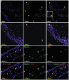
Results: Small sub-populations of nestin-expressing cells in both normal and pterygial conjunctiva epithelium were found. These were located at the superficial layer of the epithelium, and were significantly increased (P=0.007) and spread out in the pterygial conjunctiva epithelium, even though these cells were mitotically quiescent.
Conclusion: In pterygium, more nestin-positive cells were present at the superficial layer of the epithelium. With growing scientific evidence that nestin plays an important role in defining various specialized cell types, such as stem cells, cancer cells and angiogenic cells, further investigations on the roles of nestin-expressing cells in pterygium may help to uncover the mechanisms of initiation, development and the prognosis of this disease.
Keywords: distribution; human; nestin; proliferation; pterygium.
Figures


Similar articles
-
Immunophenotypic characterization of telocyte-like cells in pterygium.Mol Vis. 2018 Dec 29;24:853-866. eCollection 2018. Mol Vis. 2018. PMID: 30713424 Free PMC article.
-
Distribution of vimentin-expressing cells in pterygium: an immunocytochemical study of impression cytology specimens.Cornea. 2009 Jun;28(5):547-52. doi: 10.1097/ICO.0b013e318190931b. Cornea. 2009. PMID: 19421041
-
The study on the expression of keratin proteins in pterygial epithelium.Yan Ke Xue Bao. 2000 Mar;16(1):48-52. Yan Ke Xue Bao. 2000. PMID: 12579729
-
p53 Expression in pterygium by immunohistochemical analysis: a series report of 127 cases and review of the literature.Cornea. 2005 Jul;24(5):583-6. doi: 10.1097/01.ico.0000154404.86462.35. Cornea. 2005. PMID: 15968165 Review.
-
Immunohistochemical profile of VEGF, TGF-β and PGE₂ in human pterygium and normal conjunctiva: experimental study and review of the literature.Int J Immunopathol Pharmacol. 2012 Jul-Sep;25(3):607-15. doi: 10.1177/039463201202500307. Int J Immunopathol Pharmacol. 2012. PMID: 23058011 Review.
Cited by
-
Identification and differentiation therapy strategy of pterygium in vitro.Am J Transl Res. 2018 Aug 15;10(8):2619-2627. eCollection 2018. Am J Transl Res. 2018. PMID: 30210698 Free PMC article.
-
Immunophenotypic characterization of telocyte-like cells in pterygium.Mol Vis. 2018 Dec 29;24:853-866. eCollection 2018. Mol Vis. 2018. PMID: 30713424 Free PMC article.
References
-
- Chowers I, Pe'er J, Zamir E, Livni N, Ilsar M, Frucht-Pery J. Proliferative activity and p53 expression in primary and recurrent pterygia. Ophthalmology. 2001;108(5):985–988. - PubMed
-
- Cui D, Pan Z, Zhang S, Zheng J, Huang Q, Wu K. Downregulation of c-Myc in pterygium and cultured pterygial cells. Clin Experiment Ophthalmol. 2011;39(8):784–792. - PubMed
-
- Umemoto T, Yamato M, Nishida K, Yang J, Tano Y, Okano T. Limbal epithelial side-population cells have stem cell-like properties, including quiescent state. Stem Cells. 2006;24(1):86–94. - PubMed
LinkOut - more resources
Full Text Sources
