

Measurement of tumour size with mammography, sonography and magnetic resonance imaging as compared to histological tumour size in primary breast cancer
- PMID: 23826951
- PMCID: PMC3704854
- DOI: 10.1186/1471-2407-13-328
Measurement of tumour size with mammography, sonography and magnetic resonance imaging as compared to histological tumour size in primary breast cancer
Abstract
Background: Tumour size in breast cancer influences therapeutic decisions. The purpose of this study was to evaluate sizing of primary breast cancer using mammography, sonography and magnetic resonance imaging (MRI) and thereby establish which imaging method most accurately corresponds with the size of the histological result.
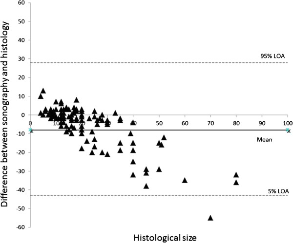
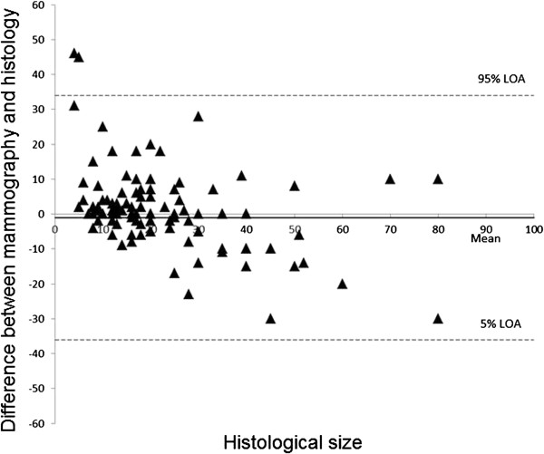
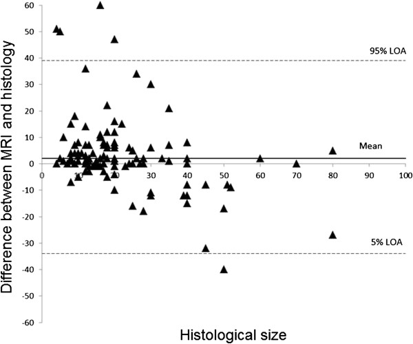
Methods: Data from 121 patients with primary breast cancer were analysed in a retrospective study. The results were divided into the groups "ductal carcinoma in situ (DCIS)", invasive ductal carcinoma (IDC) + ductal carcinoma in situ (DCIS)", "invasive ductal carcinoma (IDC)", "invasive lobular carcinoma (ILC)" and "other tumours" (tubular, medullary, mucinous and papillary breast cancer). The largest tumour diameter was chosen as the sizing reference in each case. Bland-Altman analysis was used to determine to what extent the imaging tumour size correlated with the histopathological tumour sizes.
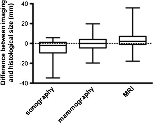
Results: Tumour size was found to be significantly underestimated with sonography, especially for the tumour groups IDC + DCIS, IDC and ILC. The greatest difference between sonographic sizing and actual histological tumour size was found with invasive lobular breast cancer. There was no significant difference between mammographic and histological sizing. MRI overestimated non-significantly the tumour size and is superior to the other imaging techniques in sizing of IDC + DCIS and ILC.
Conclusions: The histological subtype should be included in imaging interpretation for planning surgery in order to estimate the histological tumour size as accurately as possible.
Figures
References
-
- American College of Radiology (ACR) Breast Imaging Reporting and Data System Altlas (BI-RADS Atlas) 4. Reston, VA 20191, USA; 2003.
-
- Madjar H, Ohlinger R, Mundinger A, Watermann D, Frenz JP, Bader W, Schulz-Wendtland R, Degenhardt F. BIRADS-Analogue Degum Criteria for Findings in Breast Ultrasound – Consensus of the DEGUM Committee on Breast Ultrasound. Ultraschall in Med. 2006;27:374–379. doi: 10.1055/s-2006-926943. - DOI - PubMed
-
- Bosch AM, Kessels AG, Beets GL, Rupa JD, Koster D, van Engelshoven JM, von Meyenfeldt MF. Preoperative estimation of the pathological breast tumour size by physical examination, mammography and ultrasound: a prospective study on 105 invasive tumours. Eur J Radiol. 2003;48:285–292. doi: 10.1016/S0720-048X(03)00081-0. - DOI - PubMed
Publication types
MeSH terms
LinkOut - more resources
Full Text Sources
Other Literature Sources
Medical
