Review
. 2013 Nov 15;539(2):203-13.
doi: 10.1016/j.abb.2013.06.012. Epub 2013 Jul 1.

Structural basis of carotenoid cleavage: from bacteria to mammals

Affiliations
Review

Structural basis of carotenoid cleavage: from bacteria to mammals

Xuewu Sui et al. Arch Biochem Biophys. .

Abstract

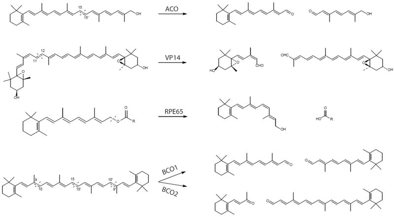
Carotenoids and their metabolic derivatives serve critical functions in both prokaryotic and eukaryotic cells, including pigmentation, photoprotection and photosynthesis as well as cell signaling. These organic compounds are also important for visual function in vertebrate and non-vertebrate organisms. Enzymatic transformations of carotenoids to various apocarotenoid products are catalyzed by a family of evolutionarily conserved, non-heme iron-containing enzymes named carotenoid cleavage oxygenases (CCOs). Studies have revealed that CCOs are critically involved in carotenoid homeostasis and essential for the health of organisms including humans. These enzymes typically display a high degree of regio- and stereo-selectivity, acting on specific positions of the polyene backbone located in their substrates. By oxidatively cleaving and/or isomerizing specific double bonds, CCOs generate a variety of apocarotenoid isomer products. Recent structural studies have helped illuminate the mechanisms by which CCOs mobilize their lipophilic substrates from biological membranes to perform their characteristic double bond cleavage and/or isomerization reactions. In this review, we aim to integrate structural and biochemical information about CCOs to provide insights into their catalytic mechanisms.

Keywords: ACO; Apocarotenoid; Carotenoid; Carotenoid oxygenase; RPE65; VP14.


Figures

Figure 1
Figure 1. Enzymatic reactions mediated by five selected carotenoid cleavage oxygenases
Dashed lines in substrates indicate cleavage sites. ACO, apocarotenoid oxygenase; VP14, viviparous 14; RPE65, retinal pigment epithelium-specific 65 kDa protein; BCO1, β,β-carotene-15, 15’-oxygenases; BCO2, β,β-carotene-9, 10-oxygenases.
Figure 2
Figure 2. Structure-based sequence alignment of selected carotenoid cleavage oxygenases
The red background indicates sequence identity and red letters stand for sequence similarity. All structural elements of VP14, ACO and RPE65 are shown over the sequence alignment. Structural elements of α-helices and β-strands are displayed as blue squiggles and arrows, respectively. The strictly conserved iron-coordinating His residues ( formula image
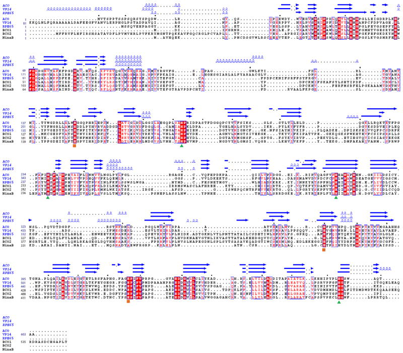
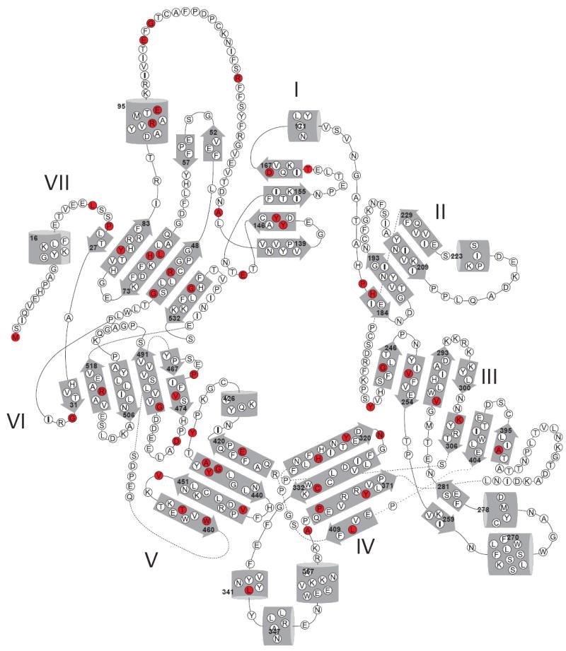
) and their fixating Glu residues ( formula image) are labeled. Dots mark every tenth residue. The sequences were aligned with with T-coffee [75] and the figure was generated with ESPript [76].
Figure 3
Figure 3. Crystal structure and topology diagram of Synechocystis ACO (left), maize VP14 (center) and bovine RPE65 (right)
(PDB accession codes: 2BIW, 3NPE and 3FSN). The ferrous catalytic iron is colored in orange. Secondary structural elements consisting of α-helices and β-sheets are colored in blue and green, respectively. The red dashed line in the RPE65 diagram represents the unmodeled loop. The blade labeling shown for the ACO topology diagram is the same for the other two topology diagrams.
Figure 4
Figure 4. Catalytic centers of ACO (A), VP14 (B) and RPE65 (C)
The iron ion is shown as an orange sphere, with the six proposed coordination sites arranged in an octahedral geometry. Four sites are occupied by the strictly conserved His residues. The second coordination sphere formed by three conserved Glu residues most likely helps orient the direct His ligands and may modulate the iron redox potential. The di-cis apocarotenoid substrate modeled in the ACO structure is displayed as orange sticks. Dioxygen and water (red stick and blue sphere, respectively) are modeled in the two remaining coordination sites of the VP14 iron center. Glu477 in VP14 points away from its putative coordinating His residue.
Figure 5
Figure 5. Surface views of the three crystal structures of ACO (A), VP14 (B) and RPE65 (C) with their hydrophobic patches for putative membrane binding
Left, hydrophobic surface portions of each enzyme are colored in yellow. Right, hydrophobic residues colored in yellow for membrane penetration are shown in each structure. The arrowhead indicates the opening of cavities that lead to the active site iron.
Figure 6
Figure 6. Tunnels lead to the active center of ACO (A), VP14 (B) and RPE65 (C)
The red and blue mesh represent tunnels connecting the membrane binding region of the protein to the active site and the active site to cytosolic-facing regions, respectively. The location of rightmost portion of the red mesh corresponds to the sites indicated by arrowheads in Fig. 5. Retinoid active site entry presumably occurs via the channel delineated by red mesh. Residues lining the tunnels are shown as sticks. Hydrophobic residues are colored in yellow, and both charged and polar residues are colored green. The catalytic iron is shown as an orange sphere.
Figure 7
Figure 7. Monooxygenase and dioxygenase catalytic mechanisms proposed for carotenoid cleavage enzymes
Except for two vacant sites, the catalytic metal irons are occupied by imidazole rings from conserved His residues. Dioxygen binding to the iron activates it for attack of the double bond in the substrate. In the monooxygenase reaction, an epoxide is formed with involvement of one O2-derived oxygen. Only one oxygen remains in the aldehyde products with the other derived from water. In the dioxygenase reaction, an unstable dioxetane intermediate is formed, and both dioxygen atoms remain in the aldehyde products.
Figure 8
Figure 8. LCA or RP-associated amino acid substitutions in RPE65
An RPE65 topology diagram reveals amino acid positions (colored in red) found substituted in patients with LCA or RP. Numbers indicate positions in the RPE65 amino acid sequence of residues in each secondary structural element. The figure is adapted from [14].

References

    1. Walter MH, Strack D. Natural product reports. 2011;28:663–692. - PubMed
    1. Blount JD. Archives of biochemistry and biophysics. 2004;430:10–15. - PubMed
    1. Fraser NJ, Hashimoto H, Cogdell RJ. Photosynthesis research. 2001;70:249–256. - PubMed
    1. Stahl W, Sies H. The American journal of clinical nutrition. 2012;96:1179S–1184S. - PubMed
    1. Carranco Jauregui ME, Calvo Carrillo L, Mde, Romo FP. Archivos latinoamericanos de nutricion. 2011;61:233–241. - PubMed

Publication types