. 2013 Jul 3;154(1):75-88.
doi: 10.1016/j.cell.2013.05.060.

Presynaptic neurexin-3 alternative splicing trans-synaptically controls postsynaptic AMPA receptor trafficking

Affiliations

Presynaptic neurexin-3 alternative splicing trans-synaptically controls postsynaptic AMPA receptor trafficking

Jason Aoto et al. Cell. .

Abstract

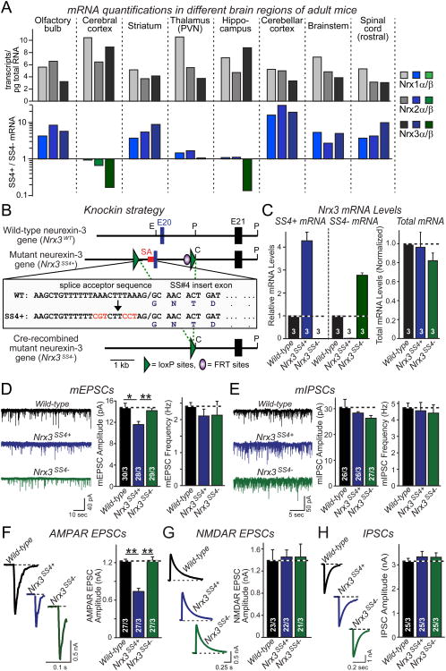
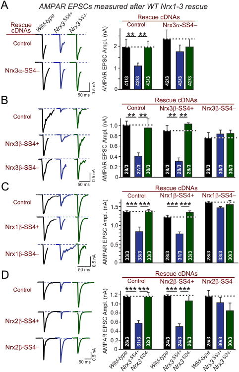
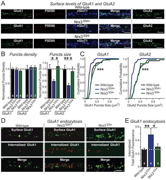
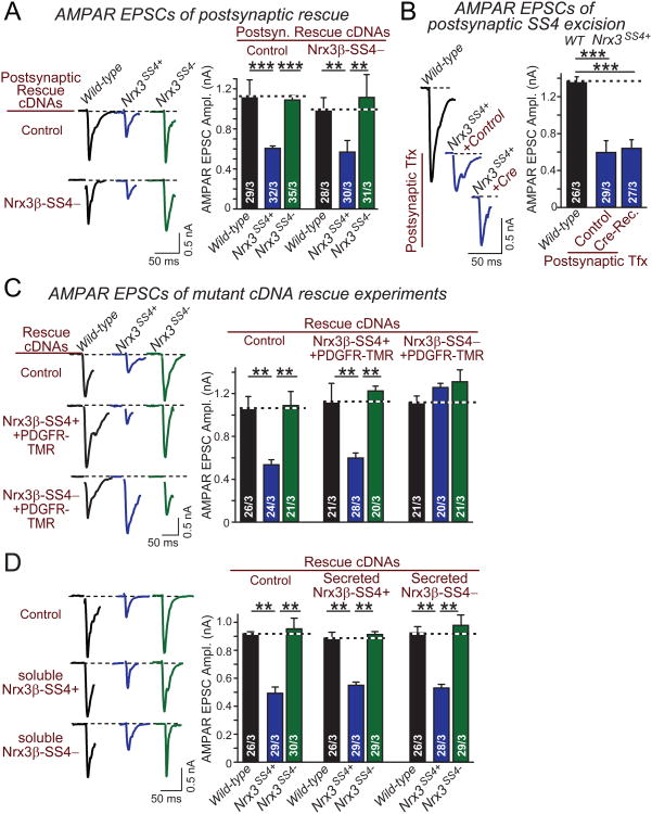
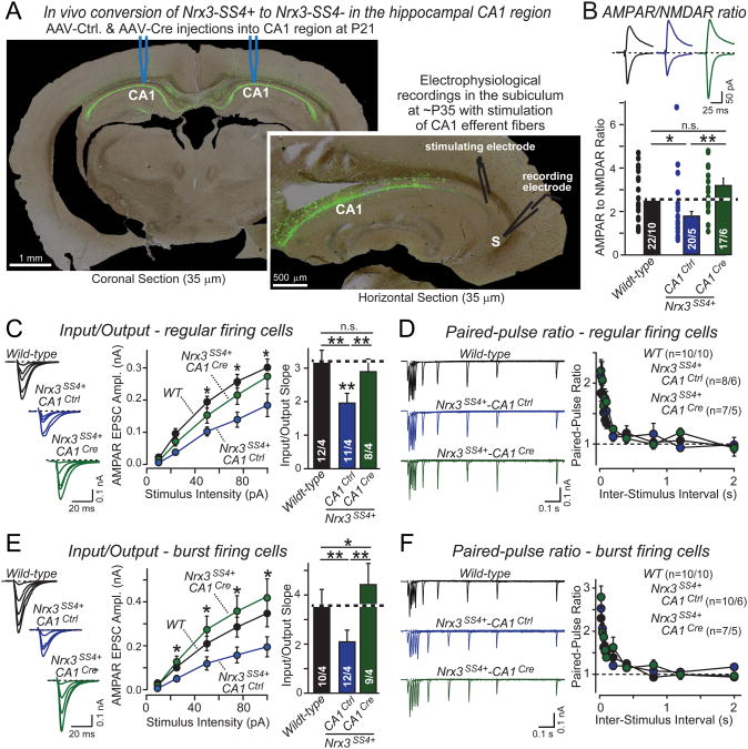
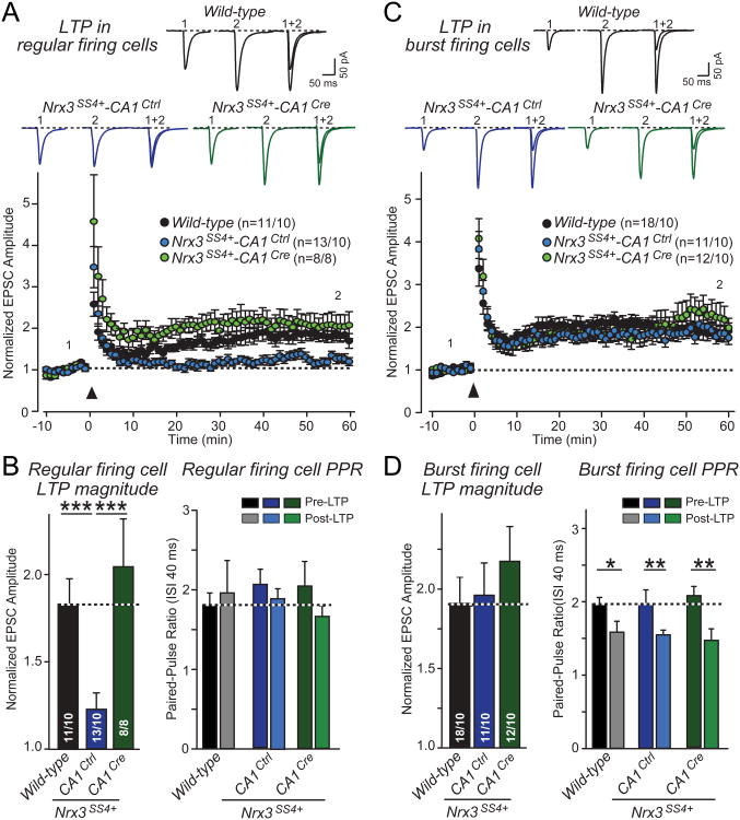
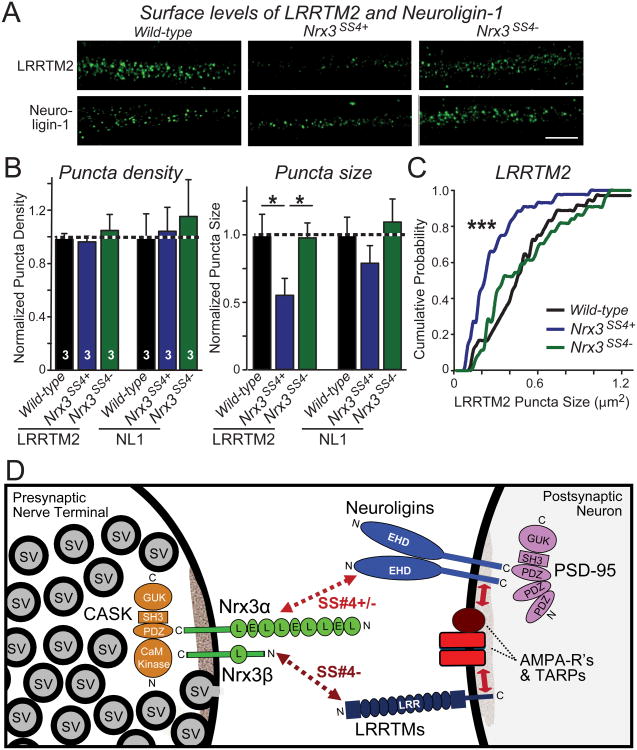
Neurexins are essential presynaptic cell adhesion molecules that are linked to schizophrenia and autism and are subject to extensive alternative splicing. Here, we used a genetic approach to test the physiological significance of neurexin alternative splicing. We generated knockin mice in which alternatively spliced sequence #4 (SS4) of neuexin-3 is constitutively included but can be selectively excised by cre-recombination. SS4 of neurexin-3 was chosen because it is highly regulated and controls neurexin binding to neuroligins, LRRTMs, and other ligands. Unexpectedly, constitutive inclusion of SS4 in presynaptic neurexin-3 decreased postsynaptic AMPA, but not NMDA receptor levels, and enhanced postsynaptic AMPA receptor endocytosis. Moreover, constitutive inclusion of SS4 in presynaptic neurexin-3 abrogated postsynaptic AMPA receptor recruitment during NMDA receptor-dependent LTP. These phenotypes were fully rescued by constitutive excision of SS4 in neurexin-3. Thus, alternative splicing of presynaptic neurexin-3 controls postsynaptic AMPA receptor trafficking, revealing an unanticipated alternative splicing mechanism for trans-synaptic regulation of synaptic strength and long-term plasticity.


Figures

Figure 1
Figure 1. Genetic switching of alternative splicing of Nrx3 at SS4 into constitutive ‘on’ or ‘off’ states alters AMPAR-mediated synaptic transmission
A. Absolute expression (# transcripts/pg total RNA) of Nrx1α/β, Nrx2α/β, and Nrx3α/β mRNAs in the indicated brain regions (top), and relative levels of SS4+ and SS4− mRNAs in the same brain regions as determined by quantitative rt-PCR (bottom; n = 4 mice; PVN, paraventricular nucleus). B. Knock-in strategy to generate conditional Nrx3-SS4 mutant mice. The imperfect splice acceptor sequence (SA; sequence in inset) of the alternatively spliced SS4 exon 20 (E20)was replaced by homologous recombination in murine ES cells with a perfect splice acceptor sequence (red letters); in addition, exon 20 was flanked by LoxP sites. The resulting mutant Nrx3 gene constitutively expresses Nrx3-SS4+ mRNA (Nrx3SS4+). Cre-recombinase deletes the exon, causing constitutive expression of Nrx3-SS4− mRNA (Nrx3SS4−). Single letters indicate locations of restriction enzyme sites (C, ClaI; E, EcoRI; P, PstI). C. Quantification of Nrx3-SS4+, Nrx3-SS4−, and total Nrx3 mRNA levels in hippocampal neurons cultured from WT or Nrx3SS4+ mice; Nrx3SS4+ neurons were infected with lentiviruses expressing inactive cre-recombinase (retaining the Nrx3SS4+ genotype) or active cre-recombinase (converting Nrx3SS4+ into Nrx3SS4− neurons). mRNA levels were normalized to those observed in WT neurons which express predominantly Nrx3-SS4−mRNA (see panel A; n=3 independent cultures). D & E. Representative traces (left) and summary graphs of the amplitude and frequency (right) of mEPSCs (D) and mIPSCs (E), monitored in cultured hippocampal WT, Nrx3SS4+, and Nrx3SS4− neurons obtained as described for C. F-H. Representative traces (left) and summary graphs (right) of evoked AMPAR- (F) and NMDAR-mediated EPSCs (G) and of evoked IPSCs (H) in cultured hippocampal WT, Nrx3SS4+ and Nrx3SS4− neurons. Data in C-H are means ± SEM; numbers in bars give number of independent experiments (C) or number of total cells/independent experiments analyzed (D-H). For D-H, statistical significance was calculated by single-factor ANOVA (** p<0.01). See also Fig. S1.
Figure 2
Figure 2. Reduced AMPAR-mediated synaptic responses in Nrx3SS4+synapses are rescued by SS4− but not SS4+ neurexins
A-D. Representative traces (left) and summary graphs (right) of evoked AMPAR-mediated peak EPSC amplitudes measured in hippocampal neurons that were cultured from WT or Nrx3SS4+ mice; Nrx3SS4+ neurons were infected with lentiviruses expressing inactive cre-recombinase (which leaves the genotype unchanged) or active cre-recombinase (which converts the Nrx3SS4+ into the Nrx3SS4− genotype). Neurons were superinfected with a second lentivirus expressing either no neurexin (Control) or the indicated Nrx1-3 isoforms and splice variants. Data are means ± SEM; numbers in bars represent total number of cells/experiments analyzed. Statistical significance was calculated by single-factor ANOVA (*, p<0.05; **, p<0.01; ***, p<0.001).
Figure 3
Figure 3. GluA1 and GluA2 AMPAR surface levels are reduced in hippocampal Nrx3SS4+neurons due to increased internalization
A. Representative images of hippocampal neurons cultured from WT or Nrx3SS4+ mice; Nrx3SS4+ neurons were infected with lentiviruses expressing either inactive (retaining the Nrx3SS4+ genotype) or active cre-recombinase (switching the Nrx3SS4+ to the Nrx3SS4− genotype). Neurons were surface-labeled for GluA1 or GluA2 AMPARs (see Fig. S2A for experimental protocol); GluA1-stained neurons were permeabilized and also stained for the excitatory synaptic makers PSD95 and vGluT1 (green: surface GluA1; red: PSD95; blue: vGluT1; scale bars, 5 μm). B. Quantifications of the normalized density and size of synaptic puncta labeled for surface GluA1 or GluA2 in WT, Nrx3SS4+, or Nrx3SS4− neurons. For more data, see Figs. S2B and S2C. C. Cumulative distributions of Nrx3SS4+ and Nrx3SS4− GluA1 and GluA2 puncta sizes. D & E. Representative images (D) and summary graph (E) of GluA1 endocytosis analyzed in WT, Nrx3SS4+, and Nrx3SS4− neurons obtained as described above. GluA1 endocytosis was quantified by dividing the internalized GluA1 fraction (red) by the total GluA1 signal (surface fraction (green) + internalized fraction (red); scale bar, 5 μm; see Fig. S2D). Data are the means ± SEM of 4 independent experiments. Statistical analyses were performed by single-factor ANOVA (* p<0.05; ** p<0.01; *** p<0.001).
Figure 4
Figure 4. Presynaptic membrane-tethered but not secreted Nrx3β-SS4− controls post-synaptic AMPARs in a non cell-autonomous fashion
A. Representative traces (left) and summary graphs (right) of evoked AMPAR EPSCs monitored in hippocampal neurons cultured from WT or Nrx3SS4+ mice. Nrx3SS4+ neurons were sparsely transfected at DIV4-5 with plasm ids encoding EGFP alone or EGFP and Nrx3β-SS4−, and were analyzed by whole-cell patch-clamp recordings at DIV14-16. B. Same as A, except that neurons were sparsely transfected with plasmids encoding inactive (Control) or active cre-recombinase (Cre-Rec). C. Representative traces (left) and summary graphs of AMPAR-mediated EPS Camplitudes in hippocampal neurons that were cultured from WT or Nrx3SS4+ mice, and infected with lentiviruses expressing inactive cre-recombinase (which retains the Nrx3SS4+ genotype) or active cre-recombinase (which converts Nrx3SS4+ into Nrx3SS4− neurons). Neurons were superinfected with lentivirus expressing control or rescue cDNAs encodingfusion proteins of the extracellular Nrx3β-SS4+ and -SS4− sequences with the PDGF-receptor transmembrane region. D. Same as C, except that the rescue cDNAs express a naturally occurring splice variant of Nrx3β that encodes secreted Nrx3β-SS4+ and Nrx3β-SS4−. Data are means ± SEM; numbers in bars represent total cells/experiments performed. Statistical significance was calculated by single-factor ANOVA (** p<0.01; *** p<0.001).
Figure 5
Figure 5. Cre-mediated in vivo conversion of presynaptic Nrx3SS4+ to Nrx3SS4− in the hippocampal CA1 region restores impaired AMPAR-mediated synaptic responses in Nrx3SS4+ postsynaptic neurons in the subiculum
A. Representative images illustrating the selective expression of EGFP in the CA1 region of the dorsal hippocampus after stereotactic injection of AAVs and the recording configuration used for measuring synaptic transmission from the CA1 region to the subiculum. Mice were stereotactically injected at P21, and analyzed at P35. Note the complete infection of CA1 region neurons without spillover into the subiculum (S). B. AMPAR/NMDAR ratios recorded in acute slices in pyramidal neurons of the subiculum after in vivo presynaptic manipulations of Nrx3-SS4 alternative splicing in the hippocampal CA1 region (top, representative EPSC traces at −65 mV [AMPAR] and +40 mV [NMDAR]; bottom, average ratios). WT mice were stereotactically injected into the hippocampal CA1 region with AAVs encoding GFP (black traces/bars), and Nrx3SS4+ mutant mice with AAVs encoding inactive (Nrx3SS4+-CA1Ctrl; blue traces/bars) or active cre-recombinase (Nrx3SS4+-CA1Cre; green traces/bars). C. Input/output relationship of postsynaptic AMPAR EPSCs in regular firing neurons of the subiculum as a function of presynaptic Nrx3-SS4 alternative splicing. Data show sample traces (left), summary curves (middle), and average input/output slopes (right) for slices obtained as described in B. D. Paired-pulse measurements of postsynaptic AMPAR EPSCs in regular firing neurons of the subiculum as a function of presynaptic Nrx3-SS4 alternative splicing. Data show sample traces (left) and summary graphs (right) for samples obtained as described in B. E & F. Same as C and D, but recorded from burst-firing neurons in the subiculum. Data shown are means ± SEM; number number of neurons/mice analyzed are shown in the bars. Statistical analyses for summary graphs were performed by single-factor ANOVA (*, p<0.05; **, p<0.01). For more data, see Fig. S3.
Figure 6
Figure 6. Nrx3SS4+ mice exhibit impaired postsynaptic NMDAR-dependent LTP that is rescued by conversion of presynaptic Nrx3SS4+ neurons into Nrx3SS4− neurons
A. LTP in regular firing neurons. Acute slices were analyzed by whole-cell recordings in the subiculum from WT mice and from Nrx3SS4+ mice in which CA1 neurons were transduced in vivo by stereotactic injection of AAV encoding inactive (Ctrl) or active cre-recombinase (Cre). Data show representative traces (top) and summary graphs of the relative synaptic strength (bottom) monitored before and after induction of LTP. LTP was induced by 4 × 100 Hz stimuli applied for 1 sec with 10 sec intervals in current clamp configuration at resting membrane potential. Following the LTP induction, cells were held at -65 mV in voltage-clamp to measure evoked EPSCs. Representative traces shown are EPSCs during baseline (1) and 50 minutes after induction (2). B. EPSC amplitudes averaged over the last 10 min of the LTP recordings, normalized to the baseline (left), and paired-pulse ratios with a 40 ms inter-stimulus interval measured 10 min pre-LTP induction and 60 min post-LTP induction (right) from regular firing neurons in the subiculum following presynaptic manipulation of Nrx3-SS4. C & D. Same as A and B, but for burst firing neurons in the subiculum. Note that consistent with previous studies (Wozny et al., 2008), LTP in burst firing neurons causes a change in paired-pulse ratio suggesting it is presynaptic (D), whereas LTP in regular firing neurons does not cause a change in paired-pulse ratio consistent with a postsynaptic localization (B). Data shown are means ± SEM; numbers of neurons/mice examined are shown in the graphs. Statistical analyses were performed by single-factor ANOVA (* p<0.05; ** p<0.01; *** p<0.001). See also Fig. S4.
Figure 7
Figure 7. Nrx3SS4+ neurons contain decreased postsynaptic concentrations of LRRTM2: Model for the mechanism of action of Nrx3-SS4 alternative splicing
A. Representative images of hippocampal neurons stained for surface-localized LRRTM2 or neuroligin-1 (NL1). Neurons were cultured from WT or Nrx3SS4+ mice; the latter were infected with lentiviruses expressing either inactive cre-recombinase (which retains the Nrx3SS4+ genotype) or active cre-recombinase (which switches Nrx3SS4+ into Nrx3SS4−neurons). B. Quantification of the density (left) and size (right) of LRRTM2 or neuroligin-1 positive puncta in WT, Nrx3SS4+, and Nrx3SS4− neurons. Data are means ± SEM; n=3 independent culture experiments; statistical analyses were performed by Student's t-test (*, p<0.05). C. Cumulative probability distribution of the puncta size in WT, Nrx3SS4+, and Nrx3SS4− neurons. Statistical significance was assessed by the Kolmogorov-Smirnov test (***, p<0.001). See also Fig. S5. D. Model of how presynaptic Nrx3 may stabilize postsynaptic AMPARs in an SS4− dependent manner via trans-synaptic interactions with neuroligin-1 and/or LRRTMs. Postsynaptic AMPAR stability is dependent on the presynaptic expression of Nrx3-SS4−that lacks an insert at splice site 4, and preferentially interacts with neuroligins and LRRTMs. Neuroligins and LRRTMs have been shown to interact with AMPARs (Etherton et al., 2011; Soler-Llavina et al., 2011; Schwenk et al., 2012), possibly via their binding to PSD95 which in turn binds to the C-terminal sequences of TARPs that form a tight complex with AMPARs (Schnell et al., 2002). The data in panels A-C suggest that the Nrx3-LRRTM interaction is most important for controlling postsynaptic AMPAR levels, but different isoforms of neuroligin differentially interact with the two Nrx3-SS4 splice variants, and thus a major contribution of the Nrx3-neuroligin interaction in controlling postsynaptic AMPARs cannot be ruled out.

References

    1. Aoto J, Nam CI, Poon MM, Ting P, Chen L. Synaptic signaling by all-trans retinoic acid in homeostatic synaptic plasticity. Neuron. 2008;60:308–320. - PMC - PubMed
    1. Boucard AA, Chubykin AA, Comoletti D, Taylor P, Südhof TC. A splice code for trans-synaptic cell adhesion mediated by binding of neuroligin 1 to α- and β-neurexins. Neuron. 2005;48:229–236. - PubMed
    1. Boucard AA, Ko J, Südhof TC. High affinity neurexin binding to cell adhesion G-protein-coupled receptor CIRL1/latrophilin-1 produces an intercellular adhesion complex. J Biol Chem. 2012;287:9399–9413. - PMC - PubMed
    1. Chen YC, Lin YQ, Banerjee S, Venken K, Li J, Ismat A, Chen K, Duraine L, Bellen HJ, Bhat MA. Drosophila neuroligin 2 is required presynaptically and postsynaptically for proper synaptic differentiation and synaptic transmission. J Neurosci. 2012;32:16018–16030. - PMC - PubMed
    1. Chih B, Gollan L, Scheiffele P. Alternative splicing controls selective trans-synaptic interactions of the neuroligin-neurexin complex. Neuron. 2006;51:171–178. - PubMed

Publication types