. 2013 Jul 3;154(1):103-17.
doi: 10.1016/j.cell.2013.05.057.

Distinct α-synuclein strains differentially promote tau inclusions in neurons

Affiliations

Distinct α-synuclein strains differentially promote tau inclusions in neurons

Jing L Guo et al. Cell. .

Abstract

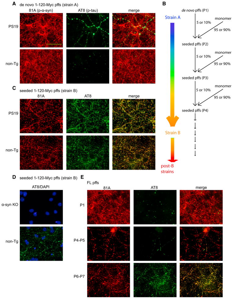
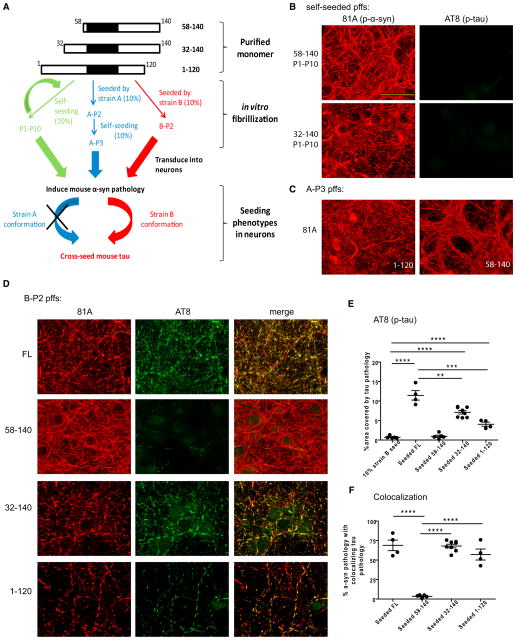
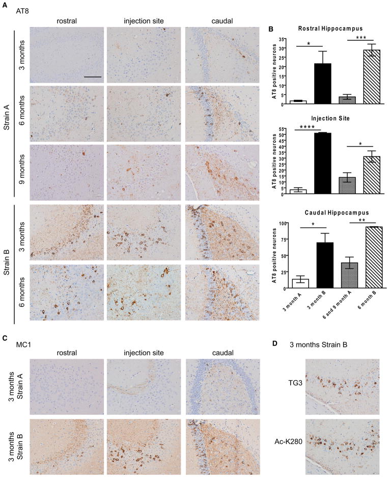
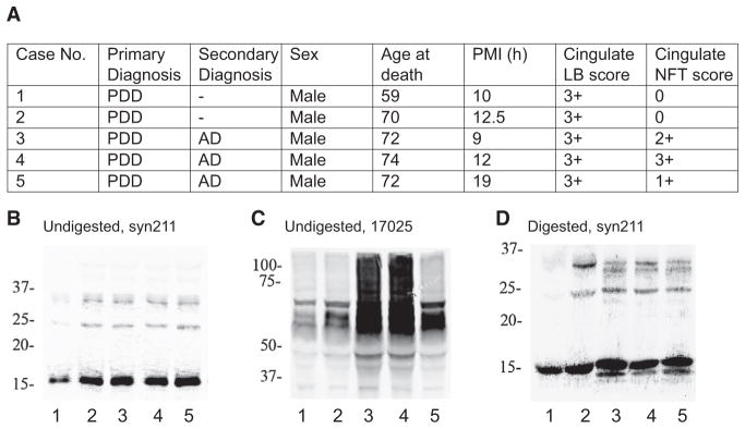
Many neurodegenerative diseases are characterized by the accumulation of insoluble protein aggregates, including neurofibrillary tangles comprised of tau in Alzheimer's disease and Lewy bodies composed of α-synuclein in Parkinson's disease. Moreover, different pathological proteins frequently codeposit in disease brains. To test whether aggregated α-synuclein can directly cross-seed tau fibrillization, we administered preformed α-synuclein fibrils assembled from recombinant protein to primary neurons and transgenic mice. Remarkably, we discovered two distinct strains of synthetic α-synuclein fibrils that demonstrated striking differences in the efficiency of cross-seeding tau aggregation, both in neuron cultures and in vivo. Proteinase K digestion revealed conformational differences between the two synthetic α-synuclein strains and also between sarkosyl-insoluble α-synuclein extracted from two subgroups of Parkinson's disease brains. We speculate that distinct strains of pathological α-synuclein likely exist in neurodegenerative disease brains and may underlie the tremendous heterogeneity of synucleinopathies.


Figures

Figure 1
Figure 1. Generation of Different Strains of α-Syn pffs with Differential Cross-Seeding of Tau in Neurons
(A) Insoluble phospho-α-syn (81A) and phospho-tau (AT8) induced by de novo 1–120-Myc pffs (strain A) in primary hippocampal neurons dissociated from PS19 or non-Tg mouse embryos. (B) Procedures of repetitive seeded fibrillization in vitro resulting in evolution of strain A pffs into B and post-B strains. (C) α-syn and tau pathologies caused by seeded 1–120-Myc pffs (strain B) in PS19 and non-Tg neurons. (D) Staining of insoluble phospho-tau (AT8, green) in α-syn knockout (α-syn KO) or non-Tg neurons transduced with strain B 1–120-Myc pffs. DAPI (blue) revealed cell nuclei. (E) Phenotypic changes in α-syn and tau pathologies induced by FL α-syn pffs of different self-seeding passages. For (A), (C), (D), and (E), neurons were fixed 18 days post-transduction, and soluble proteins were removed during fixation. Scale bar: 50 μm. See also Figure S1 and Table S2.
Figure 2
Figure 2. Ultrastructural Analysis of Strain B α-Syn pff-Induced Cytoplasmic Filaments by Immuno-EM
(A and B) 81A-immunoreactive α-syn filaments observed in strain B α-syn pff-transduced neurons labeled by nanogold. (C and D) AT8-positive tau filaments in these neurons detected by nanogold. (E–G) Double-labeling using 81A and 17025 (a pAb against tau) as primary antibodies followed by secondary antibodies conjugated to 10 nm colloidal gold (for α-syn) or 6 nm colloidal gold (for tau). (E) and (F) show filaments decorated by both α-syn (arrowheads) and tau (arrows) antibodies found in neurites, whereas (G) shows filaments almost exclusively composed of α-syn found in a cell body. Scale bar: 500 nm for (A)–(D), 100 nm for (E)–(G).
Figure 3
Figure 3. Inverse Seeding Capacity for α-Syn and Tau Pathologies Accompanied by Differential Cellular Toxicity
(A) Insoluble phospho-α-syn (81A) and phospho-tau (AT8) in non-Tg neurons at different days post-transduction with strain A or strain B FL α-syn pffs. Soluble proteins were removed during fixation. (B and C) Quantification of % area occupied by 81A- and AT8-labeled pathologies, respectively, for experiments described in (A) (results shown as mean ± standard error of the mean [SEM]. *p < 0.05; **p < 0.01; ***p < 0.0005). (D) Calculation of % of α-syn pathology with colocalizing tau pathology (area occupied by 81A/area occupied by AT8 3100%) for strain B pff-transduced neurons (results shown as mean ± SEM. ***p < 0.0005; ****p < 0.0001). (E) PS19 neurons treated with PBS or transduced with strain A or strain B FL α-syn pffs were sequentially extracted with 1% Triton X-100 lysis buffer (T) followed by 2% SDS lysis buffer (S) at different time points. Neuron lysates from T and S fractions were immunoblotted with 17025 (total tau), T14 (human tau), AT8 (phospho-tau), T49 (mouse tau), 81A (phospho-α-syn), and GAPDH antibody (loading control). (F) Non-Tg neurons at 18 days post-transduction of strain A or strain B FL α-syn pffs were sequentially extracted and immunoblotted with AT8, T49, mouse α-syn, and GAPDH antibodies. (G) LDH assay on differently treated non-Tg neurons at 14 days and 18 days post-transduction (results shown as mean ± SEM. *p < 0.05; ***p < 0.005). Scale bar: 50 μm. See also Figure S2.
Figure 4
Figure 4. Conformational Differences between the Strains Revealed by PK Digestion and a Strain-Selective Antibody
(A) Different passages (P1–P7) of self-seeded FL α-syn pffs were digested with PK, resolved on 12% Bis-Tris gel, and stained with Coomassie blue. Results from one representative series are shown. N-terminal sequencing results for the 2nd to 5th bands are presented in parentheses. (B) Quantification of the relative intensity of the 5th band to the 1st band for experiments described in (A) for three series of self-seeded FL α-syn pffs. (C) Pairs of strain A and B FL α-syn pffs made from the same batches of monomer were incubated with different concentrations of PK (1–2.5 μg/ml). Digestion products from two pairs of pffs were shown. Untreated pffs were loaded on the same gels (0 μg/ml). (D) Quantification of the ratio of cleaved species (sum of the 2nd to 5th bands) to uncleaved molecules (the 1st band) for experiments described in (C) (results shown as mean ± SEM. **p < 0.01). (E) Quantification of the ratio of the 3rd band to the 1st band for the same samples as in (D) (results shown as mean ± SEM. *p < 0.05; **p < 0.01). (F) Quantification of the ratio of the 5th band to the 1st band for the same samples as in (D) (results shown as mean ± SEM. **p < 0.01; ***p < 0.001). (G) PK-digested FL α-syn pffs were immunoblotted with antibodies recognizing different epitopes of α-syn, indicated in parentheses. (H) Deduced identities of major products from PK digestion. (I) Different amounts of strain A or B FL α-syn pffs (0.05 to 2 μg) coated on an ELISA plate were incubated with HuA1, a pAb raised against recombinant human α-syn, and 9029-03, our newly generated strain B-selective mAb. (J) Different passages of FL α-syn pffs immobilized on an ELISA plate were probed with HuA1 and 9029-03. Results from three series are shown. For (I) and (J), absorbance for 9029-03 was expressed as % of absorbance for HuA1. See also Figure S3.
Figure 5
Figure 5. The Roles of N and C Termini in Strain Conformations
(A) Schematic diagram for testing the roles of N and C termini in generating or adopting strain A and B conformation. (B) Pathology induced by de novo or self-seeded (P1–P10) 58–140 or 32–140 pffs in non-Tg neurons. (C) Insoluble α-syn aggregates resulted from 1–120 or 58–140 pffs that were initially seeded by strain A pffs. (D) Insoluble α-syn and tau aggregates induced by FL α-syn, 58–140, 32–140, or 1–120 pffs that were seeded by strain B pffs. (E) Quantification of % area occupied by AT8-labeled tau pathology in (D) (independent preparations of pffs tested: n = 5 for 10% seed control, n = 4 for FL, n = 5 for 58–140, n = 8 for 32–140, n = 4 for 1–120; results shown as scatterplot with mean ± SEM. **p < 0.005; ***p < 0.0005; ****p < 0.0001). (F) Calculation of % of α-syn pathology with colocalizing tau pathology in (D) (results shown as scatterplot with mean ± SEM, ****p < 0.0001). For (B), (C), and (D), soluble proteins were removed during fixation. Scale bar: 50 μm. See also Figure S4 and Table S3.
Figure 6
Figure 6. Strain-Specific Cross-Seeding of Tau Pathology In Vivo
(A) AT8-positive tau aggregates detected in different parts of ipsilateral hippocampus (rostral, near injection site, caudal) at 3, 6, or 9 months post-inoculation of strain A or B 1–120-Myc pffs into the hippocampus of PS19 mice. (B) Quantification of AT8-positive neurons in different parts of ipsilateral hippocampus (n = 4 for 3 months post-injection of either strain, n = 4 for 6 or 9 months post-injection of strain A, n = 3 for 6 months post-injection of strain B; results shown as mean ± SEM. *p < 0.05; **p < 0.005; ***p < 0.0005; ****p < 0.00001). (C) MC1 immunoreactivity in different parts of ipsilateral hippocampus at 3 months post-injection. (D) tau inclusions cross-seeded by strain B pffs were recognized by TG3 and Ac-K280. All sections were counterstained with hematoxylin to reveal cell nuclei. Scale bar: 100 μm. See also Figure S5.
Figure 7
Figure 7. Conformational Variations of Pathological α-Syn in Synucleinopathy Brains
(A) Demographics of PDD cases used for pathological α-syn extraction from the cingulate gyrus. Primary and secondary diagnoses are based on neuropathological examination of postmortem brains. PMI: postmortem interval. Pathological scores of LBs and NFTs in the contralateral cingulate gyrus are shown, with “3+” being the highest score. (B) Sarkosyl-insoluble fraction immunoblotted with mAb Syn211 showed abundant α-syn for all five cases. (C) Sarkosyl-insoluble fraction from these five cases showed varying extents of tau accumulation revealed by pAb 17025. (D) PK-digested sarkosyl-insoluble fraction showed different banding patterns of PK-resistant α-syn for PDD cases with or without abundant coexisting AD pathologies.

Comment in

References

    1. Abeliovich A, Schmitz Y, Fariñas I, Choi-Lundberg D, Ho WH, Castillo PE, Shinsky N, Verdugo JM, Armanini M, Ryan A, et al. Mice lacking alpha-synuclein display functional deficits in the nigrostriatal dopamine system. Neuron. 2000;25:239–252. - PubMed
    1. Aguzzi A, Heikenwalder M, Polymenidou M. Insights into prion strains and neurotoxicity. Nat Rev Mol Cell Biol. 2007;8:552–561. - PubMed
    1. Baba M, Nakajo S, Tu PH, Tomita T, Nakaya K, Lee VM, Trojanowski JQ, Iwatsubo T. Aggregation of alpha-synuclein in Lewy bodies of sporadic Parkinson’s disease and dementia with Lewy bodies. Am J Pathol. 1998;152:879–884. - PMC - PubMed
    1. Ballatore C, Lee VM, Trojanowski JQ. Tau-mediated neurodegeneration in Alzheimer’s disease and related disorders. Nat Rev Neurosci. 2007;8:663–672. - PubMed
    1. Braak H, Del Tredici K, Rüb U, de Vos RA, Jansen Steur EN, Braak E. Staging of brain pathology related to sporadic Parkinson’s disease. Neurobiol Aging. 2003;24:197–211. - PubMed

Publication types