. 2013 Jul 3;154(1):157-68.
doi: 10.1016/j.cell.2013.05.048.

Observing a DNA polymerase choose right from wrong

Affiliations

Observing a DNA polymerase choose right from wrong

Bret D Freudenthal et al. Cell. .

Abstract

DNA polymerase (pol) β is a model polymerase involved in gap-filling DNA synthesis utilizing two metals to facilitate nucleotidyl transfer. Previous structural studies have trapped catalytic intermediates by utilizing substrate analogs (dideoxy-terminated primer or nonhydrolysable incoming nucleotide). To identify additional intermediates during catalysis, we now employ natural substrates (correct and incorrect nucleotides) and follow product formation in real time with 15 different crystal structures. We are able to observe molecular adjustments at the active site that hasten correct nucleotide insertion and deter incorrect insertion not appreciated previously. A third metal binding site is transiently formed during correct, but not incorrect, nucleotide insertion. Additionally, long incubations indicate that pyrophosphate more easily dissociates after incorrect, compared to correct, nucleotide insertion. This appears to be coupled to subdomain repositioning that is required for catalytic activation/deactivation. The structures provide insights into a fundamental chemical reaction that impacts polymerase fidelity and genome stability.


Figures

Figure 1
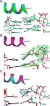
Figure 1. The Pol β Ground State Pre-catalytic Ternary Complex
(A) Pol β pre-catalytic active site organization with dUMPNPP analogue and Mg2+ in the catalytic and nucleotide metal binding sites (2FMS). The primer terminus (O3′), incoming nucleotide, and key active site residues are shown. The proposed proton movements during catalysis are highlighted by red dashed arrows and O3′ attack of Pα is represented with a black arrow. The scissile bond between Pα and Pβ is semi-transparent and the resulting pyrophosphate (PPi) is indicated. The catalytic (Mgc) and nucleotide binding (Mgn) magnesium ions are shown as red spheres. (B) Ground state closed ternary complex structure of pol β generated with dCTP; Ca2+ ions are shown in orange in the metal binding sites. A Fo-Fc omit map (green) contoured at 3σ is shown for the incoming dCTP and Ca2+. (C) Coordination sphere and distances (Å) for the Ca2+ ions (orange) in the ground state are indicated. Coordinating water molecules (small blue spheres) are also shown. (D) Overlay of the ground state ternary complex generated with either Mg2+ or Ca2+ is shown in salmon and green, respectively.
Figure 2
Figure 2. Observing Pol β Phosphodiester Bond Formation after a 20 s Soak in MgCl2
(A) A Fo-Fc omit map (green) contoured at 3σ is shown for the primer terminus, incoming dCTP, PPi, and Mg2+ ions. Density corresponding to both the product and reactant state during phosphodiester bond formation can be modeled and is indicated (arrows). The key active site residues are shown in stick format. Mg2+ and water molecules are shown as red and blue spheres, respectively. (B) 2Fo-Fc map contoured at 1.5σ is shown after refinement with both the product and reactant state (see alternate refinement models in Figure S1). (C) Structural overlay of the ground state (2FMS) and reactant state are shown in green and yellow, respectively. Mg2+ from the ground and reactant states are shown in red and green, respectively. (D) A 90° rotation of the model is shown with a 2Fo-Fc density map (grey) contoured at 1.5σ for the primer terminus, Mg2+, PPi, and dCMP. The lack of significant positive or negative density with a Fo-Fc omit map contoured at 3σ indicates the intermediate structure represents a good model. The transition state can be modeled into the density with key distances indicated; also see Figures S1 and S2.
Figure 3
Figure 3. A Transient Third Metal Binding Site in the Closed Pol β Product Complex
(A) Pol β product complex generated after a 40 s soak in MgCl2 is shown with a Fo-Fc omit map (green) contoured at 3σ (see panel C for Mgp omit map). The incorporated dCMP, templating guanine (Gt), α-helix N, key active site residues, and PPi are indicated. The Mg2+, Na2+, and water molecules are shown as red, purple, and blue spheres, respectively. (B) Structural overlay of the ground state (2FMS) and product ternary complex (40 s soak) are shown in green and yellow, respectively. The Mg2+ ions for the ground and product state are shown in green and red, respectively. (C) Fo-Fc omit map (green) contoured at 3σ for the product Mg2+ (Mgp) and coordinating water molecules is shown. The coordination distances (Å) for the Na2+ and Mg2+ in the catalytic, nucleotide or product metal binding sites are indicated. See Figure S4 for a stereo composite omit map. (D) A Fo-Fc omit map for the pol β product complex after a 45 min soak in MgCl2 is shown contoured at 3σ. The product metal and coordinating waters have dissociated; i.e., are no longer visible (also see Figures S3, S4, and S5).
Figure 4
Figure 4. Pol β Pre-catalytic Mismatch Ternary Ground State
The templating base, primer terminus (O3′), incoming nucleotide, active site residues, and α-helix N are indicated. (A) Structural overlay of the pre-catalytic matched A:dUMPNPP (2FMS) and mismatched G:dAMPCPP (3C2M) ternary pol β complexes are shown in green and cyan, respectively. The Mn2+ and Mg2+ ions are shown in purple and red spheres, respectively. Water molecules are colored blue. The key movements of the primer terminus and templating strand are highlighted with red arrows. The templating nucleotide (Nt) is indicated and labeled n and its upstream neighbor is labeled n-1. (B) The ground state ternary complex after a 2.5 min soak in MnCl2 is shown in purple with a Fo-Fc omit map (green) contoured to 3σ for the incoming dATP, primer terminus, and metal ions. An anomalous density map (purple) contoured to 5σ is shown for the catalytic and nucleotide Mn2+ ions. (C) Structural overlay of the pre-catalytic pol β ternary complex (3C2M) obtained with a non-hydrolyzable dAMPCPP analogue and the ground state ternary complex obtained with dATP after a 2.5 min soak in MnCl2 are shown in cyan and magenta, respectively.
Figure 5
Figure 5. Observing a Misincorporation Event
The pol β reactant/product states are shown (magenta carbons). For reference, the pol β pre-catalytic ternary mismatch complex (3C2M) and binary open conformation (3ISB) are shown in cyan and brown, respectively. A Fo-Fc omit map (green) contoured at 3σ is shown for the primer terminus, incorporated nucleotide, Mn2+, and PPi. The anomalous density map (purple) contoured to 5σ is shown for the Mn2+ ions. Key active site residues, the primer terminus (O3′), templating base (Gt), and the incoming dATP are indicated. The Mn2+ ions are shown in purple and water molecules in blue. The reactant state can be modeled into the omit map density after a (A) 5 and (B) 10 min soak in MnCl2. (C) After a 30 min soak, only the product state is observed and the catalytic Mn2+ can be modeled into multiple locations. The polymerase is in a partially open state based on the relative position of α-helix N compared to the open (tan) and closed (cyan) states. (D) The overnight soak shows that the polymerase is completely open, and Mn2+ and PPi are not observed (also see Figure S6)
Figure 6
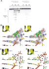
Figure 6. Insight into the Role of the Product-Associated Metal and Pyrophosphorolysis
(A) Pyrophosphorolysis was assayed under single-turnover conditions with either a matched (lanes 2-7) or mismatched (lanes 9-14) primer terminus in nicked DNA utilizing Mg2+ or Mn2+. Lanes 1 and 8 show the 16-mer primers (P) only. Controls (-M, no metal) for the matched DNA substrate (lanes 2-3) and the mismatched DNA substrate (lanes 9-10) do not exhibit a reverse reaction. For the matched primer terminus, addition of 2.5 mM PPi results in the accumulation of shorter products with increasing time; 1 (1′) and 5 min (5′). (B) Product nicked closed ternary complex generated by soaking an open binary nicked pol β complex in PPi and MgCl2 for 30 min. Stereo view of the 2Fo-Fc (grey) and Fo-Fc (green; Mg2 and associated waters) is shown contoured at 1.5 and 2.5σ, respectively. The primer terminus and its complementary templating guanine (Gt), α-helix N, key active site residues, and PPi are shown and labeled in panel C for reference. The Mg2+, Na2+, and water molecules are shown as red, purple, and blue spheres, respectively. (C) The coordination distances and ligands for the Mg2+ and Na2+ are indicated for the catalytic, nucleotide, or product metal binding site in the nick closed ternary complex. (D) Overlay of the closed nicked product complex generated either by annealing prior to crystallization and soaking in PPi or in situ after a 40 s soak of the 1-nt gap binary complex in dCTP and MgCl2 is shown in yellow or green, respectively. The annealed and in situ Mg2+, Na2+, and water molecules are shown in red, purple, and blue or green, pink, and grey spheres, respectively. The product metal can be observed in the ground state after initiating the reverse reaction.
Figure 7
Figure 7. Mechanistic Model for Correct/Incorrect Incorporation and Pyrophosphorolysis
The templating base, primer terminus, PPi, active site aspartate residues, and metals are indicated. The arrows refer to the events depicted in the models. (A) Correct incorporation involves (1) deprotonation of O3′, (2) nucleophilic attack on Pα by O3′, (3) protonation of PPi, and (4) inversion of Pα. (B) The panel is rotated 90° compared to panels A and C. Incorrect incorporation involves an activation step (Act) to position O3′ to coordinate the catalytic metal, (1) deprotonation of O3′, (2) nucleophilic attack on Pα by O3′, (3) protonation of PPi, and (4) 120° rotation of the phosphate backbone. The closed to open polymerase transition during the course of the reaction is indicated. The PPi product of this reaction is shown in semi-transparent stick format. (C) The pyrophosphorolysis reaction involves (1) deprotonation of the PPi oxygen, (2) nucleophilic attack on the phosphate of the primer terminus, (3) protonation of O3′, and (4) inversion of Pα. The product of this reaction is shown in semi-transparent stick format.

Comment in

  • Watching a DNA polymerase in action.
    Freudenthal BD, Beard WA, Wilson SH. Freudenthal BD, et al. Cell Cycle. 2014;13(5):691-2. doi: 10.4161/cc.27789. Epub 2014 Jan 14. Cell Cycle. 2014. PMID: 24424116 Free PMC article. No abstract available.

References

    1. Adams PD, Afonine PV, Bunkoczi G, Chen VB, Davis IW, Echols N, Headd JJ, Hung LW, Kapral GJ, Grosse-Kunstleve RW, et al. PHENIX: A comprehensive Python-based system for macromolecular structure solution. Acta Cryst D. 2010;66:213–221. - PMC - PubMed
    1. Batra VK, Beard WA, Shock DD, Krahn JM, Pedersen LC, Wilson SH. Magnesium induced assembly of a complete DNA polymerase catalytic complex. Structure (Camb) 2006;14:757–766. - PMC - PubMed
    1. Batra VK, Beard WA, Shock DD, Pedersen LC, Wilson SH. Structures of DNA polymerase β with active site mismatches suggest a transient abasic site intermediate during misincorporation. Mol Cell. 2008;30:315–324. - PMC - PubMed
    1. Beard WA, Shock DD, Batra VK, Pedersen LC, Wilson SH. DNA polymerase β substrate specificity: Side chain modulation of the “A-rule”. J Biol Chem. 2009;284:31680–31689. - PMC - PubMed
    1. Beard WA, Shock DD, Yang XP, DeLauder SF, Wilson SH. Loss of DNA polymerase β stacking interactions with templating purines, but not pyrimidines, alters catalytic efficiency and fidelity. J Biol Chem. 2002;277:8235–8242. - PubMed

Publication types