. 2013 Jul 3;13(1):73-86.
doi: 10.1016/j.stem.2013.05.003.

The nuclear lamina regulates germline stem cell niche organization via modulation of EGFR signaling

Affiliations

The nuclear lamina regulates germline stem cell niche organization via modulation of EGFR signaling

Haiyang Chen et al. Cell Stem Cell. .

Abstract

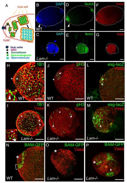
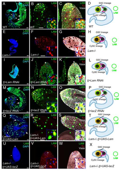
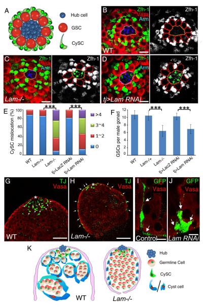
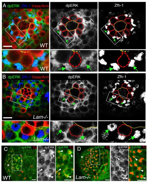
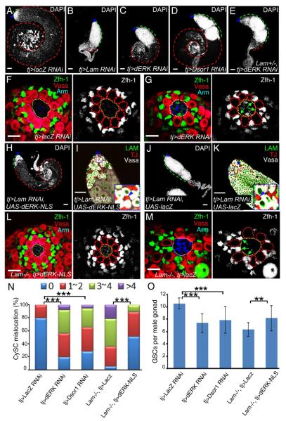
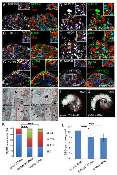
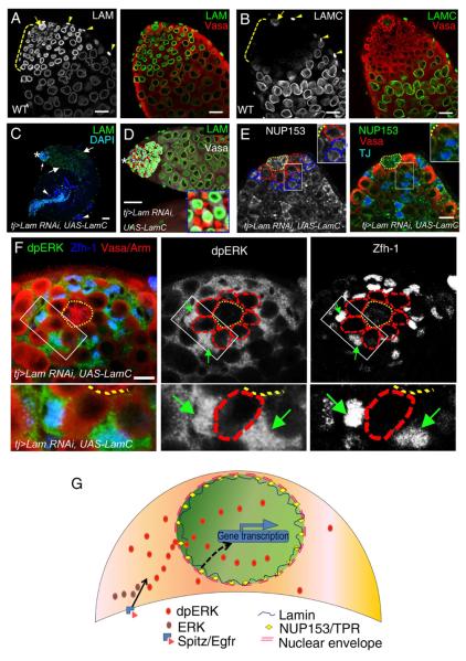
Stem cell niche interactions have been studied extensively with regard to cell polarity and extracellular signaling. Less is known about the way in which signals and polarity cues integrate with intracellular structures to ensure appropriate niche organization and function. Here, we report that nuclear lamins function in the cyst stem cells (CySCs) of Drosophila testes to control the interaction of CySCs with the hub. This interaction is important for regulation of CySC differentiation and organization of the niche that supports the germline stem cells (GSCs). Lamin promotes nuclear retention of phosphorylated ERK in the CySC lineage by regulating the distribution of specific nucleoporins within the nuclear pores. Lamin-regulated nuclear epidermal growth factor (EGF) receptor signaling in the CySC lineage is essential for proliferation and differentiation of the GSCs and the transient amplifying germ cells. Thus, we have uncovered a role for the nuclear lamina in the integration of EGF signaling to regulate stem cell niche function.


Figures

Figure 1
Figure 1. Effects of LAM deletion on the differentiation of GSCs and the transit-amplifying germ cells in 3rd instar larvae (L3) male gonads
A. The hub cells form the niche for both GSCs and CySCs. The asymmetric division of a GSC produces a new GSC and a gonialblast that displace away from the hub along with the pair of cyst cells produced through the asymmetric divisions of a pair of CySCs. Further differentiation of gonialblasts involves spermatogonial mitosis (transient amplification) followed by the growth of spermatocytes, spermatocyte meiosis, and the formation of sperm bundles (not shown). Thick arrows indicate the progression of differentiation and development. BG. Defects of germ cell differentiation in Lam-null L3 male gonads. DAPI (B and C), Notch (D and E), and Vasa (F and G) staining of wild-type (B, D, F) and LamD395/Df(2L)cl-h1 (Lam−/−) (C, E, G) L3 male gonads. Vasa (red) labels germ cells. Notch (green) labels GSCs, gonialblasts, and spermatogonia. Arrowheads mark the transition between spermatogonia and spermatocytes in the wild-type gonad, which is missing in the Lam-null gonads. H and I. L3 Male gonads labeled by the monoclonal antibody 1B1, which stains the spectrin in spectrosomes and fusomes (green). The wild-type gonad (H) contained both spherical spectrosomes (in GSCs, gonialblasts, and two-cell stage spermatogonia, arrow) and branching fusomes (in spermatogonia and spermatocytes, arrowhead), while the Lam-null gonad (I) contained mostly spectrosome-like structures (arrow) and a few poorly branched fusomes (arrowhead). The inset in H shows an enlarged area with the arrow pointing to a branched fusome. The inset in I show an enlarged area with the arrow and arrowhead pointing to a spectrosome and a poorly branched fusome, respectively. J and K. A wild-type L3 male gonad (J) contains a cyst of 8 pH3+ germ cells (arrow) that is close to the hub, whereas most pH3+ germ cells in the Lam-null gonad (K) appear as singlets or pairs (arrows) and many of them are far away from the hub (asterisk). L and M. Labeling of Hubs (asterisks), GSCs, and gonialblasts by escargotM5-4-LacZ (green) revealed the accumulation of undifferentiated GSCs or gonialblasts (arrows) in the Lam-null male gonad (M) compared to the wild-type control (L). NP. Labeling of spermatogonia by BAM-GFP expression indicates the expected localization of BAM-GFP+ cells in wild-type (N) close to the hub (asterisks). In the Lam-null gonads, 52.2% (n=23) do not have BAM-GFP+ cells (O), while the remaining gonads (P) contain large clusters of BAM-GFP+ cells that are displaced away from the hub. The arrow in P marks a cluster of more than 16 BAM-GFP+ germ cells. Scale bars, 50μm.
Figure 2
Figure 2. LAM functions in the CySC lineage to support the differentiation of GSCs and the transit-amplifying germ cells
AH. Whole-mount testes from 2-day old wild-type and Lam-null animals stained with DAPI and antibodies to LAM, TJ (the CySC lineage), and Vasa (the germline). The tip of wild-type (A) and Lam-null (D) testes are enlarged in B&C and F&G, respectively. C and G show the merge of triple color label of LAM, TJ, and Vasa of the same testes shown in B and F, respectively. Small white arrows in A and E, early germ cells. Large white arrows and arrowheads in A show spermatocytes and sperm bundles, respectively. Insets in B&C and F&G show enlarged areas with blue arrows and arrowheads indicating Vasa+ germ cells and TJ + cyst cells, respectively. D and H illustrate the nuclear LAM (green circle) status in the GSC and CySC lineages in wild-type (D) and Lam-null (H) testes. For clarity, only one GSC and one CySC are shown. Lam-null in both lineages leads to the formation of small testes as shown in H. IP. Testes with the CySC lineage depletion of LAM (I–L) using the tj-Gal4 driven Lam RNAi allele (v45635), but not the control LacZ RNAi (M–P), phenocopied defects seen in Lam-null testes (see E–H). The tip of the testis treated by Lam RNAi (I) or control RNAi (M) is enlarged in J&K and N&O, respectively. K and O show the merge of triple color label of LAM, TJ, and Vasa of the same testes shown in J and N, respectively. Blue arrows and arrowheads in the enlarged insets in J&K and N&O indicate Vasa+ germ cells and TJ+ cyst cells, respectively. L and P illustrate the testes with cyst cell depletion of LAM by RNAi (L), but not contol RNAi (P), phenocopied the defects seen in Lam-null testes. QX. Expression of Lam cDNA in the CySC lineage using tj-Gal4 (Q–T), but not control LacZ cDNA (U–X), fully rescued the testis defects and male fertility in the Lam-null mutants. The tip of a wild-type (Q) and Lam-null (U) testes are enlarged in R&S and V&W, respectively. S and W show the merge of triple color label of LAM, TJ, and Vasa of the same testes in R and V, respectively. The enlarged insets in R&S and V&W show Vasa+ germ cells (lack LAM, blue arrows) and TJ+ cyst cell (LAM+ in R&S or LAM− in V&W, blue arrowheads). T and X illustrate that Lam cDNA expression, but not the control LacZ cDNA, in the CySC lineage is sufficient for rescuing the testis defects in Lam-null mutants. Asterisks, hubs. Scale bars, 50μm. See also Figure S1 and Table S1.
Figure 3
Figure 3. LAM regulates CySC localization and cyst cell morphogenesis A
A cartoon of the testis niche illustrating the organization of GSCs and CySCs with respect to the hub cells. In wild-type testes, Zfh-1-expressing CySCs surround GSCs. The thin cellular protrusions of CySCs contact the hub with the nuclei localized at the base of the GSCs away from the hub. BD. Immunostaining using antibodies against Drosophila beta-catenin (Arm, blue), Vasa (red), and Zfh-1 (green or white) in wild type (B), Lam-null (C), and Tj>Lam RNAi L3 male gonads (D). Green arrows point to the Zfh-1-expressing CySCs with their nuclei localized close to the hub in C and D, while such kind of close localization is not seen in the control testis (B). The hubs are outlined by yellow dotted lines, while GSCs are outlined by red dotted lines in the images with only Zfh-1 staining. Scale bars, 10μm. E. Quantification of the percentage of L3 male gonads with mis-localized CySCs as judged by the close proximity of the Zfh-1+ nuclei to the hub as seen in C and D. The colored bars indicate the number of CySCs showing mislocalization in each hub. LAM-null (n=36) and tj>LAM RNAi (n=36) L3 male gonads exhibit a significant increase of mis-localized CySCs compared to the wild type (n=27), Lam heterozygous (n=31), or LacZ RNAi (n=28) controls. ***, p<0.001, Wilcoxon Two Sample Tests. F. LAM-null (n=21) and tj>LAM RNAi (n=23) L3 male gonads exhibit a significant decrease in the number of GSCs associated with the hub compared to the wild type (n=16), Lam heterozygous (n=18), or LacZ RNAi (n=17) controls. ***, p<0.001, Student T tests. Error bars, standard deviation (SD). G and H. The cross section views of L3 male gonads. Comparing to wild type, the TJ+ (green, indicated by white arrows) cyst cells in the Lam-null gonad were displaced away from the early germ cells (red, Vasa) near the hub with many cyst cells mislocalized to the surface of the gonads. Asterisks, hubs. Scale bar, 50μm. I and J. The use of the Flp-out RNAi strategy to specifically mark individual cyst cells by GFP. UAS-GFP expressed in both the cytoplasm and nucleus labeled the control (I) and Lam-RNAi (J) cyst cells. Antibodies to GFP (green) and Vasa (red) were used. The long cell extentions seen in the control cyst cell (arrows in I) are missing in the Lam-RNAi cyst cell (arrows in J). Scale bars, 20μm. K. Cartoon illustration of the role of lamin-B in regulating the CySCs localization and the morphogenesis of cyst cells. See also Figure S2.
Figure 4
Figure 4. LAM regulates dpERK nuclear location in CySCs and cyst cells
A and B. dpERK is localized in both the cytoplasm and the nuclei of CySCs (blue, Zfh-1) in wild type (A) but is depleted in the nuclei of CySCs in Lam-null (B) L3 male gonads. The boxed areas in (A) and (B) are enlarged at the bottom. Green arrows indicate the CySCs with (A) or without (B) nuclear dpERK staining. Immunostaining using antibodies against dpERK (green), Arm (red, labels the hub), Vasa (red), and Zfh-1 (blue) in wild type (A) and LAM-null (B) L3 male gonads. Hub areas are outlined with yellow dotted lines, while GSCs in the black and white images are outlined with red dotted lines. Scale bars, 10μm. C and D. dpERK is enriched in the cyst cell nuclei (red, TJ) in wild type (C), but not in Lam-null (D), L3 male gonads. The boxed areas in (C) and (D) are enlarged to the right. Arrowheads, cyst cells with (C) or without (D) nuclear dpERK staining. Asterisks, hubs. Scale bars, 20μm. See also Figure S3.
Figure 5
Figure 5. LAM regulates GSCs maintenance and differentiation in the CySC lineage through EGFR signaling
A–E. The CySC lineage depletion of dERK (C) or DSOR1 (D) by tj-driven RNAi phenocopied the CySC lineage depletion of LAM (B) as revealed by the accumulation of DAPI-bright early germ cells compared to control LacZ RNAi (A). Cyst cell depletion of dERK in Lam+/− (E) resulted in a more severe phenotype than in Lam+/+ flies (compare E to C). Green and red dashed lines outline the DAPI-bright early and DAPI-dim late germ cells, respectively. Scale bars, 50μm. F and G. The CySC lineage depletion of dERK by tj-driven dERK RNAi (G), but not the control lacZ RNAi (F), phenocopied the CySCs mislocalization observed in Lam-null gonads. Arm (blue), Vasa (red), and Zfh-1 (green) label the hub, the germline lineage, and the CySC lineage, respectively. Green arrows in the black and white image in G point to the Zfh-1-expressing CySCs with their nuclei close to the hub. The hubs are outlined with yellow dotted lines, while GSCs (in the black and white images) are outlined with red dotted lines. Scale bars, 10μm. H–K. The effects of forced expression of dERK-NLS in the CySC lineage depleted of LAM on spermatogenesis. An example of a strongly rescued testis is shown in H and I (19 out of 42) along with the control LacZ rescued testis (J and K). The testis tips in H and J are enlarged in I and K, respectively, with the enlarged insets showing the expression of LAM (green) in germ cells (Vasa+, white) but not in cyst cells (blue arrowheads pointing to the TJ+, red cyst cells). Asterisks, hubs. Scale bars, 50μm. L and M. Forced expression of dERK-NLS (L), but not LacZ (M), in the CySC lineage, effectively rescued the CySC mislocalization defect in Lam-null L3 male gonads. Hubs, germline cells, CySCs are labeled by Arm (blue), Vasa (red), and Zfh-1 (green), respectively. Green arrows in the black and white image in M point to the Zfh-1+ CySCs with their nuclei next to the hub. The hubs are outlined with yellow dotted lines, while GSCs (in the black and white images) are outlined with red dotted lines. Scale bar, 10μm. N. Quantification of the percentages of L3 male gonads with mis-localized CySCs as judged by the close proximity of the Zfh-1+ nuclei to the hub. The colored bars indicate the number of CySCs showing mis-localization in each hub. The tj-driven dERK (n=35) or Dsor1 RNAi (n=32) caused a significant increase of gonads with mis-localized CySCs compared to the control tj-driven LacZ RNAi (n=30). The tj-driven expression of dERK-NLS in Lam-null gonads (n=25) resulted in a significant rescue of CySC localization defects compared to control tj-driven LacZ expression (n=26). ***, p<0.001, Wilcoxon Two Sample Test. O. Quantification of the number of GSCs associated with the hub. The tj-driven dERK (n=21) or Dsor1 RNAi (n=19) caused a significant decrease in the number of GSCs associated with the hub compared to the control tj-driven LacZ RNAi (n=16). The tj-driven expression of dERK-NLS in Lam-null animals (n=15) resulted in a significant increase in the number of GSCs associated with the hub compared to the control tj-driven LacZ expression (n=14). **, p<0.01, ***, p<0.001, Student T tests. Error bars, SD. See also Figure S4.
Figure 6
Figure 6. LAM regulates EGFR signaling through NUP153 and MTOR
A–F. NUP153 (A), MTOR (B), and NUP98 (C) are evenly distributed throughtout the nuclear envelope of both CySCs (TJ, red) and GSCs in adult testes treated with control RNAi (tj-Gal4>lacZ RNAi), whereas LAM depletion in CySCs (tj-Gal4>Lam RNAi) caused aggregation of NUP153 (D) and MTOR (E) without affecting NUP98 (F) in CySCs. Insets, enlarged images revealing in more detail the distribution of nucleoporins in CySCs. The bright dots in (C) and (F) are non-specific antibody labels. The hubs, GSCs, and CySCs are outlined with yellow, red, or blue dotted lines, respectively. Scale bars, 10μm. G and H. Electron micrographs of cyst cells revealing that LAM depletion did not affect the assembly of nucleopores in these cells. Both the face (G' and H', arrows) and side (G” and H”, arrowheads) views of nucleopores are shown. N, nucleus; C, cytoplasm. Scale bars, 0.1μm. I and J. Depletion of either NUP153 (I) or MTOR (J) in the CySC lineage resulted in the accumulation of DAPI-bright early germ cells (green dashed lines) at the expense of DAPI-dim differentiated germ cells (red dashed lines) in testes, similar to those seen in LAM depletion. Asterisks, hubs. Scale bars, 50μm. K. Quantification of the percentages of L3 gonads with mis-localized CySCs as judged by the close proximity of the Zfh-1+ nuclei to the hub. The colored bars show the number of CySCs near each hub. The tj-driven Nup153 (n=32) or Mtor RNAi (n=28) caused a significant increase of gonads with mis-localized CySCs compared to the control tj-driven LacZ RNAi (n=25). ***, p<0.001, Wilcoxon Two Sample Test. L. Quantification of the number of GSCs associated with the hub. The tj-driven Nup153 (n=17) or Mtor RNAi (n=21) caused a significant decrease in the number of GSCs associated with the hub compared to the control tj-driven LacZ RNAi (n=13). ***, p<0.001, Student T tests. Error bars, SD. See also Figure S5.
Figure 7
Figure 7. A shared function between A- and B-type lamins in the CySC lineage
A and B. A strong and ubiquitous expression of LAM (A) in the wild-type adult testis compared to the undetectable LAMC in the CySCs, early cyst cells, and early germ cells (B) in the tip region (brackets). LAMC is expressed in the hub and basement muscle cells. Arrows, hub cells. Arrowheads, basement muscle cells. Scale bars, 20μm. C and D. Ectopic expression of tj>LamC cDNA in cyst cells resulted in strong (34 out of 56) or weak (22 out of 56) rescues of germ cell differentiation defects caused by the CySC lineage-specific Lam-RNAi as judged by LAM (green) and DAPI (blue) staining. Small arrows, large arrows, and arrowheads in (C) indicate early germ cells, spermatocytes, and sperm bundles, respectively, in a strongly rescued testis. The tip region in (C) is enlarged in (D) with the inset showing cyst cells (red, lacking LAM) and germ cells (white, containing LAM). An example of a weak LAMC rescued testis and a control tj-driven LacZ cDNA expressed testis are shown in Figure S6E&F and S6G&H, respectively. Asterisks, hubs. Scale bar, 50μm. E. Ectopic expression of tj>LamC cDNA in the CySC lineage resulted in a partial rescue of the NUP153 localization defect caused by tj>Lam-RNAi in a strongly rescued testis. An example of a testis with control tj-driven LacZ cDNA expression is shown in Figure S6I. The hubs, GSCs, and CySCs are outlined with yellow, red, or blue dotted lines, respectively. Scale bars, 10μm. F. The nuclear dpERK localization defect caused by the CySC lineage specific Lam-RNAi was partially rescued by the ectopic expression of tj-driven LamC cDNA in a strongly rescued L3 male gonads. Boxed areas in (F) are enlarged to the bottom. Green arrows point to CySCs with nuclear dpERK staining. Images of the control expression of tj-driven LacZ cDNA are shown in Figure S6J. Arm (red), Vasa (red), and Zfh-1 (blue) label the hub, the germline lineage, and the CySC lineage respectively. The hub and GSCs are outlined with yellow and red dotted lines, respectively. Scale bar, 10μm. G. A model illustrating that LAM regulates the maintenance and differentiation of the GSC and transit-amplifying germ cells by controlling the localization of CySCs and cyst cell morphogenesis via the nucleoporin-dependent EGFR signaling. See also Figure S6.

Similar articles

Cited by

References

    1. Adachi M, Fukuda M, Nishida E. Two co-existing mechanisms for nuclear import of MAP kinase: passive diffusion of a monomer and active transport of a dimer. EMBO J. 1999;18:5347–5358. - PMC - PubMed
    1. Al-Haboubi T, Shumaker DK, Koser J, Wehnert M, Fahrenkrog B. Distinct association of the nuclear pore protein Nup153 with A- and B-type lamins. Nucleus. 2011;2:500–509. - PubMed
    1. Bank EM, Gruenbaum Y. The nuclear lamina and heterochromatin: a complex relationship. Biochem Soc Trans. 2011;39:1705–1709. - PubMed
    1. Cherry CM, Matunis EL. Epigenetic regulation of stem cell maintenance in the Drosophila testis via the nucleosome-remodeling factor NURF. Cell Stem Cell. 2010;6:557–567. - PMC - PubMed
    1. Coffinier C, Chang SY, Nobumori C, Tu Y, Farber EA, Toth JI, Fong LG, Young SG. Abnormal development of the cerebral cortex and cerebellum in the setting of lamin B2 deficiency. Proc Natl Acad Sci U S A. 2010;107:5076–5081. - PMC - PubMed

Publication types

MeSH terms