. 2013:3:2152.
doi: 10.1038/srep02152.

Pannexin 1 involvement in bladder dysfunction in a multiple sclerosis model

Affiliations

Pannexin 1 involvement in bladder dysfunction in a multiple sclerosis model

Hiromitsu Negoro et al. Sci Rep. 2013.

Abstract

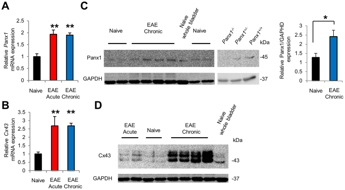
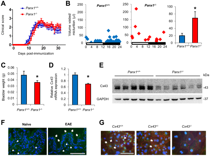
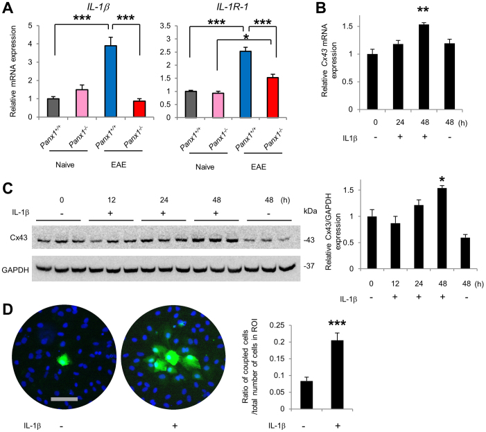
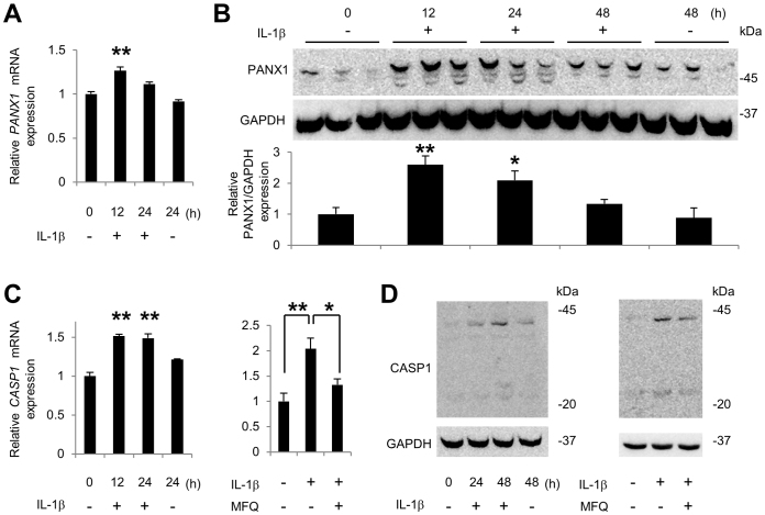
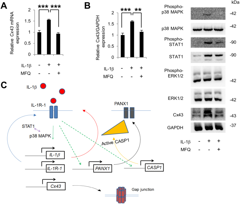
Bladder dysfunction is common in Multiple Sclerosis (MS) but little is known of its pathophysiology. We show that mice with experimental autoimmune encephalomyelitis (EAE), a MS model, have micturition dysfunction and altered expression of genes associated with bladder mechanosensory, transduction and signaling systems including pannexin 1 (Panx1) and Gja1 (encoding connexin43, referred to here as Cx43). EAE mice with Panx1 depletion (Panx1(-/-)) displayed similar neurological deficits but lesser micturition dysfunction compared to Panx1(+/+) EAE. Cx43 and IL-1β upregulation in Panx1(+/+) EAE bladder mucosa was not observed in Panx1(-/-) EAE. In urothelial cells, IL-1β stimulation increased Cx43 expression, dye-coupling, and p38 MAPK phosphorylation but not ERK1/2 phosphorylation. SB203580 (p38 MAPK inhibitor) prevented IL-1β-induced Cx43 upregulation. IL-1β also increased IL-1β, IL-1R-1, PANX1 and CASP1 expression. Mefloquine (Panx1 blocker) reduced these IL-1β responses. We propose that Panx1 signaling provides a positive feedback loop for inflammatory responses involved in bladder dysfunction in MS.


Figures

Figure 1
Figure 1. Micturition dysfunction in C57BL/6 wild type mice with EAE.
(A) Time course of neurological symptoms in mice with EAE (n = 28, 5 mice died at dpi 10, 11, 15, 16 and 21). (B, C) Representative charts of UVVM of naïve mice (left), EAE mice in acute phase (16–17 dpi, middle) and EAE mice in chronic phase (38–39 dpi, right) in (B), and average UVVM in (C). EAE mice in chronic phase had significantly lower functional bladder capacity than naïve mice. *P < 0.05 by Kruskal Wallis test followed by Dunn's post-hoc test (n = 4, 3 and 6, respectively). (C) Bladder weight was significantly increased in chronic phase. **P < 0.01 by one-way ANOVA followed by Tukey's post-hoc test (n = 6, 7 and 5, in naïve, acute and chronic phases of EAE, respectively). All error bars represent SEM.
Figure 2
Figure 2. EAE mice display increased Panx1 and Cx43 expression levels in bladder mucosa.
(A, B) Increased Panx1 mRNA expression in (A) Cx43 mRNA expression in (B) in mucosa of EAE mice in acute and chronic phases (n = 5, 3 and 3 in naïve, EAE acute phase and EAE chronic phase, respectively). **P < 0.01 by one-way ANOVA followed by Tukey's post hoc test. (C) Blot on the left: Increased Panx1 protein expression in chronic phase. *P < 0.05 by unpaired t-test (n = 4). Blot on the right: Specificity of the Panx1 antibody is demonstrated by the absence of the expected band in the protein homogenate of Panx1−/− mouse bladder. (D) Increased Cx43 protein expression in chronic phase. All error bars represent SEM. For the relative levels, the values from naïve mice were set as 1 in (A) and (B), and the value of naïve bladder was set as 1 in (C). Full-length blots are presented in Supplementary Fig. S11.
Figure 3
Figure 3. Genetic depletion of Panx1 ameliorates bladder dysfunction and prevents Cx43 upregulation in EAE mice.
(A) Clinical score of Panx1+/+ EAE and Panx1−/− EAE mice (n = 16, Panx1+/+; n = 19, Panx1−/−; Three Panx1+/+ mice died). No significant difference in neurological deficits, as determined by two-way repeated measures ANOVA (Three dead Panx1+/+ mice were excluded from the ANOVA, but an analysis before deaths was also not significant). (B) Representative charts for UVVM of Panx1+/+ EAE mice [left, Clinical Score (CS) = 2], Panx1−/− EAE mice (middle, CS = 3), and average UVVM (right). Panx1−/− EAE mice in chronic phase had significantly higher UVVM than Panx1+/+ EAE mice. *P < 0.05 by Mann-Whitney U test (n = 3, Panx1+/+; n = 8, Panx1−/−). (C) Bladder weight of Panx1−/− EAE was significantly lower than that of Panx1+/+ EAE mice. *P < 0.05 by unpaired t-test. (n = 8, Panx1+/+; n = 14, Panx1−/−). (D, E) Cx43 expression in the bladder mucosa of Panx1−/− EAE was lower than that in Panx1+/+ EAE mice; mRNA is shown in (D) and protein in (E) (full-length blots are presented in Supplementary Fig. S12). *P < 0.05 by unpaired t-test. (n = 3, Panx1+/+; n = 8, Panx1−/− in (D)). (F) Representative immunostaining of Cx43 in the bladders of Panx1+/+ naïve and EAE mice. Arrows indicate positive staining in urothelium, more evident in EAE mice, and arrow heads in submucosa. Scale bar = 100 μm. (G) Cx43 immunostaining in urothelial cells cultured from Cx43+/+, Cx43+/ and Cx43/ mice. Green, Cx43; Red, E-cadherin. Arrows indicate typical dot stain of gap junctions. Scale bar = 100 μm. All error bars indicate SEM.
Figure 4
Figure 4. IL-1β is upregulated in Panx1+/+ but not in Panx1−/− EAE mice, and IL-1β increases Cx43 expression and function in cultured urothelial cells.
(A) IL-1β mRNA expression in bladder mucosa of Panx1+/+ EAE mice was significantly increased when compared to that in naïve Panx1+/+ mice mucosa, a phenomenon that was not observed in Panx1−/− EAE mice. IL-1R-1 upregulation in Panx1−/− EAE mice was significantly lower than in Panx1+/+ EAE mice. *** P < 0.001 and * P < 0.05 by one-way ANOVA followed by Bonferroni's post hoc test (n = 5, 4, 3 and 8, respectively). (B, C) IL-1β stimulation of TRT-HU1 cells increased levels of Cx43 mRNA in (B) and its protein in (C) (full-length blots are presented in Supplementary Fig. S13) (n = 3 for each time). * P < 0.05 and ** P < 0.01 compared with time 0 h and 48 h without IL-1β stimulation using one-way ANOVA followed by Tukey's post hoc test. (D) Enhanced gap junctional communication in TRT-HU1 cells stimulated with IL-1β evaluated by Lucifer yellow (LY) microinjection. Image from one representative experiment with and without IL-1β stimulation (green, LY; blue, Hoechst 33342), and dye spread quantification within the region of interest (ROI) (n = 19 each; average cell number in ROIs was 42.2 ± 2.1 and 44.5 ± 4.0, respectively; mean ± SEM). *** P < 0.0001 by unpaired t-test. Scale bar = 100 μm. For relative levels, values of Panx1+/+ naïve mice in (A) and time 0 h in (B) and (C) were set as 1. All error bars represent SEM.
Figure 5
Figure 5. IL-1β signaling via p38 MAPK.
(A) IL-1β increased phosphorylation of p38 MAPK and STAT1 but not of ERK (full-length blots are presented in Supplementary Fig. S14). (B) IL-1β induced upregulation of Cx43 expression was blocked by SB203580 but not U0126 and was potentiated by Fludarabine treatment at 48 h (full-length blots are presented in Supplementary Fig. S14). * P < 0.05 and ** P < 0.01 compared with DMSO by one-way ANOVA followed by Dunnett's post hoc test. (C, D) IL-1β stimulation increased IL-1β, IL-1R-1 and STAT1 mRNA levels in TRT-HU1 cells in (C), a response that was blocked by SB203580 and enhanced by Fludarabine in (D) at 24 h. *** P < 0.0005 compared with time 0 h and 24 h without IL-1β by one-way ANOVA followed by Tukey's post hoc test in (C) (n = 3). * P < 0.05, ** P < 0.01 and *** P < 0.0005 compared with DMSO by one-way ANOVA followed by Dunnett's post hoc test in (D) (n = 3). All error bars indicate SEM.
Figure 6
Figure 6. IL-1β stimulation increases pannexin 1 and caspase-1 expression in urothelial cells.
(A, B) IL-1β stimulation of TRT-HU1 cells increased levels of Panx1 mRNA in (A) and its protein in (B) (full-length blots are presented in Supplementary Fig. S15). ** P < 0.01 and * P < 0.05 compared with time 0 and 24 in (A) or 48 in (B) without IL-1β by one-way ANOVA followed by Tukey's post hoc test (n = 3). (C, D) IL-1β stimulation of TRT-HU1 cells increased levels of CASP1 mRNA in (C) and its protein in (D), a response that was inhibited by the Panx1 channel blocker mefloquine (MFQ) at 24 h in (C) and at 48 h in (D) (full-length blots are presented in Supplementary Fig. S15). ** P < 0.01 compared with time 0 and 24 without IL-1β by one-way ANOVA followed by Tukey's post hoc test in (C), left panel (n = 3). * P < 0.05 and ** P < 0.01 by one-way ANOVA followed by Dunnett's post hoc test in (C), right panel (n = 3). All error bars indicate SEM.
Figure 7
Figure 7. Mefloquine inhibits IL-1β induced Cx43 upregulation.
(A, B) Treatment of TRT-HU1 cells with mefloquine (MFQ) inhibited IL-1β induced increase of Cx43 mRNA in (A) and its protein in (B), an event that was accompanied by decreased phosphorylation of p38 MAPK and STAT1 but not of ERK (full-length blots are presented in Supplementary Fig. S16). *** P < 0.0001 by one-way ANOVA followed by Dunnett's post hoc test in (A) (n = 3), and *** P < 0.005 and ** P < 0.01 by one-way ANOVA followed by Dunnett's post hoc test in (B) (n = 3). All error bars indicate SEM. (C) A putative mechanism for Panx1 regulation of Cx43 expression through a positive feedback loop with IL-1β signaling. IL-1R-1 stimulation by IL-1β increases Cx43 expression and upregulates IL-1β and IL-1R-1 via p38 MAPK and STAT1 activation; through a still unknown pathway, IL-1R-1 stimulation also upregulates PANX1 and CASP1. In turn, PANX1 signaling activates CASP1, which results in maturation of IL-1β thereby closing the functional loop between PANX1 and IL-1β that regulates Cx43 expression.

References

    1. McCombe P. A., Gordon T. P. & Jackson M. W. Bladder dysfunction in multiple sclerosis. Expert Rev. Neurother. 9, 331–340 (2009). - PubMed
    1. de Seze M., Ruffion A., Denys P., Joseph P. A. & Perrouin-Verbe B. The neurogenic bladder in multiple sclerosis: review of the literature and proposal of management guidelines. Mult. Scler. 13, 915–928 (2007). - PubMed
    1. Lassmann H. Models of multiple sclerosis: new insights into pathophysiology and repair. Curr. Opin. Neurol. 21, 242–247 (2008). - PubMed
    1. Constantinescu C. S., Farooqi N., O'Brien K. & Gran B. Experimental autoimmune encephalomyelitis (EAE) as a model for multiple sclerosis (MS). Br. J. Pharmacol. 164, 1079–1106 (2011). - PMC - PubMed
    1. Al-Izki S., Pryce G., Giovannoni G. & Baker D. Evaluating potential therapies for bladder dysfunction in a mouse model of multiple sclerosis with high-resolution ultrasonography. Mult. Scler. 15, 795–801 (2009). - PubMed

Publication types

MeSH terms