. 2013 Aug 23;288(34):24302-15.
doi: 10.1074/jbc.M113.496703. Epub 2013 Jul 3.

Transcriptional properties of mammalian elongin A and its role in stress response

Affiliations

Transcriptional properties of mammalian elongin A and its role in stress response

Junya Kawauchi et al. J Biol Chem. .

Abstract

Elongin A was shown previously to be capable of potently activating the rate of RNA polymerase II (RNAPII) transcription elongation in vitro by suppressing transient pausing by the enzyme at many sites along DNA templates. The role of Elongin A in RNAPII transcription in mammalian cells, however, has not been clearly established. In this report, we investigate the function of Elongin A in RNAPII transcription. We present evidence that Elongin A associates with the IIO form of RNAPII at sites of newly transcribed RNA and is relocated to dotlike domains distinct from those containing RNAPII when cells are treated with the kinase inhibitor 5,6-dichloro-1-β-d-ribofuranosylbenzimidazole. Significantly, Elongin A is required for maximal induction of transcription of the stress response genes ATF3 and p21 in response to several stimuli. Evidence from structure-function studies argues that Elongin A transcription elongation activity, but not its ubiquitination activity, is most important for its function in induction of transcription of ATF3 and p21. Taken together, our data provide new insights into the function of Elongin A in RNAPII transcription and bring to light a previously unrecognized role for Elongin A in the regulation of stress response genes.

Keywords: ATF3; Elongin A; Gene Regulation; Mammal; RNA Polymerase II; Stress Response; Transcription Elongation Factors.


Figures
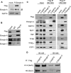
FIGURE 1.
FIGURE 1.
Elongin A complexes immunopurified under different salt conditions. A, nuclear extracts were prepared from HeLa cells stably transfected with pCI-neo-FLAG-hElongin A (F-Elongin A) or with empty pCI-neo (mock) and subjected to Western blotting using anti-FLAG, anti-Elongin A, or anti-RAP74 antibody. Note that dual bands (arrowheads) in FLAG-hElongin A cell extract in the Elongin A blot indicate both endogenous and FLAG-tagged hElongin A. B, nuclear extracts from hElongin A-expressing cells were subjected to immunoprecipitation (IP) using the control IgG or anti-FLAG antibody, and the resultant immunocomplexes were assayed for the Elongin trimer using anti-FLAG, anti-Elongin A, anti-Elongin B, or anti-Elongin C antibody. C, nuclear extracts from HeLa cells stably expressing FLAG-hElongin A, FLAG-RAP74, or the control mock vector were immunoprecipitated by anti-FLAG antibody or control IgG under low (80 mm) or high (150 mm) salt conditions. Resulting immunocomplexes containing Elongin A or RAP74 were assayed by Western blotting using anti-Rpb1, -TBP, -TFIIB, -TFIIEα, and -FCP1 antibodies. D, immunocomplexes in C were also analyzed by Western blot using monoclonal H14 or H5 antibody recognizing the RNAPII CTD phosphorylated on Ser5 or Ser2, respectively.
FIGURE 2.
FIGURE 2.
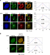
Colocalization of Elongin A and RNAPIIO and their relocation in the presence of DRB. A, HeLa cells stably expressing FLAG-tagged Elongin A grown in the absence or presence of 100 μm DRB for 3 h were immunostained with anti-FLAG (Elongin A), monoclonal H14 (RNAPIIO (IIO) at Ser5), or H5 (RNAPIIO at Ser2) antibody as described under “Experimental Procedures.” Magnified views are shown at the upper left-hand corner. Right panels indicate the results of van Steensel's cross-correlation analysis. Each value of Pearson's correlation coefficient (CCF) (dx, 0) is shown. B, HeLa cells stably expressing FLAG-tagged RAP74 were treated with DRB and immunostained using anti-FLAG (RAP74) and monoclonal H5 (RNAPIIO at Ser2) antibodies as in A. Cross-correlation analysis was performed as well (right panels).
FIGURE 3.
FIGURE 3.
Colocalization of Elongin A with newly synthesized RNA. A, HeLa cells stably expressing FLAG-tagged Elongin A were cultured in the presence of 2 mm bromouridine (BrU) at 37 °C for 20 min and then immunostained with anti-BrdU and anti-FLAG antibodies. Magnified views are also shown. Colocalizations were analyzed as in Fig. 2. The result of the van Steensel's cross-correlation analysis is shown. CCF, correlation coefficient. B, Western blot of Elongin A in the wild type (+/+) and Elongin A-deficient MEFs (+/− and −/−) are shown in the top panel. MEFs from the wild type and Elongin A-null mouse (−/−) were immunostained with monoclonal H5 (RNAPIIO (Pol IIO) at Ser2) and anti-Elongin A antibodies (lower left panel). In the right panel, MEFs were treated as in A and immunostained with anti-BrdU or anti-Elongin A antibody.
FIGURE 4.
FIGURE 4.
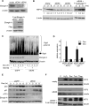
Mammalian Elongin A is required for the induction of several stress response genes. A, whole cell extract of Elongin A, siElA4, or siElA6 was subjected to Western blotting using anti-Elongin A antibody (upper panel). In the lower panel, an expression vector encoding FLAG-tagged rat Elongin A (F-rat Elongin A) was reintroduced into the siElA4 or siElA6 cells, and cells stably expressing rat Elongin A were selected by hygromycin. Cell extracts were analyzed by Western blotting using anti-Elongin A or anti-FLAG antibody. B, wild type, siElA6, siElA4, or siElA6 cells with FLAG-rat Elongin A were subjected to heat shock at 43 °C for 30 min and returned to 37 °C. After the indicated times of incubation, whole cell extracts were prepared and subjected to Western blot analysis. Relative intensities of each band are indicated. C, HeLa cells stably expressing siElA6 were treated with MG132 for 1 h followed by 1 μm doxorubicin stimulation. Cells were harvested at the indicated time points, and polyubiquitination (poly Ub) of Rpb1 was assessed by Western blot with H14 antibody. The amount of β-actin is shown as a loading control. D, Elongin A-knocked down cells were treated with 1 μm doxorubicin, and total RNA was extracted after 2-h stimulation. ATF3 and p21 mRNA levels were measured by quantitative RT-PCR. The means of three independent experiments with error bars (S.E.) are shown. E, cells treated as in D were harvested at the indicated time points, and levels of p53, p21, and ATF3 proteins were assessed by Western blotting. The level of β-actin is also shown. F, HeLa cells stably expressing siElA6 or the control cells (C) were treated for 3 h with 50 μm H2O2, 1 μm doxorubicin (Dox), 1 μm tunicamycin (Tuni), or 2 μm thapsigargin (Thap), and the induction of the ATF3 gene was measured by Western blot.
FIGURE 5.
FIGURE 5.
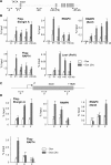
Mammalian Elongin A is associated with the entire ATF3 gene under doxorubicin treatment. A, diagram of the ATF3 gene. Positions of the major two promoters are indicated (P1 and P2; Ref. 40). Boxes denote exons. Double headed arrows denote the locations of regions amplified by qPCR. B, HeLa cell lines stably expressing FLAG-tagged Elongin A were treated with or without 1 μm doxorubicin (Dox) for 2 h. Then a ChIP assay combined with qPCR was carried out as described under “Experimental Procedures” using anti-FLAG (Elongin A), anti-RNAPII, H5 (anti-RNAPIIO Ser2-phosphorylated), and anti-Leo1 antibodies. RAP74 association was also investigated using FLAG-RAP74 stable cell lines and anti-FLAG antibody. Primer sets used for qPCR are depicted in A. Data represent the means of three independent experiments with error bars representing S.E. C, diagram of the HSP70 gene. Double headed arrows denote the locations of regions amplified by qPCR. D, ChIP combined with qPCR was performed as in B with the indicated primer sets in C.
FIGURE 6.
FIGURE 6.
Elongin A association is increased on the p21, c-myc, and c-jun genes after doxorubicin (Dox) treatment. A FLAG (Elongin A) ChIP assay was performed for the five indicated genes. The positions of primer sets are depicted above each bar graph. Data represent the means of three independent experiments with error bars representing S.E.
FIGURE 7.
FIGURE 7.
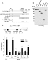
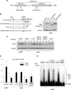
The BC box of mammalian Elongin A is essential for ATF3 induction. A and B, structures and expressions of FLAG-tagged rat Elongin A mutants are shown (upper and lower right panels). SII, SOCS, and RNAPII denote the SII homology domain, SOCS box, and RNAPII binding domain, respectively (22). C, wild type or siElA6 HeLa cells were co-transfected with reporter plasmid pLuc-ATF3 (−384) along with expression vectors encoding various mutants of rat Elongin A. Cells were then treated with doxorubicin (Dox), and the reporter activity was assayed as described under “Experimental Procedures.” The bar graph represents the -fold induction of activity after treatment (solid columns) compared with that without treatment (open columns). The means of five independent experiments with error bars (S.E.) are shown. LUC, luciferase; WB, Western blot; CRE, cAMP response element. NLS, nuclear localization signal.
FIGURE 8.
FIGURE 8.
The C terminus of mammalian Elongin A is essential, but the Cul5 box is dispensable for ATF3 induction. A, amino acid sequence of SOCS box in human Elongin A from 546 to 590. Locations of BC box and Cul5 box are depicted. Arrows indicate the positions of amino acid substitution to abolish each functional domain. B, structures and expressions of several variants of Elongin A are shown. Elongin A full length and L550P/C554F were derived from rat Elongin A. Elongin A V571A/P572A and 1–674 were derived from mouse Elongin A. Each construct was reintroduced into the Elongin A-knocked down cells with retrovirus vectors. The levels of each variant expression were assessed by Western blot using anti-FLAG antibody. The amount of β-actin is shown as a loading control. The asterisks indicate the positions of amino acid substitution detailed in A. C, inductions of ATF3 by doxorubicin (Dox) were analyzed by Western blot. Elongin A mutants shown in B were reintroduced into Elongin A-knocked down HeLa cells. Cells were harvested at 3 h following treatment with 1 μm doxorubicin. As a control, HeLa cells stably expressing siGFP with empty vector reintroduction were also investigated. D, Elongin A mutants shown in C were transfected with ATF3 reporter into HeLa cells expressing siElA6, and then cells were treated with doxorubicin. Reporter activity was measured as in Fig. 6C. Data represent the means of three independent experiments with error bars representing S.E. E, cells as in B were treated with MG132 followed by 1 μm doxorubicin stimulation. After 8 h, polyubiquitination (poly Ub) of Rpb1 was assessed by Western blot as in Fig. 4C.

Similar articles

Cited by

References

    1. Shilatifard A., Conaway R. C., Conaway J. W. (2003) The RNA polymerase II elongation complex. Annu. Rev. Biochem. 72, 693–715 - PubMed
    1. Sims R. J., 3rd, Belotserkovskaya R., Reinberg D. (2004) Elongation by RNA polymerase II: the short and long of it. Genes Dev. 18, 2437–2468 - PubMed
    1. Wada T., Takagi T., Yamaguchi Y., Ferdous A., Imai T., Hirose S., Sugimoto S., Yano K., Hartzog G. A., Winston F., Buratowski S., Handa H. (1998) DSIF, a novel transcription elongation factor that regulates RNA polymerase II processivity, is composed of human Spt4 and Spt5 homologs. Genes Dev. 12, 343–356 - PMC - PubMed
    1. Yamaguchi Y., Takagi T., Wada T., Yano K., Furuya A., Sugimoto S., Hasegawa J., Handa H. (1999) NELF, a multisubunit complex containing RD, cooperates with DSIF to repress RNA polymerase II elongation. Cell 97, 41–51 - PubMed
    1. Saunders A., Core L. J., Lis J. T. (2006) Breaking barriers to transcription elongation. Nat. Rev. Mol. Cell Biol. 7, 557–567 - PubMed

Publication types

MeSH terms