Review
. 2013 Dec;2(12):1544-63.
doi: 10.1002/adhm.201300063. Epub 2013 Jul 5.

Combinatorial biomatrix/cell-based therapies for restoration of host tissue architecture and function

Affiliations
Review

Combinatorial biomatrix/cell-based therapies for restoration of host tissue architecture and function

David Antonio Cantu et al. Adv Healthc Mater. 2013 Dec.

Abstract

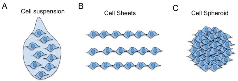
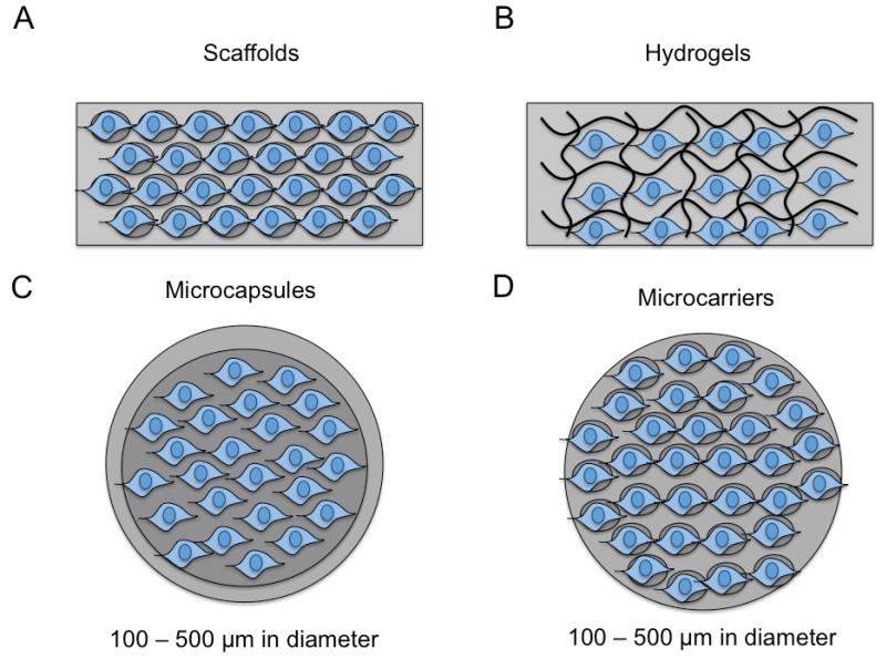
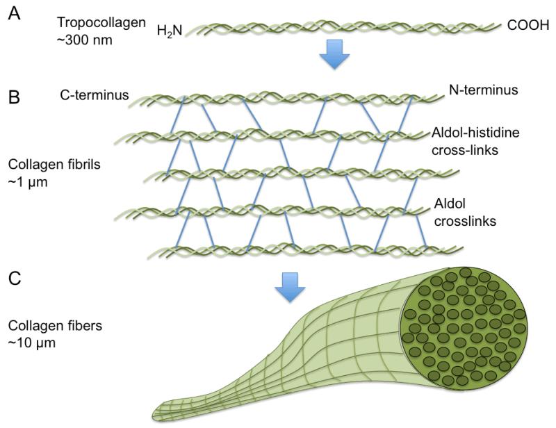
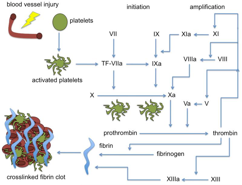
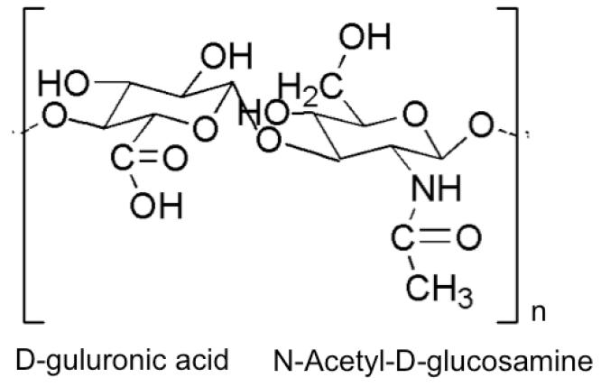
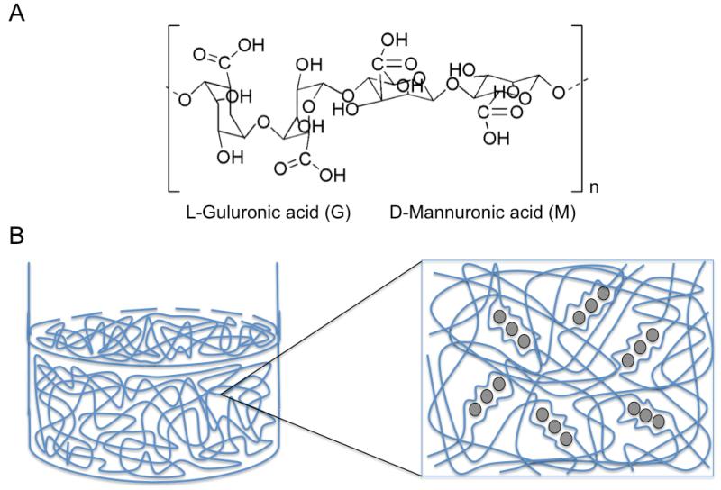
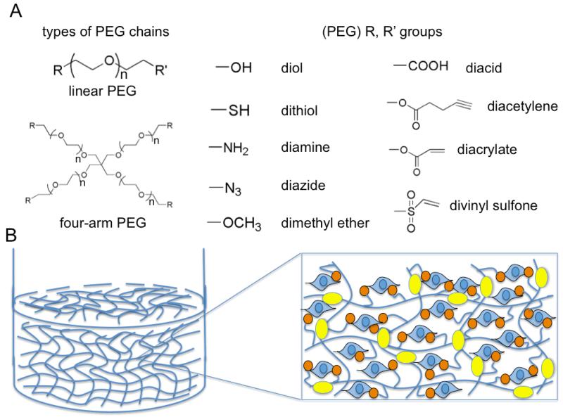
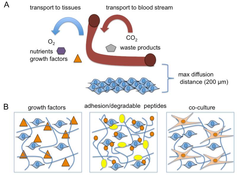
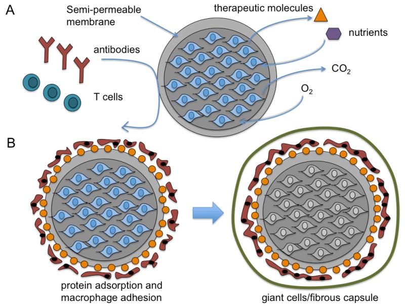
This Progress Report reviews recent advances in the utility of extracellular matrix (ECM)-mimic biomaterials in presenting and delivering therapeutic cells to promote tissue healing. This overview gives a brief introduction of different cell types being used in regenerative medicine and tissue engineering while addressing critical issues that must be overcome before cell-based approaches can be routinely employed in the clinic. A selection of five commonly used cell-associated, biomaterial platforms (collagen, hyaluronic acid, fibrin, alginate, and poly(ethylene glycol)) are reviewed for treatment of a number of acute injury or diseases with emphasis on animal models and clinical trials. This article concludes with current challenges and future perspectives regarding foreign body host response to biomaterials and immunological reactions to allogeneic or xenogeneic cells, vascularization and angiogenesis, matching mechanical strength and anisotropy of native tissues, as well as other non-technical issues regarding the clinical translation of biomatrix/cell-based therapies.

Keywords: biomaterials; cell presentation; cell-based therapy; extracellular matrix; foreign body response.


Figures

Figure 1
Figure 1
Schematic of scaffold-free, cell-based therapies. A) Cell suspension for systemic or direct injection of trypsin-harvested cells; B) Cell sheets for surgical implantation of 2D cell layers; C) Spheroids for direct injection or surgical implantation of 3D cell clusters.
Figure 2
Figure 2
Schematic of biomaterial-centered, cell-based therapies. A) Scaffolds (macroscale) with porous structure seeded with cells prior to implantation; B) Hydrogels (macroscale) with crosslinked structure (chemical, ionic, physical) that gels in vitro or in situ containing encapsulated cells ; C) Microcapsules consisting of hydrogel-core/shells, liquid-core/shells, cell-core/shells that contain cells surrounded by a semi-permeable membrane that can be injected into specific tissue sites; D) Microcarriers (similar to scaffolds) with a porous structure seeded will cells prior to injection.
Figure 3
Figure 3
Schematic of collagen organization. A) Tropocollagen organization of triple helical polypeptides that consists of a repeating Gly-X-Y (where X is often proline and Y is often hydroxyproline) amino acid sequence to form a right handed coiled-coil ; B) Collagen helices self-assemble into collagen fibrils via aldol-histidine and aldol crosslinking; C: Collagen fibrils further organize into via end-to-end self-assembly into collagen fibers.
Figure 4
Figure 4
Schematic of the coagulation cascade for fibrin clot generation. Blood vessel injury leads to platelet activation involving several mediators lead to thrombin conversion of fibrinogen to fibrin. Fibrin polymerization along with platelet aggregation leads to blot clot stabilization and hemostasis.
Figure 5
Figure 5
Basic structure of hyaluronic acid, which requires additional crosslinking or additional modification and reaction with poly(ethylene glycol) to form stable hydrogel structures.
Figure 6
Figure 6
Alginate polymer overview. A) Basic structure of alginate that form block co-polymers repeating G and M monomers; B) Addition of divalent cations such as calcium or barium ions (grey circles) in aqueous solution causes an exchange of sodium ions from guluronic acids (G) units. The divalent cations cause the guluronic (G) units to undergo a conformational change (stiff egg-box structure) that permits the stacking of G units between adjacent alginate chains, which induces a sol-gel transition for hydrogel or microcapsule formation. The concentration of the sodium, alginate wt%, and number of G units in the alginate structure dictate the overall stability and viscoelastic properties of the alginate gels.
Figure 7
Figure 7
Poly(ethylene glycol) (PEG) synthetic polymer overview. A) Types of PEG chains and example functional groups that can be conjugated to the ends of PEG chains; B) Bioactive modification of PEG for maintenance of viability and function (via cell attachment of anchorage-dependent cells) using cell adhesive peptides derived from adhesive protein domains and enzyme-degradable peptide sequences that permit cell or host-mediated remodeling of the hydrogel network. Cell adhesive peptides (orange circles with linkers) can be incorporated into the hydrogel network by post-grafting cell-adhesive moieties to a polymerized hydrogel network or by co-polymerization with acrylated- or thiolated-peptides using Michael-type reactions or acrylate-acrylate or thiol-acrylate photopolymerization. Enzyme-sensitive peptide sequences (yellow ovals) can also be conjugated to acrylated-PEG effectively inserting a degradable moiety between two PEG monoarcylate chains or thiolated enzyme-degradable peptides can be incorporated at crosslink junctions using multi-arm PEG during photopolymerization.
Figure 8
Figure 8
Vascularization overview and solutions to improve vascularization for cell-encapsulating tissue engineered constructs. A) Transport of oxygen, nutrients, and growth factors into tissues and transport of carbon dioxide and waste products into the blood stream. Maximum diffusion distance in most tissues is 200 μm and is significant limitation with most macro-scale scaffolds or polymers for most medical applications that lack pre-vascularization or are not anastomosed to an existing vascular bed. B) Inclusion of growth factors, adhesion peptides (orange circles) and enzyme-degradable peptide sequences (yellow oval), or co-culturing with endothelial cells can all improve the vascularization of specific constructs when implanted in vivo.
Figure 9
Figure 9
Microcapsules and foreign body reaction. A) Schematic of alginate microcapsules containing cells that secrete therapeutic molecules with necessary transport that block large antibodies and T cells due to the MWCO of the semi-permeable membrane effectively evading immune recognition; B: Blood protein adsorption to the capsule surface can facilitate macrophage adhesion, which leads to downstream foreign body giant cell formation and fibrous encapsulation with eventual necrosis of the microencapsulated cells.

Similar articles

Cited by

References

    1. Grim SA, Clark NM. Nature. 2011;90:333. - PubMed
    1. Daley GQ, Scadden DT. Cell. 2008;132:544. - PubMed
    1. Lechler RI, Sykes M, Thomson AW, Turka LA. Nature. 2005;11:605. - PubMed
    1. Ziche M, Donnini S, Morbidelli L. Curr. Drug Targ. 2004;5:485. - PubMed
    1. Anitua E, Sanchez M, Orive G, Andia I. TRENDS Pharma. Sci. 2007;29:37. - PubMed

Publication types

Substances

LinkOut - more resources