Review
. 2013:54:79-90.
doi: 10.1042/bse0540079.

Role of small nuclear RNAs in eukaryotic gene expression

Affiliations
Review

Role of small nuclear RNAs in eukaryotic gene expression

Saba Valadkhan et al. Essays Biochem. 2013.

Abstract

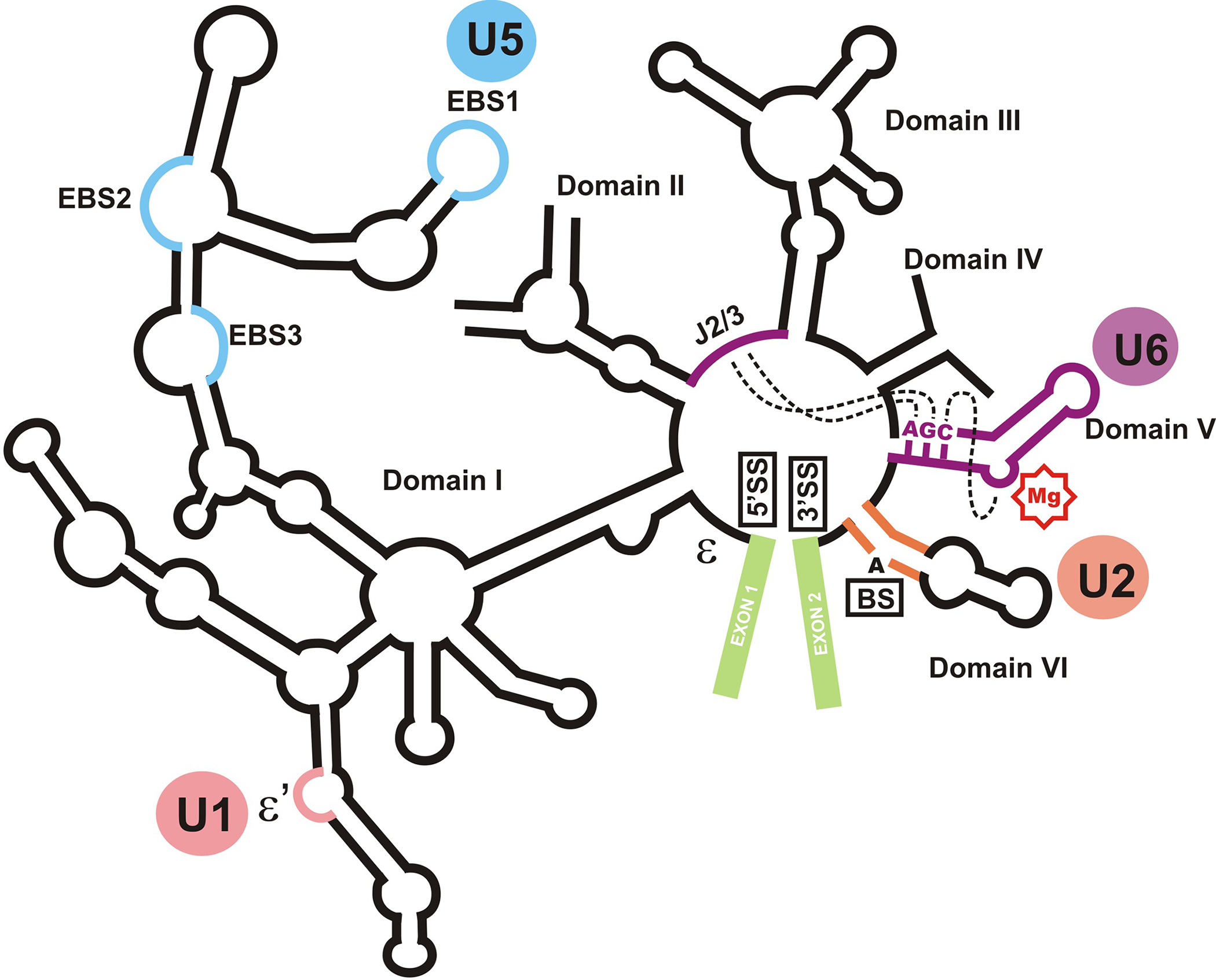
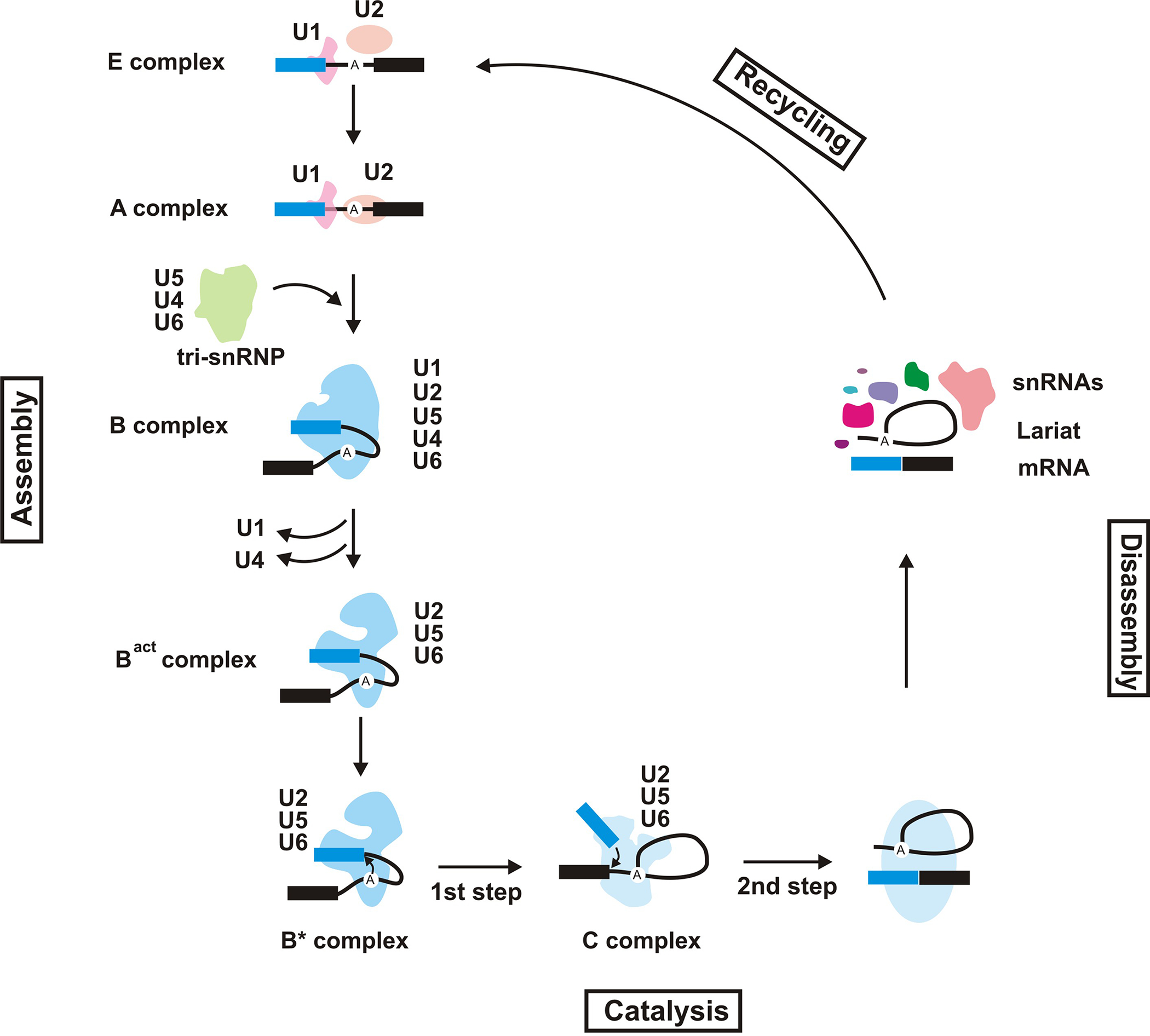
Eukaryotic cells contain small, highly abundant, nuclear-localized non-coding RNAs [snRNAs (small nuclear RNAs)] which play important roles in splicing of introns from primary genomic transcripts. Through a combination of RNA-RNA and RNA-protein interactions, two of the snRNPs, U1 and U2, recognize the splice sites and the branch site of introns. A complex remodelling of RNA-RNA and protein-based interactions follows, resulting in the assembly of catalytically competent spliceosomes, in which the snRNAs and their bound proteins play central roles. This process involves formation of extensive base-pairing interactions between U2 and U6, U6 and the 5' splice site, and U5 and the exonic sequences immediately adjacent to the 5' and 3' splice sites. Thus RNA-RNA interactions involving U2, U5 and U6 help position the reacting groups of the first and second steps of splicing. In addition, U6 is also thought to participate in formation of the spliceosomal active site. Furthermore, emerging evidence suggests additional roles for snRNAs in regulation of various aspects of RNA biogenesis, from transcription to polyadenylation and RNA stability. These snRNP-mediated regulatory roles probably serve to ensure the co-ordination of the different processes involved in biogenesis of RNAs and point to the central importance of snRNAs in eukaryotic gene expression.


Figures

Fig. 1.
Fig. 1.
U6 and U2 snRNAs and the mRNA at the time of first and second steps of splicing. The location of U6, U2 and the U6 intramolecular stemloop is shown. The intron is shown by a thick light blue line connecting the two exons (shown). Position of the 5’ splice site (5’SS), 3’ splice site (3’SS) and branch site are shown. Solid arrows point to the site of the nucleophilic attack during the two steps of splicing. The first step involves a nucleophilic attack by the 2’ hydroxyl group of a specific adenosine residue in the intron, the branch site adenosine (the bulged A), on the 5’ splice site. This leads to a transesterification reaction in which the 2’ oxygen of the branch site adenosine replaces the 3’ oxygen of the last nucleotide of the upstream exon. The result of this reaction is the release of the first exon and the formation of an unusual 2’−5’ linkage between the branch site adenosine and the first nucleotide of the intron (right panel). During the second step, the free 3’ hydroxyl moiety of the newly released exon is activated for a similar nucleophilic attack on the 3’ splice site, resulting in ligation of the two exons and release of the intron as a branched lariat. Base-pairing interactions are shown by short black lines. The location of the 2’−5’ linkage formed after the first step of splicing at the branch site is shown.
Fig. 2.
Fig. 2.
The structural organization of the group II self-splicing intron aI5γ. The location of domains I through VI, the two exons (shown in green), the splice sites and the branch site (5’SS, 3’SS and BS, respectively) are shown. The position of J2/3 and the AGC sequence are indicated. The metal binding site of domain V is shown by a red “Mg” sign. Dashed lines connects regions which are juxtaposed to form the catalytic core. The circles denote the functional equivalent of each domain or subdomain in the spliceosome. ε and ε’ sites, which are involved in an interaction important in recognition of the 5’ splice site are shown.
Fig. 3.
Fig. 3.
The spliceosomal cycle. The spliceosomal complexes formed during a splicing cycle are shown. The snRNAs present in each splicing complex is indicated. The exons on the pre-mRNA are shown as blue and black rectangles, with the intron drawn as a thin line connecting the two. The position of the branch site adenosine is marked.
Fig. 4.
Fig. 4.
The known RNA-RNA interactions at the spliceosomal catalytic core at the time of the first step of splicing. The central domain of human U6 and the 5’ domain of human U2, which contain the sequences necessary for splicing in vivo, are shown. The basepairing interactions between U2 and U6 which form helices I, II and III are shown. The exons in pre-mRNA are drawn as rectangles, with the intron as a thin solid line. The sequence of the branch site of intron is shown. The conserved stemloop I of U5 snRNA is shown at the bottom, with the thin dashed lines marking non-canonical basepairs between the exonic sequences next to the splice sites and the U5 loop I. The ACAGAGA and AGC domains are highlighted in yellow. The dotted, curved red line joining circled nucleotides points to a tertiary interaction detected in activated spliceosomes. The dotted red line connects the binding sites for two functionally-required metal ions (shown by red “Mg” signs) which may be located near each other in activated spliceosomes.
Fig. 5.
Fig. 5.
Structural and functional similarities between the catalytically crucial domain V of group II introns and U6 snRNA. The central domains of human U6 and 5’ domain of human U2 snRNAs are shown. The pre-mRNA is shown as a lighter green line, with the sequence of the branch site indicated. The ACAGAGA and AGC sequences in U6 and domain V are highlighted in yellow. The sites of phosphorothioate interference, which may point to metal binding sites, are shown with red marks.

References

    1. Will CL, Luhrmann R: Spliceosome structure and function. In The RNA World. Edited by Gesteland RF, Cech TR, Atkins JF. Cold Spring Harbor Laboratory Press; 2006:369–400.
    1. Chorev M, Carmel L (2012) The function of introns. Front Genet, 3:55. - PMC - PubMed
    1. Roy SW, Irimia M (2009)Splicing in the eukaryotic ancestor: form, function and dysfunction. Trends Ecol. Evol. (Amst.) 24:447–455. - PubMed
    1. Rodríguez-Trelles F, Tarrío R, Ayala FJ (2006) Origins and evolution of spliceosomal introns. Annu. Rev. Genet 40:47–76. - PubMed
    1. Valadkhan S (2010) Role of the snRNAs in spliceosomal active site. RNA Biol, 7:345–353. - PubMed

Substances