Review
. 2013 Sep;17(9):1053-66.
doi: 10.1517/14728222.2013.815726. Epub 2013 Jul 6.

CD8(+) T cells in multiple sclerosis

Affiliations
Review

CD8(+) T cells in multiple sclerosis

Aleksandar Denic et al. Expert Opin Ther Targets. 2013 Sep.

Abstract

Introduction: CD8(+) T cells were originally considered to exert a suppressive role in demyelinating disease because of bias toward the CD4(+) T cell-mediated experimental autoimmune encephalomyelitis, the most common multiple sclerosis (MS) model. However, recent studies of human MS lesion samples and cerebrospinal fluid (CSF) provided compelling evidence about the pathogenic role of CD8(+) T cells. In this article, we discuss the theoretical roles of different CD8(+) T-cell subsets in MS.

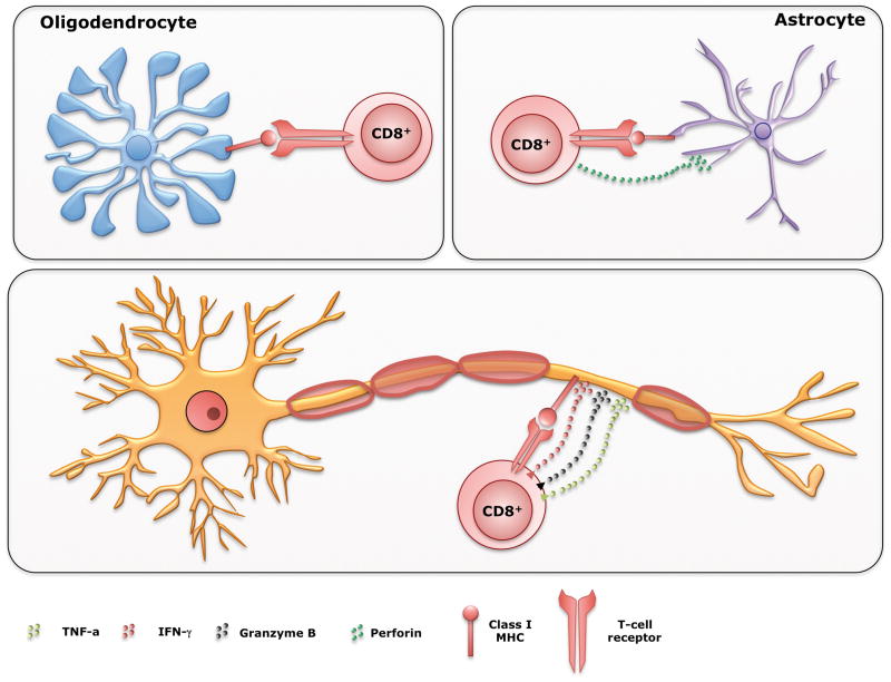
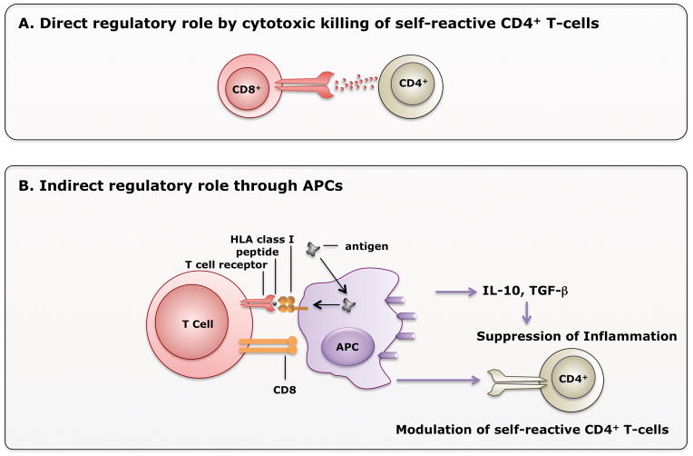
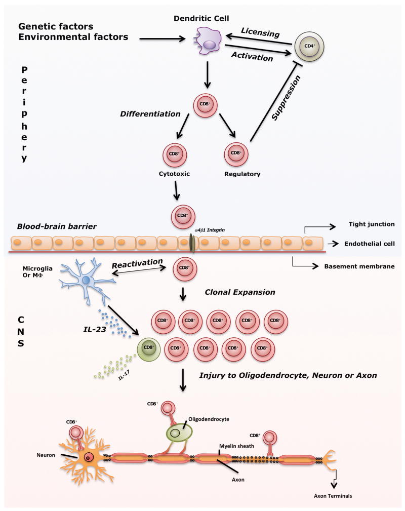
Areas covered: A revised focus from CD4(+) to CD8(+) T cell-mediated demyelinating disease is summarized. Clonal expansion of CD8(+) T cells in MS lesions and in vitro evidence that CD8(+) T cells injure every central nervous system (CNS) cell type and transect axons are discussed. The role of CD8(+) T cells in two animal models of MS and of regulatory, interleukin (IL)-17-secreting CD8(+) T cells is reviewed. Lastly, an overview about the pathogenic and/or beneficial role of various CD8(+) T-cell subsets is offered.

Expert opinion: Growing evidence supports the pathogenic role of CD8(+) T cells. Clonally expanded CD8(+) T cells within MS lesions may damage the nervous system. Revealing the specific antigen is critical to design novel efficient treatments with minimal adverse effects. Increasing evidence exists for the role of regulatory, IL-17-secreting CD8(+) T cells in MS.


Figures

Figure 1
Figure 1. CD8+ T cells can kill all CNS cell types
+ T cells can mediate pathology in MS lesions by killing all cell types in the CNS. Under inflammatory conditions, astrocytes, oligodendrocytes, and neurons/axons express MHC Class I molecules and can be recognized and killed by cytotoxic CD8+ T cells. Upon recognition of MHC Class I, CD8+ T cells in a polarized manner, release granules containing perforin and/or granzyme B, molecules that have cytolitic effect on a target cell. In addition, in the inflammatory setting IFN-γ and TNF-α are also released, affecting the homeostasis of the neurons and axons that express their corresponding receptors, leading them to apoptosis.
Figure 2
Figure 2. CD8+ T cells are responsible for axonal loss and clinical deficits
A) Regardless of the initial trigger that causes demyelination, CD8+ T cells induce injury to axons, lead to progressive axonal loss in the environment with bare, unprotected axons due to the chronic demyelination. B) When CD8+ T cells are absent, demyelination may still occur, but due to sodium channel redistribution there is relative preservation of axons, functional preservation and increased potential for remyelination.
Figure 3
Figure 3. Mechanisms of Regulatory CD8+ T cell-mediated suppression of disease
A) In the direct mechanism, CD8+ T cells exert direct cytotoxic role by recognizing classical Class I molecule or non-classical HLA-E molecule on CD4+ T cells. [84] B) CD8+ T cells may indirectly suppress inflammation by acting through antigen presenting cells (APCs). APCs then secrete anti-inflammatory cytokines IL-10 or TGF-β, or modulate self-reactive CD4+ T cells.
Figure 4
Figure 4. From unknown trigger to CNS injury; a summarized view of the role of CD8+ T cells in MS pathogenesis
Several environmental factors in combination with genetic factors activate dendritic cells (DCs), which then present epitopes and influence priming of CD4+ T cells. Subsequently, primed CD4+ T cells license DCs to activate CD8+ T cells by cross-presenting epitopes. Activated CD8+ T cells then differentiate into cytotoxic (CTL) and regulatory cells. Under normal conditions, regulatory CD8+ T cells prevent immune response against self-antigens as shown in Figure 3. However, when this protective mechanism fails autoreactive CTLs migrate through blood-brain barrier with the help of α4β1 integrin (molecular target of natalizumab). Within the CNS, resident microglia or macrophages that express co-stimulatory and MHC class I molecules likely are involved in reactivation of CD8+ T cells. As a result of reactivation, CD8+ T cells clonally expand, meet hypothetical target antigens presented on MHC class I on neurons, axons or oligodendrocyte and induce injury. In the inflammatory milieu of an MS lesion, IL-6 and IL-1β as well as IL-23 produced by macrophages, polarize some CD8+ T cells into IL-17-secreting cells which can comprise up to 10% of all CD8+ T cells detected in MS lesions. Thus, CD8+ IL-17-secreting T cells may influence exacerbations in MS.

References

    1. Noseworthy JH, Lucchinetti C, Rodriguez M, et al. Multiple sclerosis. N Engl J Med. 2000;343:938–52. - PubMed
    1. Kappos L, Comi G, Panitch H, et al. Induction of a non-encephalitogenic type 2 T helper-cell autoimmune response in multiple sclerosis after administration of an altered peptide ligand in a placebo-controlled, randomized phase II trial. The Altered Peptide Ligand in Relapsing MS Study Group. Nat Med. 2000;6:1176–82. - PubMed
    1. Bielekova B, Goodwin B, Richert N, et al. Encephalitogenic potential of the myelin basic protein peptide (amino acids 83–99) in multiple sclerosis: results of a phase II clinical trial with an altered peptide ligand. Nat Med. 2000;6:1167–75. - PubMed
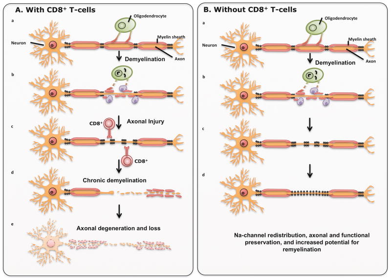
    1. Rivera-Quinones C, McGavern D, Schmelzer JD, et al. Absence of neurological deficits following extensive demyelination in a class I-deficient murine model of multiple sclerosis. Nat Med. 1998;4:187–93. Important study that clearly implicates CD8+ T cells as pathogenic, leading to axonal loss and clinical deficits in a murine model of demyelination. - PMC - PubMed
    1. Howe CL, Adelson JD, Rodriguez M. Absence of perforin expression confers axonal protection despite demyelination. Neurobiol Dis. 2007;25:354–9. - PMC - PubMed

Publication types