

Other cestodes: sparganosis, coenurosis and Taenia crassiceps cysticercosis
- PMID: 23829923
- PMCID: PMC4080899
- DOI: 10.1016/B978-0-444-53490-3.00027-3
Other cestodes: sparganosis, coenurosis and Taenia crassiceps cysticercosis
Abstract
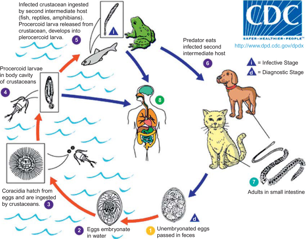
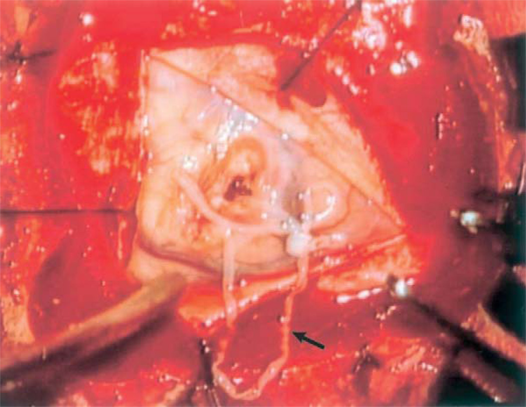
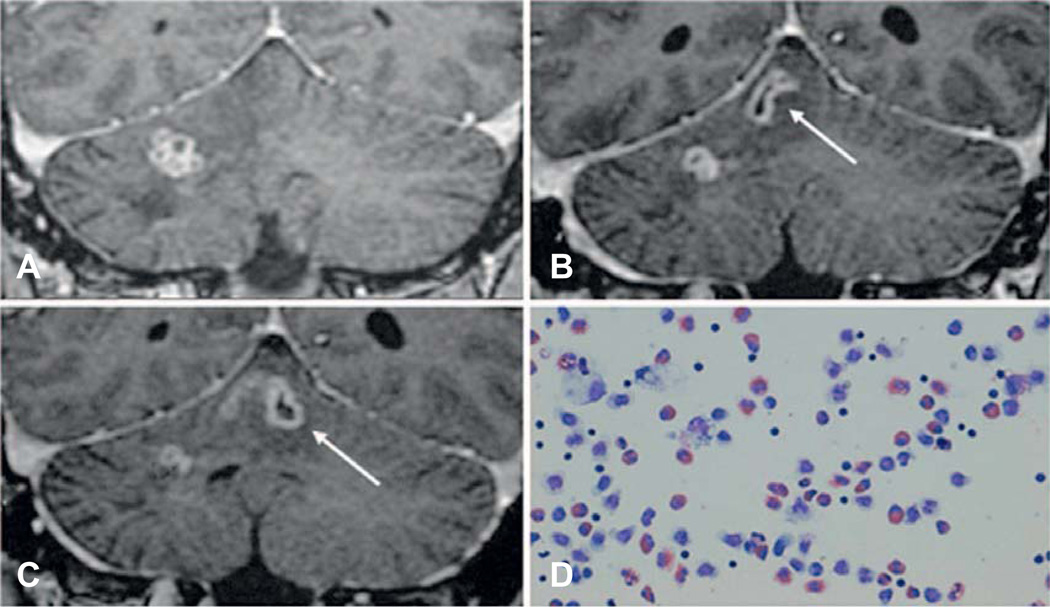
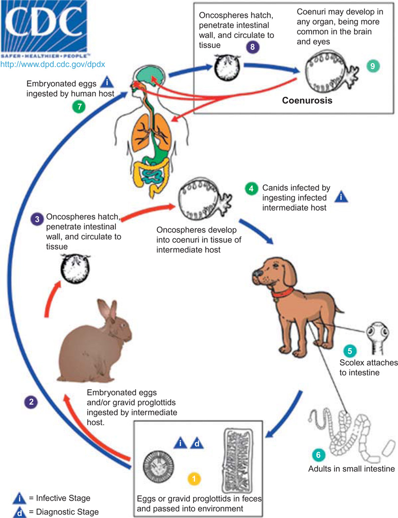
Many cestodes are capable of invading the central nervous system (CNS), and several are highly prevalent in the developing world. Neurocysticercosis due to Taenia solium and echinococcosis due to Echinoccocus granulosus are two of the most common parasitic infections affecting humans, but other less well-known parasites can also infect the nervous system. Coenurosis, caused by Taenia spp. such as T. multiceps, T. serialis, or T. brauni; sparganosis, caused by Spirometra spp., and neurocysticercosis caused by T. crassiceps are three less frequent zoonotic conditions that should be considered in the differential diagnosis of patients presenting with CNS infection - especially if they have lived in or traveled through areas where these infections are endemic. Diagnosis of these infections is typically made through a combination of serological testing, histopathology, and neuroimaging.
Keywords: Central nervous system; Spirometra spp.; Taenia crassiceps; Taenia multiceps; cestodiasis; coenurosis; cysticercosis; parasites; sparganosis; zoonoses.
Copyright © 2013 Elsevier B.V. All rights reserved.
Figures

References
-
- Ambekar S, Prasad C, Dwarakanath S, et al. MRS findings in cerebral coenurosis due to Taenia multiceps. J Neuroimaging. 2013;23:149–151. - PubMed
-
- Anantaphruti MT, Nawa Y, Vanvanitchai Y. Human sparganosis in Thailand: an overview. Acta Trop. 2011;118:171–176. - PubMed
-
- Anders K, Foley K, Stern E, et al. Intracranial sparganosis: an uncommon infection Case report. J Neurosurg. 1984;60:1282–1286. - PubMed
-
- Arocker-Mettinger E, Huber-Spitzy V, Auer H, et al. Taenia crassiceps in the anterior chamber of the human eye. A case report. Klin Monbl Augenheilkd. 1992;201:34–37. - PubMed
-
- Baer JG, Scheidegger S. Not available. Schweiz Z Pathol Bakteriol. 1946;9:61–66. - PubMed
Publication types
MeSH terms
Grants and funding
LinkOut - more resources
Full Text Sources
Other Literature Sources
Miscellaneous
