. 2013 Aug;15(8):986-98.
doi: 10.1016/j.jcyt.2013.04.003.

Optimizing the transduction efficiency of capsid-modified AAV6 serotype vectors in primary human hematopoietic stem cells in vitro and in a xenograft mouse model in vivo

Affiliations

Optimizing the transduction efficiency of capsid-modified AAV6 serotype vectors in primary human hematopoietic stem cells in vitro and in a xenograft mouse model in vivo

Liujiang Song et al. Cytotherapy. 2013 Aug.

Abstract

Background aims: Although recombinant adeno-associated virus serotype 2 (AAV2) vectors have gained attention because of their safety and efficacy in numerous phase I/II clinical trials, their transduction efficiency in hematopoietic stem cells (HSCs) has been reported to be low. Only a few additional AAV serotype vectors have been evaluated, and comparative analyses of their transduction efficiency in HSCs from different species have not been performed.

Methods: We evaluated the transduction efficiency of all available AAV serotype vectors (AAV1 through AAV10) in primary mouse, cynomolgus monkey and human HSCs. The transduction efficiency of the optimized AAV vectors was also evaluated in human HSCs in a murine xenograft model in vivo.

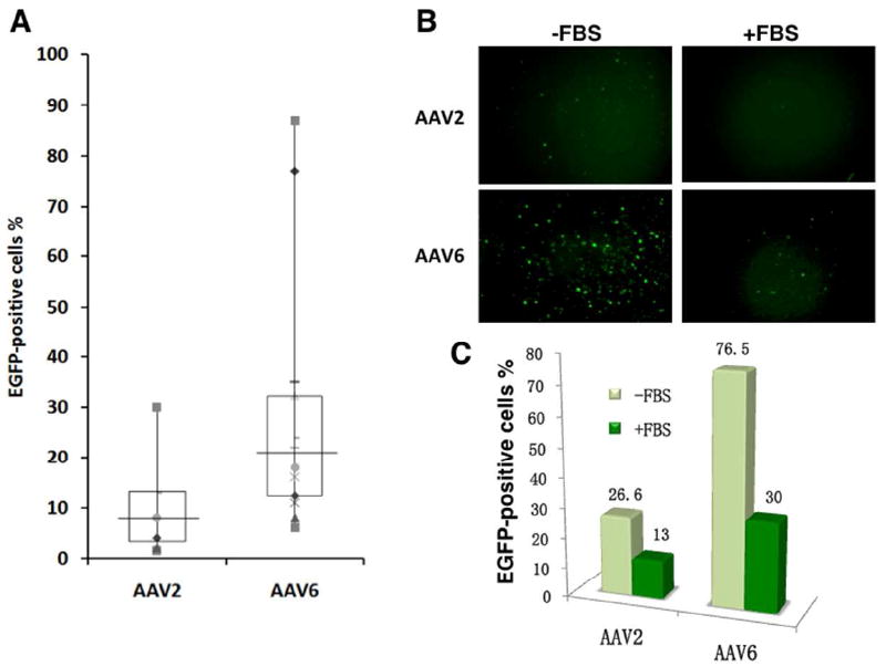
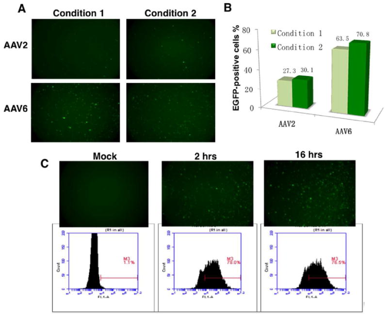
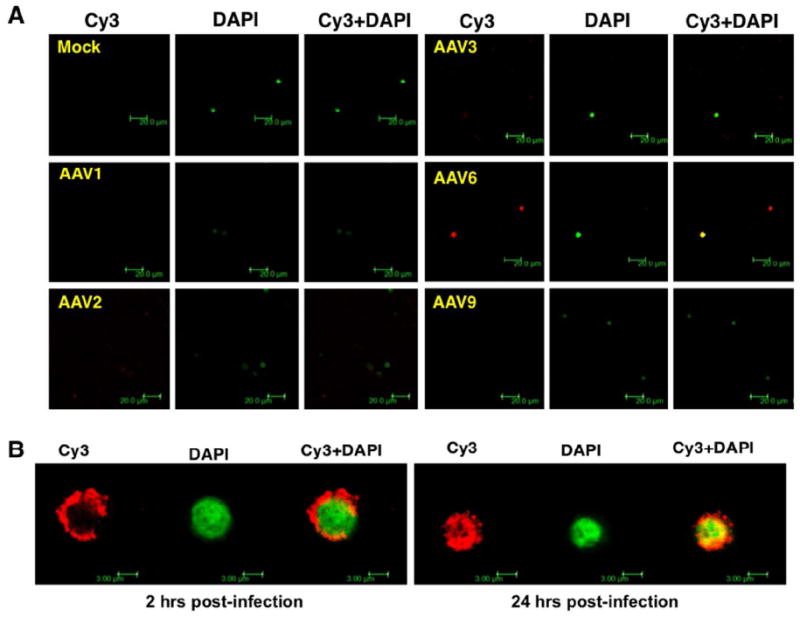
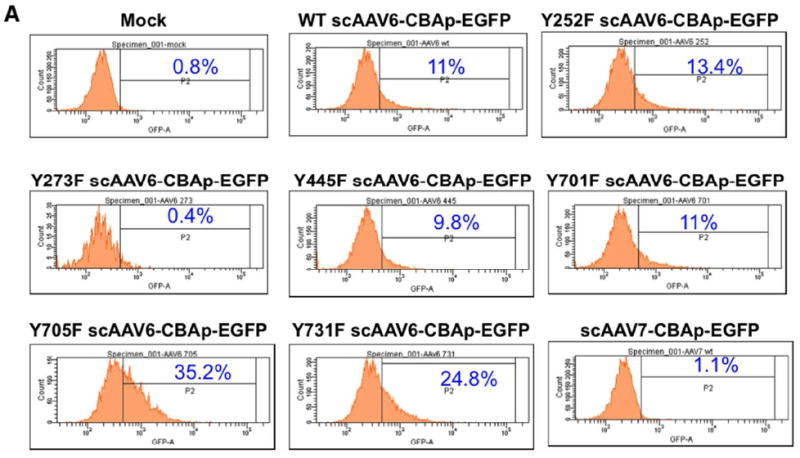
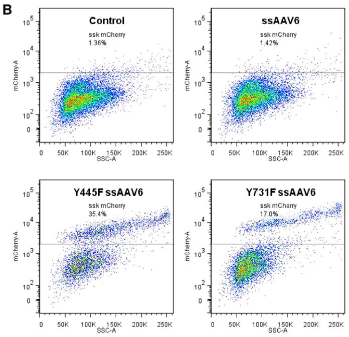
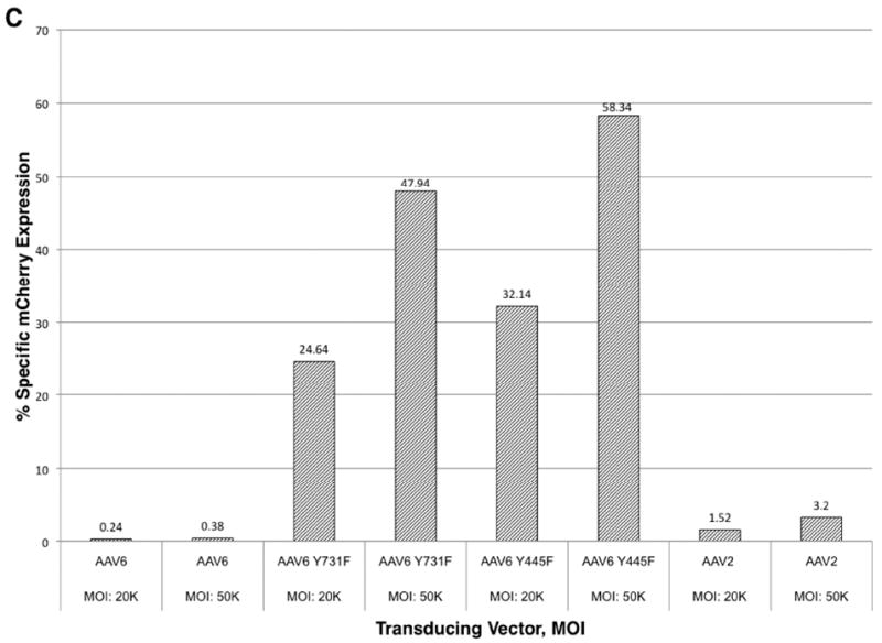
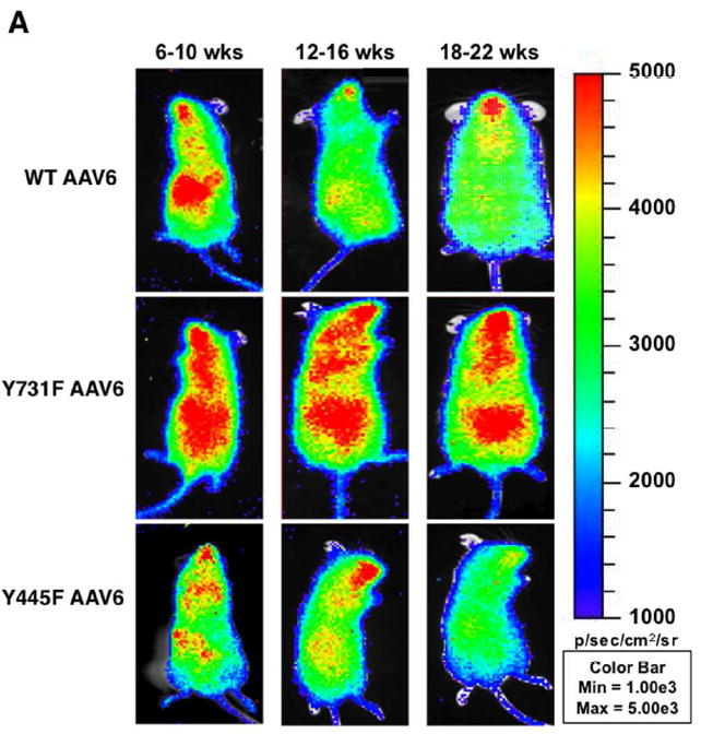
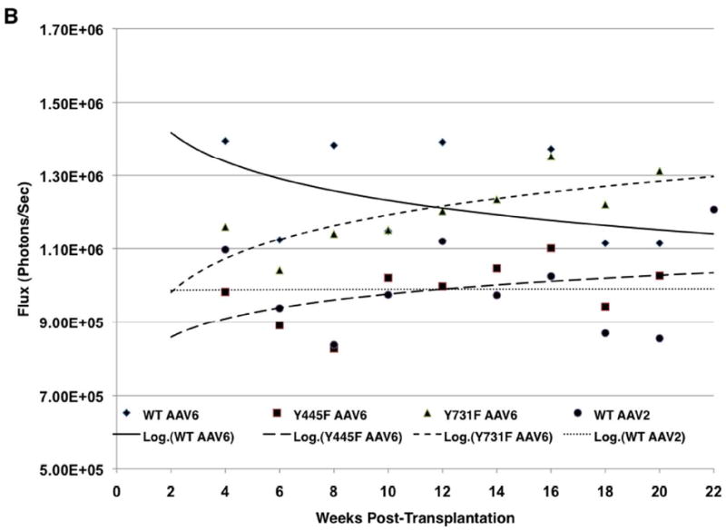
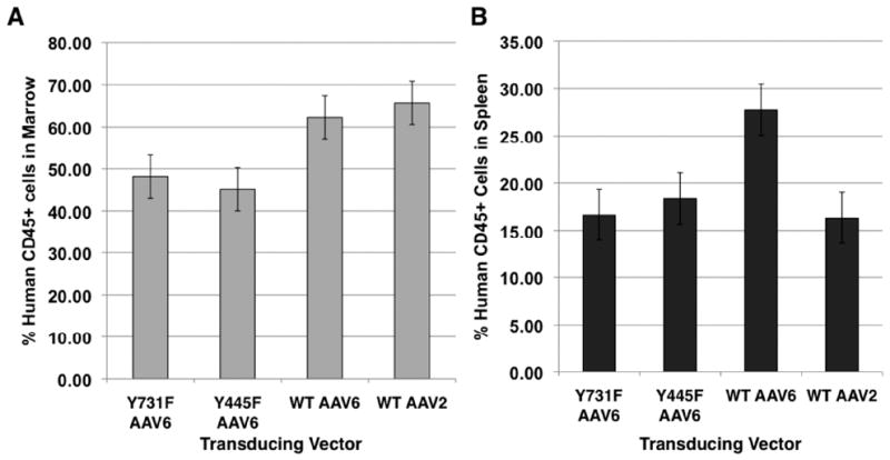
Results: We observed that although there are only six amino acid differences between AAV1 and AAV6, AAV1, but not AAV6, transduced mouse HSCs well, whereas AAV6, but not AAV1, transduced human HSCs well. None of the 10 serotypes transduced cynomolgus monkey HSCs in vitro. We also evaluated the transduction efficiency of AAV6 vectors containing mutations in surface-exposed tyrosine residues. We observed that tyrosine (Y) to phenylalanine (F) point mutations in residues 445, 705 and 731 led to a significant increase in transgene expression in human HSCs in vitro and in a mouse xenograft model in vivo.

Conclusions: These studies suggest that the tyrosine-mutant AAV6 serotype vectors are the most promising vectors for transducing human HSCs and that it is possible to increase further the transduction efficiency of these vectors for their potential use in HSC-based gene therapy in humans.

Keywords: AAV vectors; gene expression; gene transfer; hematopoietic stem cells.


Figures

Figure 1
Figure 1
Comparative analyses of the transduction efficiencies of scAAV1 through AAV10 serotype vectors in primary human CD34+ cells. (A) Equivalent numbers of cells from a single donor were infected with of scAAV serotype vectors at 2×10⁴ vgs/cell under identical culture conditions in IMDM containing 10% FBS, 10 ng/ml of hIL6, 10 ng/ml of hIL3 and 1 ng/ml of rhSCF for 16 hrs. Transgene expression was evaluated 48 hrs post-transduction by fluorescence microscopy and the data were quantitated by flow cytometry (B).
Figure 2
Figure 2
Comparative analyses of the transduction efficiencies of scAAV2 and AAV6 serotype vectors in primary human CD34+ cells from multiple donors. (A) Equivalent numbers of cells from various donors were infected with 2×10⁴ vgs/cell of scAAV2 or AAV6 vectors and transgene expression was evaluated as described above. The boxes represent the upper quartile and lower quartile of the values; the horizontal lines indicate the median values; and different markers represent individual experiments. (B, C) Equivalent numbers of CD34+ cells from the same lots were transduced with scAAV2 or scAAV6 vectors as described above, either in the absence or the presence of 10% FBS. Transgene expression was evaluated 72 hrs post-transduction by fluorescence microscopy, and quantitated by flow cytometry.
Figure 3
Figure 3
Transduction efficiencies of scAAV2 and AAV6 serotype vectors in primary human CD34+ cells under different culture conditions. (A, B) Equivalent numbers of CD34+ cells were transduced with scAAV2 or scAAV6 vectors in the presence of 10 ng/ml of rhFlt3, 10 ng/ml of rhTPO and 1 ng/ml of rhSCF for 2 hrs (Condition 1), or in the presence of 10 ng/ml of rhIL6, 10 ng/ml of rhIL3 and 1 ng/ml for 2 hrs (Condition 2). Transgene expression was evaluated 72 hrs post-transduction by fluorescence microscopy, and quantitated as described above. (C) Equivalent numbers of CD34+ cells were either mock-infected, or infected with scAAV6 vectors for either 2 or 16 hrs. Transgene expression was evaluated 72 hrs post-infection by flow cytometry as described above.
Figure 4
Figure 4
Confocal microscopy for intracellular trafficking of scAAV serotype vectors in human CD34+ cells. Cells were seeded in 24 well plates on poly-L-lysine-coated coverslips, and were either mock-infected, or infected with Cy3-labeled scAAV1, scAAV2, scAAV3, scAAV6, and scAAV9 vectors for 2 hrs (A). Cells were fixed with 4% paraformaldehyde for 8 min at room temperature and nuclei were stained with DAPI for 10 min at room temperature. Images were acquired with a Leica TCS-SP5 laser scanning confocal microscope under an oil-immersed ×63 objective lens. A z-stack was collected with step size of 0.5 μm until Cy3-signal intensity became faint under the laser. The co-localization analyses were performed by velocity software. To facilitate comparisons, the color from DAPI counterstaining was converted into green. The scale bar was calibrated to show the size of the cells (Magnification, × 107.1) (B). Representative confocal images for intracellular trafficking of scAAV6 vectors in human CD34+ cells at 2 and 24 hrs post-transduction. Images were acquired as described above (Magnification, ×806.4).
Figure 5
Figure 5
Transduction efficiencies of WT and tyrosine-mutant AAV6 serotype vectors in primary human CD34+ cells. (A) Equivalent numbers of CD34+ cells were either mock-transduced, or transduced with WT and tyrosine-mutant scAAV6-CBAp- EGFP vectors at 5×10³ vgs/cells, and transgene expression was evaluated 72 hrs post-infection by flow cytometry as described above. scAAV7-CBAp-EGFP vectors were used as a negative control. (B, C) Equivalent numbers of CD34+ cells were either mock-transduced, or transduced with WT and tyrosine-mutant ssAAV6-CBAp-mCherry vectors at 2×10⁴ or 5×10⁴ vgs/cells. WT ssAAV2 vectors were used as appropriate controls. Transgene expression was evaluated at 46 hrs postinfection by flow cytometry, and quantitated as described above.
Figure 5
Figure 5
Transduction efficiencies of WT and tyrosine-mutant AAV6 serotype vectors in primary human CD34+ cells. (A) Equivalent numbers of CD34+ cells were either mock-transduced, or transduced with WT and tyrosine-mutant scAAV6-CBAp- EGFP vectors at 5×10³ vgs/cells, and transgene expression was evaluated 72 hrs post-infection by flow cytometry as described above. scAAV7-CBAp-EGFP vectors were used as a negative control. (B, C) Equivalent numbers of CD34+ cells were either mock-transduced, or transduced with WT and tyrosine-mutant ssAAV6-CBAp-mCherry vectors at 2×10⁴ or 5×10⁴ vgs/cells. WT ssAAV2 vectors were used as appropriate controls. Transgene expression was evaluated at 46 hrs postinfection by flow cytometry, and quantitated as described above.
Figure 6
Figure 6
Transduction efficiencies of WT and tyrosine-mutant ssAAV6 serotype vectors in primary human CD34+ cells in NOD/SCID xenograft mice in vivo. (A) Whole-body bioluminescent imaging was performed as described previously [21]. Representative mice from different time intervals post-transplantation are shown for each capsid type. Serial in vivo bioluminescent imaging showing luciferase expression in NOD/SCID mice transplanted with human CD34+ cells transduced with rAAV Human cord blood CD34+ cells were transduced with the indicated vectors (WT AAV2: n=3; WT AAV6: n=3; Y445F AAV6: n=8; Y731F: n=10). (B). Xeno-transplanted mice were imaged biweekly following luciferin administration. Results are shown for 3-10 mice per group. Each mouse was transplanted with cells pooled from 1-5 CB samples. Luciferase expression is depicted as flux (photons/second) over time.
Figure 6
Figure 6
Transduction efficiencies of WT and tyrosine-mutant ssAAV6 serotype vectors in primary human CD34+ cells in NOD/SCID xenograft mice in vivo. (A) Whole-body bioluminescent imaging was performed as described previously [21]. Representative mice from different time intervals post-transplantation are shown for each capsid type. Serial in vivo bioluminescent imaging showing luciferase expression in NOD/SCID mice transplanted with human CD34+ cells transduced with rAAV Human cord blood CD34+ cells were transduced with the indicated vectors (WT AAV2: n=3; WT AAV6: n=3; Y445F AAV6: n=8; Y731F: n=10). (B). Xeno-transplanted mice were imaged biweekly following luciferin administration. Results are shown for 3-10 mice per group. Each mouse was transplanted with cells pooled from 1-5 CB samples. Luciferase expression is depicted as flux (photons/second) over time.
Figure 7
Figure 7
Engraftment of human cells in the marrow (A) and spleen (B) of NOD/SCID mice. Mononuclear cells from the marrow and spleen of transplant recipients were analyzed for human CD45 expression at the time of harvest 16-22 weeks post-transplantation. The presence of human CD45+ cells indicated human cell engraftment.

References

    1. Cavazzana-Calvo M, Payen E, Negre O, et al. Transfusion independence and HMGA2 activation after gene therapy of human beta-thalassaemia. Nature. 2010;467(7313):318–22. - PMC - PubMed
    1. Mueller C, Flotte TR. Clinical gene therapy using recombinant adeno-associated virus vectors. Gene Therapy. 2008;15(11):858–63. - PubMed
    1. Mingozzi F, High KA. Therapeutic in vivo gene transfer for genetic disease using AAV: progress and challenges. Nature Reviews Genetics. 2011;12(5):341–55. - PubMed
    1. Zhou SZ, Cooper S, Kang LY, et al. Adeno-associated virus 2-mediated high efficiency gene transfer into immature and mature subsets of hematopoietic progenitor cells in human umbilical cord blood. The Journal of Experimental Medicine. 1994;179(6):1867–75. - PMC - PubMed
    1. Goodman S, Xiao X, Donahue RE, et al. Recombinant adeno-associated virus-mediated gene transfer into hematopoietic progenitor cells. Blood. 1994;84(5):1492–500. - PubMed

Publication types