. 2013 Jul 11;93(1):181-90.
doi: 10.1016/j.ajhg.2013.05.028. Epub 2013 Jul 3.

Recessive TRAPPC11 mutations cause a disease spectrum of limb girdle muscular dystrophy and myopathy with movement disorder and intellectual disability

Affiliations

Recessive TRAPPC11 mutations cause a disease spectrum of limb girdle muscular dystrophy and myopathy with movement disorder and intellectual disability

Nina Bögershausen et al. Am J Hum Genet. .

Abstract

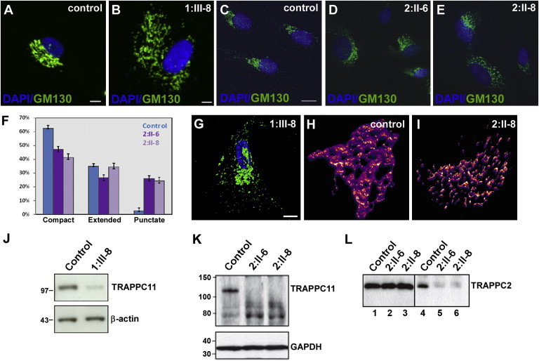
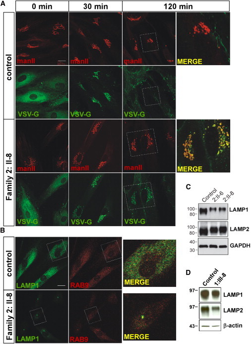
Myopathies are a clinically and etiologically heterogeneous group of disorders that can range from limb girdle muscular dystrophy (LGMD) to syndromic forms with associated features including intellectual disability. Here, we report the identification of mutations in transport protein particle complex 11 (TRAPPC11) in three individuals of a consanguineous Syrian family presenting with LGMD and in five individuals of Hutterite descent presenting with myopathy, infantile hyperkinetic movements, ataxia, and intellectual disability. By using a combination of whole-exome or genome sequencing with homozygosity mapping, we identified the homozygous c.2938G>A (p.Gly980Arg) missense mutation within the gryzun domain of TRAPPC11 in the Syrian LGMD family and the homozygous c.1287+5G>A splice-site mutation resulting in a 58 amino acid in-frame deletion (p.Ala372_Ser429del) in the foie gras domain of TRAPPC11 in the Hutterite families. TRAPPC11 encodes a component of the multiprotein TRAPP complex involved in membrane trafficking. We demonstrate that both mutations impair the binding ability of TRAPPC11 to other TRAPP complex components and disrupt the Golgi apparatus architecture. Marker trafficking experiments for the p.Ala372_Ser429del deletion indicated normal ER-to-Golgi trafficking but dramatically delayed exit from the Golgi to the cell surface. Moreover, we observed alterations of the lysosomal membrane glycoproteins lysosome-associated membrane protein 1 (LAMP1) and LAMP2 as a consequence of TRAPPC11 dysfunction supporting a defect in the transport of secretory proteins as the underlying pathomechanism.


Figures

Figure 1
Figure 1
Pedigrees and Molecular Characterization of the TRAPPC11 Mutations (A) Pedigree structure of families 1, 2, and 3. (B) Representative wild-type (WT) and mutant electropherograms from family 1. The c.2938G>A mutation is indicated by a black arrow. (C) Representative WT and mutant electropherograms from families 2 and 3 demonstrating the homozygous c.1287+5G>A mutation (indicated by a black arrow) in relation to the exon 12 and intron 12 boundary. (D) RT-PCR analysis confirmed the presence of altered C11 splicing in lymphocytes obtained from one carrier parent, two affected individuals, and two controls. Numbers 1, 2, 3, and 4 correspond to different transcripts illustrated in (E). (E) Schematic representation of these RT-PCR products after gel isolation and Sanger sequencing. In control cells, the full-length transcript (1) predominates over an alternatively spliced minor transcript lacking exon 11 (2). In cells from affected individuals, the minor transcript lacks only exon 12 (3), whereas the predominant transcript (4) lacks both exons 11 and 12, resulting in an in-frame deletion of 58 amino acids (p.Ala372_Ser429del). (F) Schematic representation of TRAPPC11 and the location of the foie gras and gryzun domains. The two mutations identified in this study are indicated by black arrows.
Figure 2
Figure 2
TRAPPC11 Mutations Alter Golgi Morphology, Protein Stability and TRAPP Assembly (A–E) Immunostaining of fibroblasts from affected individuals and controls with GM130 antibody and DAPI show that the affected individuals have disrupted Golgi morphology compared to healthy controls. Scale bars represent 10 μm. (F) Quantitation of the Golgi phenotype seen in the cells from (C)–(E). A minimum of 300 cells for each sample were quantitated over three independent experiments. (G) High-resolution confocal image of a fibroblast from individual III-8 of family 1 illustrating the scattered Golgi structure. The scale bar represents 10 μm. (H and I) 3D reconstruction of a z stack of the Golgi from a control cell and from individual II-8 of family 2. (J and K) Immunoblot analysis of cell lysates from individual 1:III-8 (J) or 2:II-6 and 2:II-8 (K) fibroblasts show a reduction of full-length C11 in comparison to control cells. Different mobility of C11 results from different types of gel and buffer used for the experiments shown in (J) and (K). Note the presence of possible C11 fragments in (K) suggesting a destabilization of the protein as a result of the c.1287+5G>A mutation. (L) Lysates from fibroblasts from individuals II-6 and II-8 of family 2 (lanes 1–3) and control were prepared, and equal amounts (0.5 mg) were incubated with anti-C11 IgG. The immune complexes were collected onto beads (lanes 4–6), eluted, and probed for the TRAPP protein C2. Lanes 1–3 show equal amounts of C2 in the starting material. The C2 and C11 antibodies are noncommercial, generated by the group of M.S. in rabbits against full-length His-tagged C2 and a peptide derived from the carboxy-terminal region of C11. Antibodies were used as described.
Figure 3
Figure 3
Altered VSV-G Trafficking and LAMP1 Localization in Cells from Affected Individuals (A) Fibroblasts from a control and individual II:8 of family 2 were infected with virus expressing VSV-G-ts045-GFP (VSV-G) for 1 hr and then incubated overnight at 37°C. The cells were then shifted to 40°C for 6 hours at which point cyclohexamide was added and the samples were transferred to 32°C. Samples were removed at 0, 30, and 120 min following transfer to 32°C and stained with the Golgi marker mannosidase II (manII). The scale bar represents 10 μm. (B) Immunostaining with the late endosome/lysosome markers Rab9 and LAMP1 (Abcam, Cambridge, UK) demonstrated a normal diffuse pattern of both proteins in control fibroblasts but strong perinuclear localization in cells from affected individuals. The scale bar represents 10 μm. (C) Equal amounts of lysate prepared from control, individual II:6 and individual II:8 of family 2 were probed for LAMP1, LAMP2 (Abcam), and GAPDH as a loading control. (D) Equal amounts of cell lysate from fibroblasts of individual III:8 of family 1 and control fibroblasts were incubated with monoclonal antibodies against LAMP1, LAMP2 (H4A3 and H4B4, respectively; Santa Cruz Biotechnology, Santa Cruz, USA), and β-actin as a loading control.

References

    1. Rosales X.Q., Tsao C.Y. Childhood onset of limb-girdle muscular dystrophy. Pediatr. Neurol. 2012;46:13–23. - PubMed
    1. Bushby K. Diagnosis and management of the limb girdle muscular dystrophies. Pract. Neurol. 2009;9:314–323. - PubMed
    1. Pegoraro, E., and Hoffman, E. (2000, updated 2012). Limb-Girdle Muscular Dystrophy Overview. In GeneReviews™ [Internet], Pagon RA, Bird TD, Dolan CR, et al., ed. (University of Washington, Seattle, WA, USA), 1993-. http://www.ncbi.nlm.nih.gov/books/NBK1408/ - PubMed
    1. Minetti C., Sotgia F., Bruno C., Scartezzini P., Broda P., Bado M., Masetti E., Mazzocco M., Egeo A., Donati M.A. Mutations in the caveolin-3 gene cause autosomal dominant limb-girdle muscular dystrophy. Nat. Genet. 1998;18:365–368. - PubMed
    1. Richard I., Broux O., Allamand V., Fougerousse F., Chiannilkulchai N., Bourg N., Brenguier L., Devaud C., Pasturaud P., Roudaut C. Mutations in the proteolytic enzyme calpain 3 cause limb-girdle muscular dystrophy type 2A. Cell. 1995;81:27–40. - PubMed

Publication types

MeSH terms