Review
. 2013 Dec;23(6):1090-7.
doi: 10.1016/j.conb.2013.06.008. Epub 2013 Jul 5.

Advanced optical imaging techniques for neurodevelopment

Affiliations
Review

Advanced optical imaging techniques for neurodevelopment

Yicong Wu et al. Curr Opin Neurobiol. 2013 Dec.

Abstract

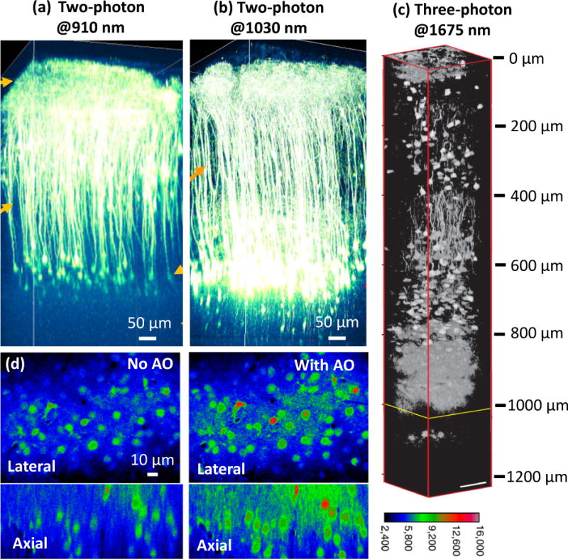
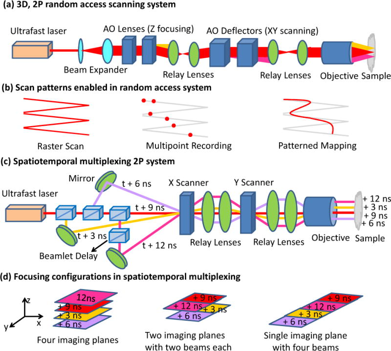
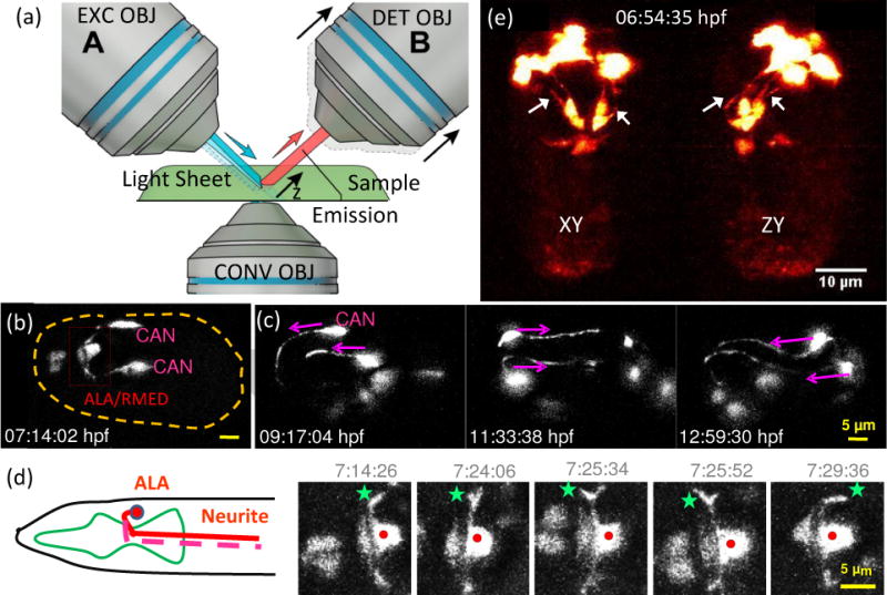
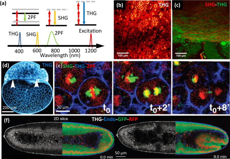
Over the past decade, developmental neuroscience has been transformed by the widespread application of confocal and two-photon fluorescence microscopy. Even greater progress is imminent, as recent innovations in microscopy now enable imaging with increased depth, speed, and spatial resolution; reduced phototoxicity; and in some cases without external fluorescent probes. We discuss these new techniques and emphasize their dramatic impact on neurobiology, including the ability to image neurons at depths exceeding 1mm, to observe neurodevelopment noninvasively throughout embryogenesis, and to visualize neuronal processes or structures that were previously too small or too difficult to target with conventional microscopy.


Figures

Fig. 1
Fig. 1
(a–c) Deep in vivo fluorescence imaging with NIR excitation. Two-photon fluorescence imaging of cortical pyramidal neurons with (a) 910-nm and (b) 1030-nm excitation in an adult eYFP-labelled mouse brain. (c) Three-photon fluorescence imaging with 1675-nm excitation of RFP-labelled pyramidal neurons in a mouse brain. (d) AO correction improves calcium imaging. Left: OGB-1 AM labeled neurons 155 μm below the brain surface without AO correction. Right: The same neurons with AO correction. Panels a-b are reprinted from R. Kawakami, et al. [1] with permission from Science; panel c is adapted from N. G. Horton, et al. [3] with permission from Nature; panel d is adapted from N. Ji, et al. [6] with permission from PNAS.
Fig. 2
Fig. 2
Higher-speed two-photon microscopy. Optical schematic (a) and scanning patterns (b) for 3D random-access scanning. The system allows conventional raster scanning for structural imaging, discrete point sampling for multiunit recording, and pattern mapping for functional imaging. In the high-speed 3D random-access mode, 2000 points can be scanned in 40 ms, enabling volumetric calcium imaging in hundreds of neurons in vivo. Compensating elements must be used because acousto-optical devices generate high spatial and temporal dispersion. Optical schematic (c) and focusing configurations (d) possible in a spatially- and temporally-multiplexed two-photon system. Ultrafast laser pulses are emitted every 12 ns and divided into four beams with 3 ns relative delay that are simultaneously focused at different positions within a sample. Different imaging configurations include a scan of four imaging planes, a single plane scan with four beams, or a scan of two imaging planes with two beams each. When used with a resonant scanning mirror, fast (250 Hz/plane at 500×500 pixels) calcium imaging in four axial planes is possible.
Fig. 3
Fig. 3
Light sheet fluorescence microscopy enables high-speed, long-term neurodevelopmental imaging during embryogenesis. (a) Inverted selective plane illumination microcopy (iSPIM) schematic. Two long working distance, water immersion objective lenses enable orthogonal SPIM excitation and detection. The excitation objective (EXC OBJ) introduces a light sheet at the sample, and the resulting fluorescence emission is collected by the detection objective (DET OBJ). High-speed volumetric imaging is achieved by sweeping the light sheet and focal plane through the sample, along the detection axis. The bottom objective provides an additional view of the sample and allows other optical modalities. iSPIM enables conventional mounting of specimens. (b–d) Visualization of neuron migration and neurite outgrowths in C.elegans embryos with iSPIM: (b) Maximum-intensity projections of ceh-10p:GFP, highlighting ALA/RMED and CAN neurons before twitching. Scale bar, 5 μm. (c) Time series of the CAN neurite outgrowth through the entire twitching period. (d) The cartoon shows the ALA neuron in the adult worm. Both neurites of ALA project towards the posterior end of the animal. The time-series images display a higher magnification view of the red box in (b). Red dot: ALA soma, green star: left neurite outgrowth. The images show the neuronal outgrowth of ALA through twitching and reveal when the bilateral neurites project towards the posterior end of the embryo. (e) Dual-view iSPIM (diSPIM) imaging highlighting GFP-tagged AIY neurons. Arrows indicate AIY neurites, clearly visible in both lateral projection (left image) and axial projection (right image). Panel c-d are adapted from Y. Wu, et al. [35] with permission from PNAS.
Fig. 4
Fig. 4
Label free imaging. (a) Energy diagrams and wavelengths in two-photon fluorescence (2PM), second harmonic generation (SHG), and third harmonic generation (THG) imaging. (b) THG image of striatum in a mouse brain (coronal section), showing white-matter fibers and neurons. The bright grainy structures are axon bundles that run perpendicular to the image plane. (c) Merged THG (green) and SHG (red) signal of a mouse corpus callosum. White-matter structures are visible in both SHG and THG images, but gray matter is only visible in the THG image. THG signals originate mostly from the myelin sheaths surrounding axons, whereas SHG signals are produced by polarized microtubules inside the axons. (d) Sagittal THG image of a zebrafish embryo during the 512 cell stage. The THG signal, generated mostly from the lipids in the plasma membrane, highlights cell contours and reveals the yolk-blastoderm interface as indicated by the white arrowheads. (e) Temporal sequence (0, 2 and 8 minutes) of βactin:H2B/mcherry transgenic zebrafish embryo, highlighting mitosis, simultaneously with SHG (green), 2PM (red) and THG (blue). (f) Gastrulating Drosophila embryos with contrast derived from THG (white) and 2PM (mRFP-red, EGFP-green, and autofluorescence-blue) imaging. Panels b-c are adapted from S. Witte, et al. [50] with permission from PNAS; panel d-e are adapted from N. Olivier, et al. [51] with permission from Science; and panel f is adapted from P. Mahou, et al. [52] with permission from Nature.

References

    1. Kawakami R, et al. Visualizing hippocampal neurons with in vivo two-photon microscopy using a 1030 nm picosecond pulse laser. Sci Rep. 2013;3:1014. - PMC - PubMed
    1. Kobat D, Horton NG, Xu C. In vivo two-photon microscopy to 1.6-mm depth in mouse cortex. J Biomed Opt. 2011;16(10):106014. - PubMed
    1. Horton Nicholas G, Demirhan Kobat KW, Clar Catharine G, Wise Frank W, Schaffer Chris B, Xu Chris. In vivo three-photon microscopy of subcortical structures within an intact mouse brain. Nature Photonics. 2013;7:205–209. Three-photon fluorescence microscopy with a high-pulse-energy laser at ~1650 nm has enabled the in vivo high-resolution imaging of subcortical structures within an intact mouse brain with a penetration depth of ~1.4 mm. - PMC - PubMed
    1. Wang YM, et al. Deep-tissue focal fluorescence imaging with digitally time-reversed ultrasound-encoded light. Nature Communications. 2012;3 - PMC - PubMed
    1. Wang C, Ji N. Pupil-segmentation-based adaptive optical correction of a high-numerical-aperture gradient refractive index lens for two-photon fluorescence endoscopy. Opt Lett. 2012;37(11):2001–3. - PubMed