

Single-particle cryo-EM of calcium release channels: structural validation
- PMID: 23831288
- PMCID: PMC3805725
- DOI: 10.1016/j.sbi.2013.06.003
Single-particle cryo-EM of calcium release channels: structural validation
Abstract
Few tools are available to determine the structure of large integral membrane proteins such as intracellular Ca(2+) release channels, RyRs and IP3Rs. Single particle cryo-EM can readily determine the structure of such channels to intermediate resolution, and can be used to quantitatively assess conformational variability. However, due to the, often low, image contrast of these cryospecimens, methods for validation are critical to insure the accuracy of such structures, and to put limits on their interpretability. The low-resolution structure of RyR has been well established for some time, but high-resolution has been slow to emerge. The structure of IP3R channel by cryo-EM had a number of false-starts, but improved validation methods have recently lead to a demonstrably accurate reconstruction.
Copyright © 2013 Elsevier Ltd. All rights reserved.
Figures
References
-
- Tusnady GE, Dosztanyi Z, Simon I. Transmembrane proteins in the Protein Data Bank: identification and classification. Bioinformatics. 2004;20:2964–2972. http://dx.doi.org/10.1093/bioinformatics/bth340. - DOI - PubMed
-
- Ludtke SJ, Serysheva II, Hamilton SL, Chiu W. The pore structure of the closed RyR1 channel. Structure (Camb) 2005;13:1203–1211. http://dx.doi.org/10.1016/j.str.2005.06.005. - DOI - PMC - PubMed
-
- Samso M, Wagenknecht T, Allen PD. Internal structure and visualization of transmembrane domains of the RyR1 calcium release channel by cryo-EM. Nat Struct Mol Biol. 2005;12:539–544. http://dx.doi.org/10.1038/nsmb938. - DOI - PMC - PubMed
-
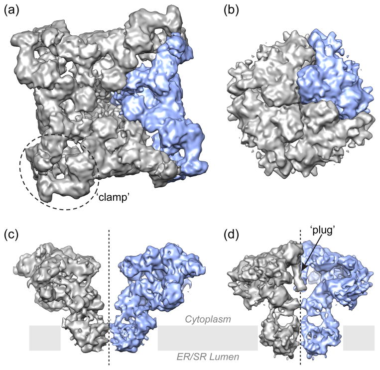
- Ludtke SJ, Tran TP, Ngo QT, Moiseenkova-Bell VY, Chiu W, Serysheva II. Flexible architecture of IP3R1 by Cryo-EM. Structure. 2011;19:1192–1199. http://dx.doi.org/10.1016/j.str.2011.05.003 Reports a 3D cryo-EM structure of a fully functional type 1 IP3R channel from rat recebellum in the closed state. High structural variance has been identified in the cytoplasmic region. This study puts to rest the long-standing debates about the molecular architecture of IP3R1 channel. - DOI - PMC - PubMed
-
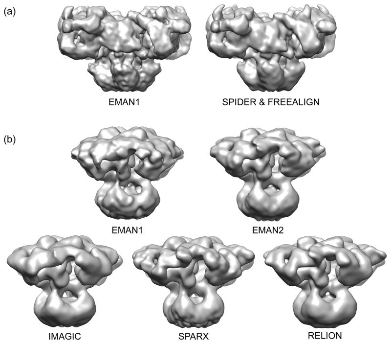
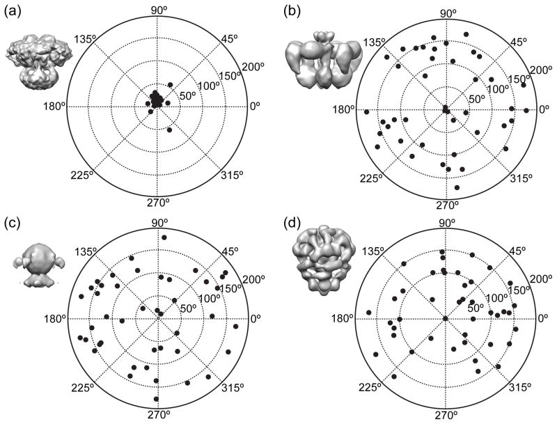
- Murray SC, Flanagan J, Popova OB, Chiu W, Ludtke SJ, Serysheva II. Validation of Cryo-EM Structure of IP3R1 Channel. Structure. 2012 in press. 3D structure of tetrameric IP3R1 channel obtained using single-particle cryo-EM (see ref. 4 in the current review) has been validated by use five reconstruction algorithms (EMAN1, EMAN2, Relion, Imagic, SPARX), tilt-pair analysis and class-average/map comparisons. The map resolution and feature resolvability have been re-assessed using the “gold standard” criterion. - PMC - PubMed
Publication types
MeSH terms
Substances
Grants and funding
LinkOut - more resources
Full Text Sources
Other Literature Sources
Miscellaneous
