. 2013:3:2157.
doi: 10.1038/srep02157.

Continuous DOPA synthesis from a single AAV: dosing and efficacy in models of Parkinson's disease

Affiliations

Continuous DOPA synthesis from a single AAV: dosing and efficacy in models of Parkinson's disease

Erik Cederfjäll et al. Sci Rep. 2013.

Abstract

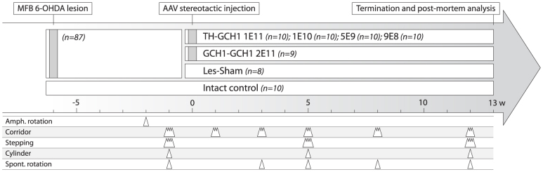
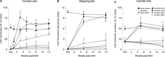
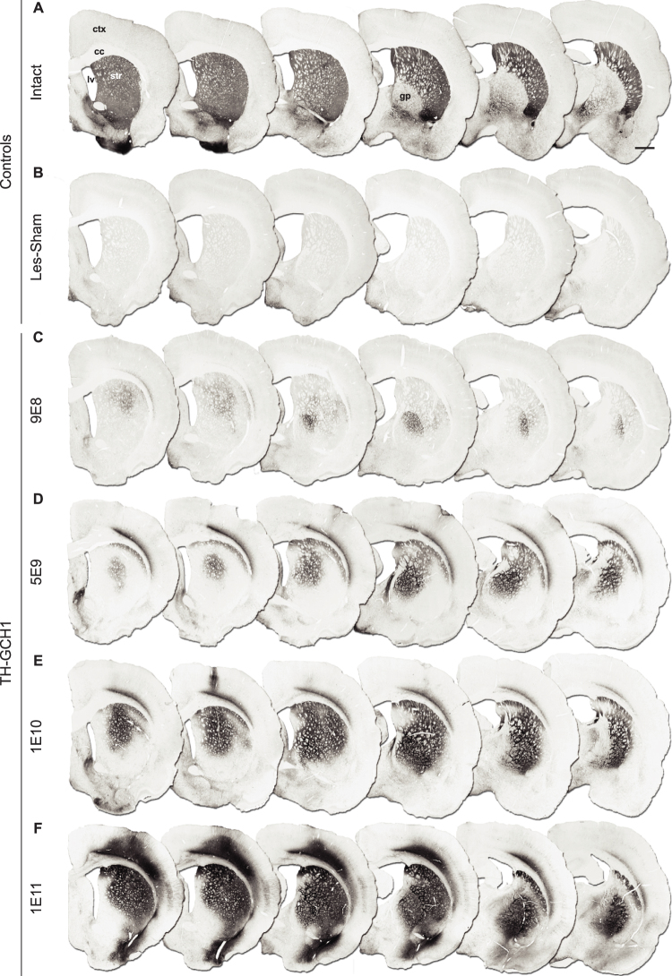
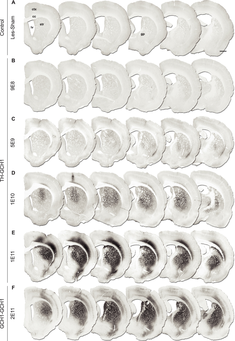
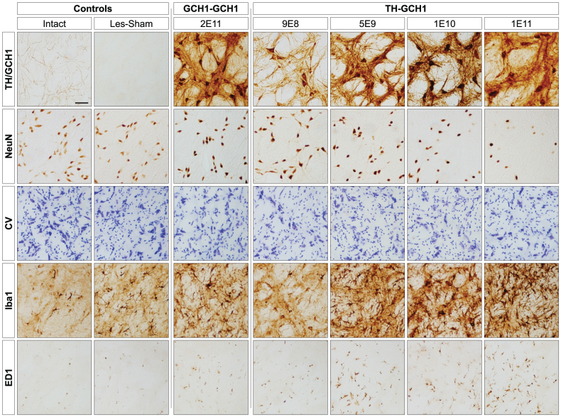
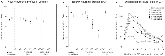
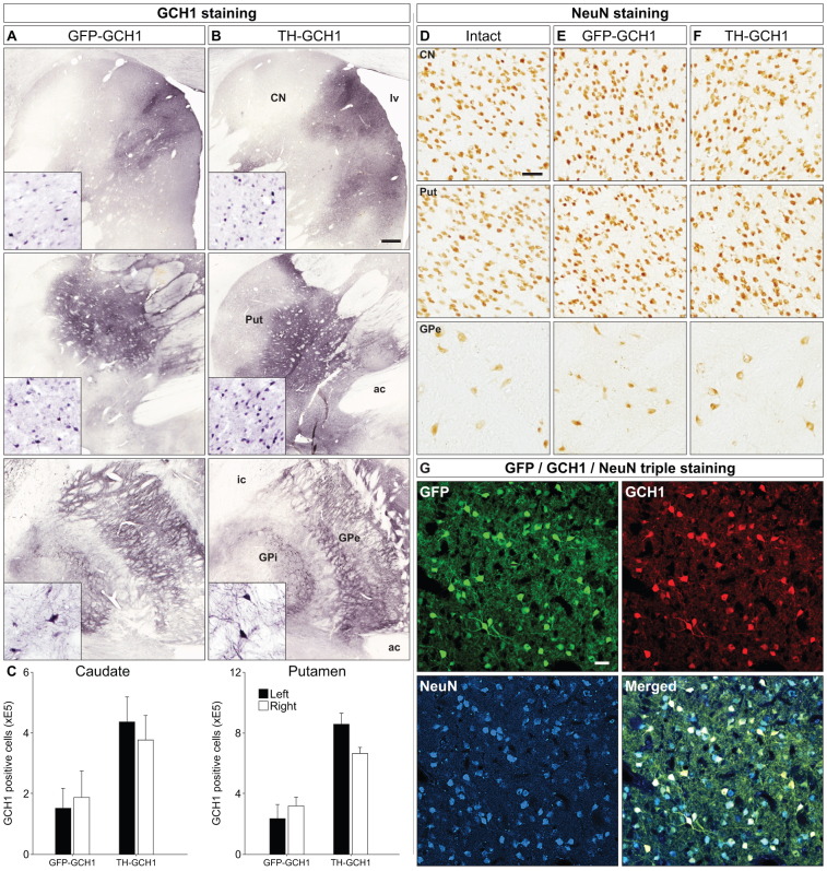
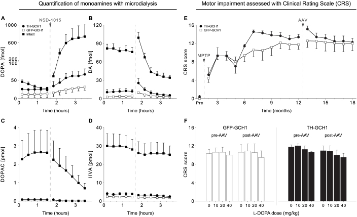
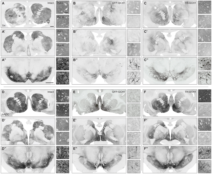
We used a single adeno-associated viral (AAV) vector co-expressing tyrosine hydroxylase (TH) and GTP cyclohydrolase 1 (GCH1) to investigate the relationship between vector dose, and the magnitude and rate of recovery in hemi-parkinsonian rats. Intrastriatal injections of >1E10 genomic copies (gc) of TH-GCH1 vector resulted in complete recovery in drug-naïve behavior tests. Lower vector dose gave partial to no functional improvement. Stereological quantification revealed no striatal NeuN+ cell loss in any of the groups, whereas a TH-GCH1 dose of >1E11 gc resulted in cell loss in globus pallidus. Thus, a TH-GCH1 dose of 1E10 gc gave complete recovery without causing neuronal loss. Safety and efficacy was also studied in non-human primates where the control vector resulted in co-expression of the transgenes in caudate-putamen. In the TH-GCH1 group, GCH1 expression was robust but TH was not detectable. Moreover, TH-GCH1 treatment did not result in functional improvement in non-human primates.


Conflict of interest statement

DK and TB are co-inventors of a patent application on the construct reported in this paper. TB is a founding-director of a company that intends to develop this for commercial purposes. DK receives financial compensation in the form of retainer fees, milestones and royalty payments. JHK is a Founding Scientist and currently an SAB member of Ceregene Inc. EC, NN, GS, EN and YC have no conflict of interest.

Figures

Figure 1
Figure 1. Experimental design for rodent study.
Fifty-seven rats with complete lesions that fulfilled the criteria of > 6 net ipsiversive rotations over 90 min upon challenge with D-amphetamine (2.5 mg/kg) were included in the study. The animals were behaviorally characterized using corridor, stepping and cylinder tests and allocated to one of 6 groups based on their performance either to receive different doses of the TH-GCH1 vector (TH-GCH1:1E11, 1E10, 5E9 and 9E8 gc) or followed as one of two control groups (GCH1-GCH1:2E11 gc or Les-Sham). In addition, an intact control group (n = 10) was included in the study. Animals were followed over a 12-week period with behavioral tests to assess motor function as indicated in the time line. Thirteen weeks after AAV treatment, animals from each experimental group were sacrificed for either histology and stereology (n = 6) or biochemical analysis (n = 4, data not shown).
Figure 2
Figure 2. Recovery of motor function in drug-naive behavior tests over the 12-week follow up after AAV treatment.
Due to the ease in longitudinal assessments, the corridor test (A) was administered at several time points, which revealed an early partial recovery in the TH-GCH1:1E11 group at 1 week, a transient overcompensation (strong contralateral bias) at 3 weeks and a stable response with slight bias in left retrievals at 5 weeks and onwards. Animals in the TH-GCH1:1E10 group displayed a similar behavior, however, at a slower rate and without the overcompensation at 3 weeks post-AAV. The TH-GCH1:5E9 group showed only partial recovery but were significantly different from Les-Sham at the 12 week time point. Animals receiving the TH-GCH1:9E8 and GCH1-GCH1:2E11 did not show any recovery compared with Les-Sham. Behavioral recovery in the stepping (B) and cylinder (C) tests were comparable in the 1E11 and 1E10 groups, while the response in the 5E9 group was less pronounced or not detectable, respectively. Statistics: Repeated measures ANOVA (A) Time F(3,20) = 65.85 p < 0.001; Time × Group F(3,20) = 16.64 p < 0.001; (B) Time F(2,9) = 129.05 p < 0.001; Time × Group F(2,9) = 39.26 p < 0.001; (C) Time F(2,12) = 73.82 p < 0.001; Time × Group F(2,12) = 43.86 p < 0.001; One-way ANOVA at each time point followed by post hoc comparison with Tukey's HSD when Levene's test was significant (p < 0.05) otherwise by Dunnett's T3 test. * = different from Les-Sham p < 0.05, # = different from intact controls p < 0.05. Error bars represent mean SEM.
Figure 3
Figure 3. Dose-dependent increase in TH transgene expression.
Intrastriatal injections of an AAV vector expressing TH and GCH1 at 9E8 gc results in a small number of TH positive neuronal profiles in the striatum and in globus pallidus (C). Increasing the vector dose to 5E9 gc (D) resulted in larger transduction area in the striatum and further spread to globus pallidus and cortex. Injection of 1E10 and 1E11 gc gave more widespread transduction not only in the striatum but also more extensive expression in the globus pallidus and overlying cortex (E,F). ctx = cortex, cc = corpus callosum, gc = genomic copies, gp = globus pallidus, lv = lateral ventricle, str = striatum. Scale bar in a represent 0.2 mm in all panels.
Figure 4
Figure 4. Dose-dependent increase in GCH1 transgene expression.
Note that the antibody used here does not cross react with the rodent protein and thus only shows the transgenic human GCH1 expressed from the AAV vector. Intrastriatal injections of 9E8 gc TH-GCH1 vector (B) resulted in very few GCH1 positive neurons in the striatum and globus pallidus, whereas the transduction efficiency increased with vector doses of 5E9 gc (C), 1E10 (D) and 1E11 gc (E). The two high doses resulted in complete coverage of striatum and globus pallidus (D,E). The transduction spread was similar in the GCH1-GCH1 control vector group (E,F). ctx = cortex, cc = corpus callosum, gc = genomic copies, gp = globus pallidus, lv = lateral ventricle, str = striatum. Scale bar in a represent 0.2 mm in all panels.
Figure 5
Figure 5. High magnification images of neurons in the globus pallidus (GP).
Transgene immunoreactivity (row 1, TH staining depicted in all panels except GCH1-GCH1 which is a GCH1 staining) was present in GP in all vector-treated animals filling cell bodies, dendritic processes and axon terminals. Loss of NeuN positive cells in GP was seen in animals injected with 1E11 gc of the TH-GCH1 vector (row 2, far right) but also to a lesser extent in the 1E10 group. Nissl stained specimens from adjacent sections confirmed loss of neurons (row 3). In the same region, there was an increase in the microglial marker Iba1 (row 4) seen as a dose-dependent increase in arborized reactive cells. ED1 (equivalent to human CD68) immunoreactivity indicated presence of macrophages. Scale bar in top left panel represent 0.2 mm in all panels.
Figure 6
Figure 6
Quantification of NeuN-positive neurons with stereological analysis in both striatum (A) and globus pallidus (B). Vector-treatment did not result in any decrease in cell number in the striatum, regardless of dose (A). However, quantification of NeuN positive cells in globus pallidus revealed a significant decrease in animals treated with TH-GCH1 vector dose of 1E11 gc or higher. Inspection of the distribution of NeuN positive cell counts in the globus pallidus (C) suggested that the decrease was primarily in the anterior part of the structure. The TH-GCH1 vector at doses below 1E10 or GCH1-GCH1 vector treatments showed no apparent loss of either striatal or pallidal NeuN positive cells. Statistics: One way ANOVA: (A) F(7,36) = 0.88, (B) F(7,36) = 12.18 p < 0.001, followed by Tukey's HSD post hoc since Levene's test was not significant. * = different from Les-Sham p < 0.001, # = different from intact controls p < 0.001. Error bars represent mean SEM.
Figure 7
Figure 7. Transgene expression in MPTP-treated monkeys.
Transgene expression from both AAV vectors encoding GFP-GCH1 (A) and TH-GCH1 (B) were visualized with the GCH1 antibody, which showed GCH1 immunoreactivity in the putamen (Put, row 1), caudate nucleus (CN, row 2) and globus pallidus (GP, row 3). Interestingly, both the external and internal segment of GP had GCH1 immunoreactive cells and projections (insets). Stereological quantification of GCH1 positive neurons was performed in both putamen and caudate nucleus bilaterally (C). NeuN stainings were performed in adjacent sections and are shown from Put, CN and GPe (E-F). Co-expression of GFP and GCH1 in the transduced neurons was confirmed with confocal microscopy (G). Whereas the GCH1 staining was robust, the expression of the TH transgene could not be confirmed in the TH-GCH1 injected monkeys. ac = anterior commissure, CN = caudate nucleus, GPi/GPe = globus pallidus internal/external segment, lv = lateral ventricle, Put = putamen. Scale bar in (B) represent 0.5 mm and (D) and (G) represents 50 μm. GCH1 insets in (A–B) have the same magnification as (D). Cell counts in (C) represent mean SEM.
Figure 8
Figure 8. Six MPTP lesioned stably parkinsonian cynomolgus macaques received bilateral AAV injections in the caudate-putamen with either TH-GCH1 (n = 3) or GFP-GCH1 (n = 3) and were followed for 6 months.
After the last assessment point at 18 months, microdialysis was performed on all vector-treated animals and two intact controls by placing bilateral probes in the putamen (A–D). In the analysis, we only included cases where probe placement could be histologically confirmed (TH-GCH1 n = 4, GFP-GCH1 n = 4, Intact n = 2). After one hour equilibration, five baseline samples á 20 minutes were collected. Baseline levels of DA was decreased by 96% in GFP-GCH1 when compared with intact controls in response to the MPTP lesions (B). During sampling of the fifth baseline sample, all animals received an iv injection of the central AADC inhibitor NSD-1015 (100 mg/kg in saline). Inhibition of AADC resulted in an accumulation of DOPA in intact animals (A), whereas this increase was blunted in the vector control group and only marginally higher in the TH-GCH1 treated monkeys. Simultaneously, DA (B) and its immediate metabolite DOPAC (C) were markedly reduced in intact animals. This could not be detected in the vector-treated animals. HVA levels (D) remained at a constant level throughout the experiment. The animals were assessed behaviorally using the non-human primate equivalent of the clinical rating scale (CRS), both pre- and post-AAV treatment longitudinally under baseline conditions (E) or following L-DOPA administration (F) (10, 20 or 40 mg/kg dose). Error bars represent mean SEM.
Figure 9
Figure 9. Residual TH and AADC expression in the MPTP lesioned monkey brain.
Pre-commissural and post-commissural caudate-putamen, and the substantia nigra stained for TH (A–C) or AADC (D–F) from the intact (A,D), GFP-GCH1 (B,E) and TH-GCH1 (C,F) groups. Insets are high-power images from the left and right hemisphere respectively. MPTP was administered via the right intracarotid artery and by systemic doses until stable parkinsonian impairments were obtained, which varied between the individual monkeys. Two out of six monkeys had spared dopaminergic innervation on the contralateral side to the intracarotid injections (as shown in C), whereas the remaining four were severely lesioned in both hemispheres (as shown in B,E). Since the number of TH and AADC positive cells in ventral tier of substantia nigra in B and C was greatly reduced, the high power insets are from the dorsal tier. TH expression observed in B and C was not vector-derived. CN = caudate nucleus, ic = internal capsule, Put = putamen, SN = substantia nigra. Scale bars in A (that also applies to A′) and A′′ represent 2 mm. Scale bar in the high-power inset Put-L represent 0.5 mm, whereas it represents 0.25 mm in SN-L and SN-R.

References

    1. Fahn S. Parkinson disease, the effect of levodopa, and the ELLDOPA trial. Earlier vs Later L-DOPA. Arch Neurol 56, 529–535 (1999). - PubMed
    1. Nutt J. G. & Holford N. H. The response to levodopa in Parkinson's disease: imposing pharmacological law and order. Ann Neurol 39, 561–573 (1996). - PubMed
    1. Obeso J. A., Olanow C. W. & Nutt J. G. Levodopa motor complications in Parkinson's disease. Trends Neurosci 23, S2–7 (2000). - PubMed
    1. Kurlan R. et al. Duodenal delivery of levodopa for on-off fluctuations in parkinsonism: preliminary observations. Ann Neurol 20, 262–265 (1986). - PubMed
    1. Nilsson D. et al. Long-term intraduodenal infusion of a water based levodopa-carbidopa dispersion in very advanced Parkinson's disease. Acta Neurol Scand 97, 175–183 (1998). - PubMed

Publication types