. 2013 Aug;15(8):895-904.
doi: 10.1038/ncb2790. Epub 2013 Jul 7.

Role of telomere dysfunction in cardiac failure in Duchenne muscular dystrophy

Affiliations

Role of telomere dysfunction in cardiac failure in Duchenne muscular dystrophy

Foteini Mourkioti et al. Nat Cell Biol. 2013 Aug.

Abstract

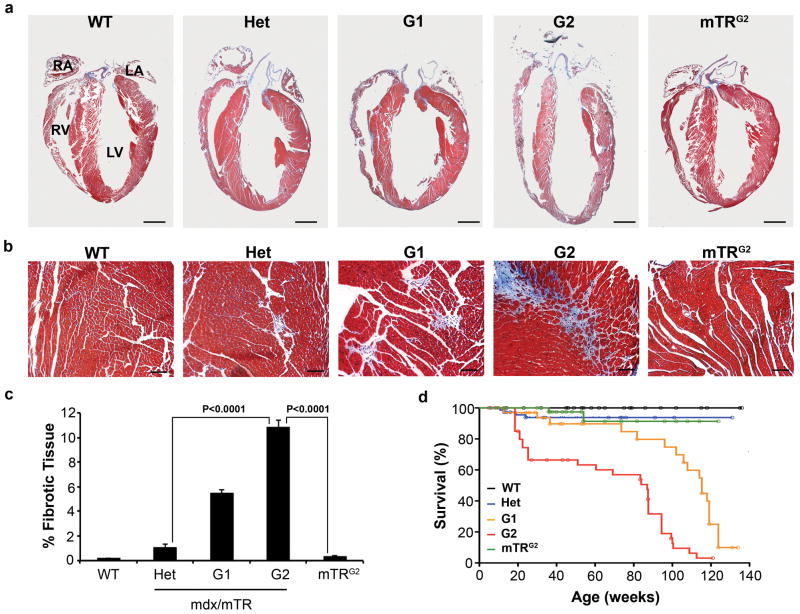
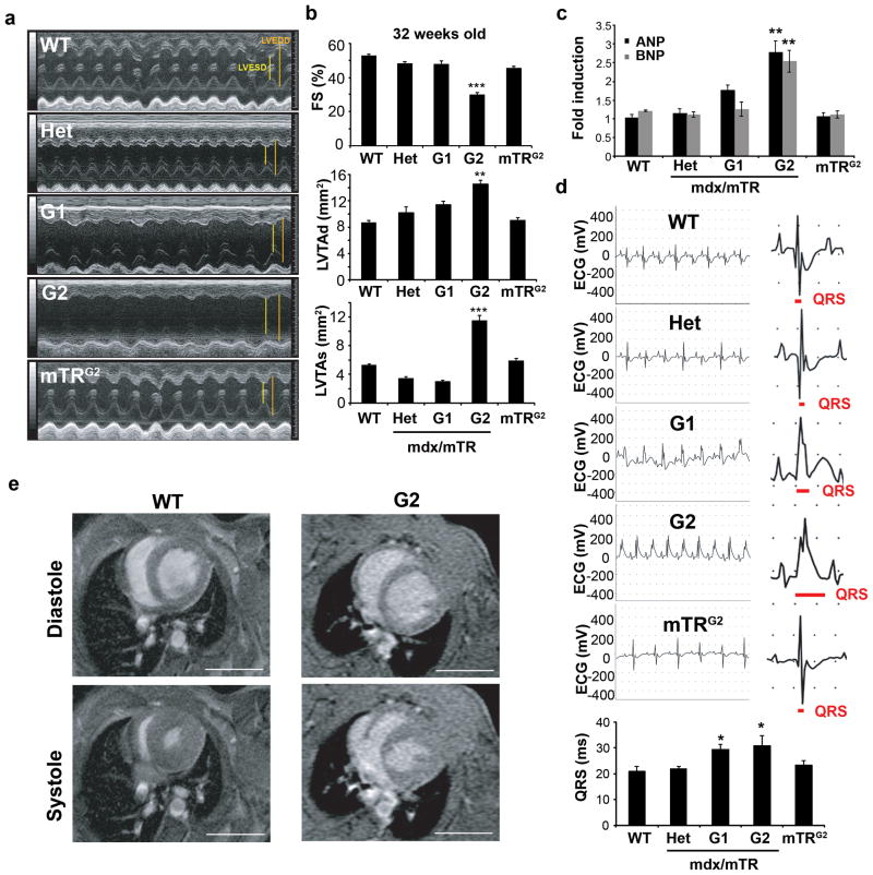
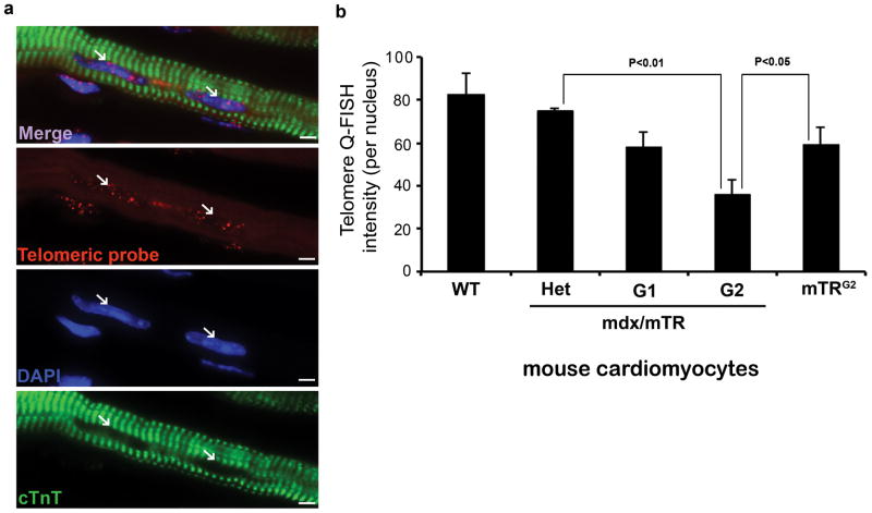
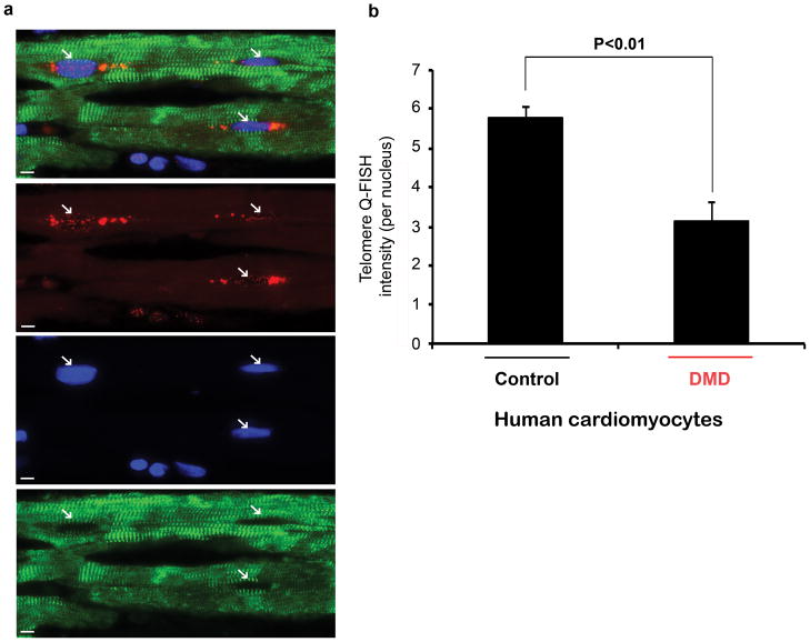
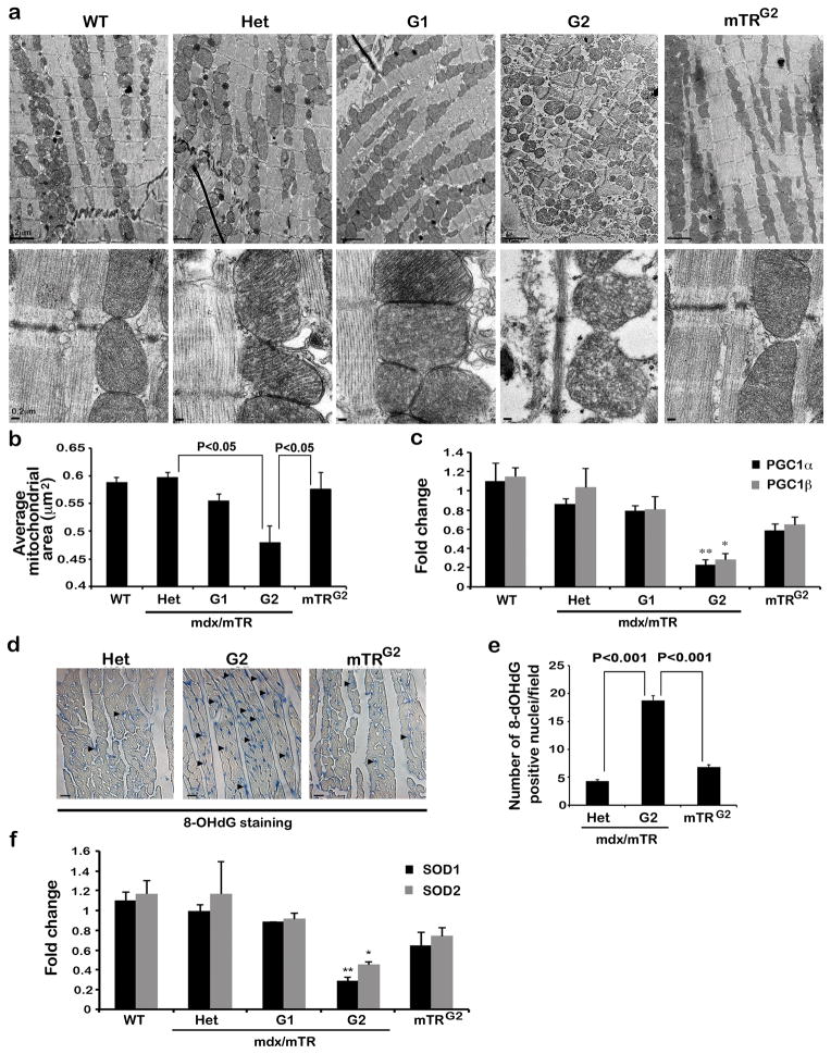
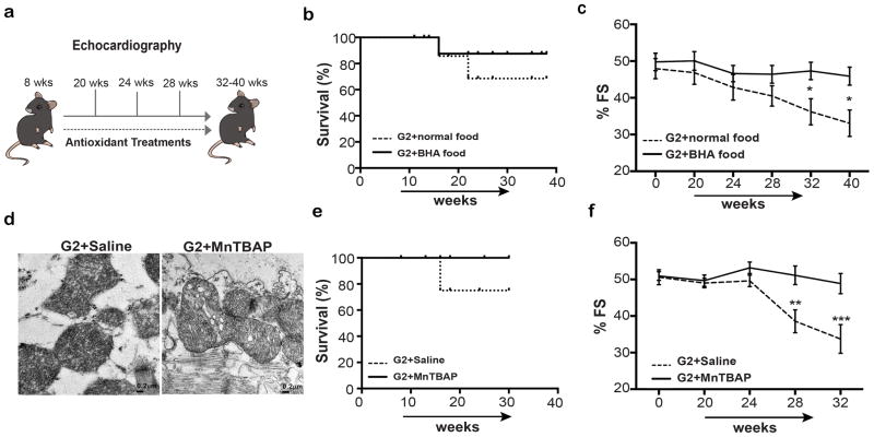
Duchenne muscular dystrophy (DMD), the most common inherited muscular dystrophy of childhood, leads to death due to cardiorespiratory failure. Paradoxically, mdx mice with the same genetic deficiency of dystrophin exhibit minimal cardiac dysfunction, impeding the development of therapies. We postulated that the difference between mdx and DMD might result from differences in telomere lengths in mice and humans. We show here that, like DMD patients, mice that lack dystrophin and have shortened telomeres (mdx/mTR(KO)) develop severe functional cardiac deficits including ventricular dilation, contractile and conductance dysfunction, and accelerated mortality. These cardiac defects are accompanied by telomere erosion, mitochondrial fragmentation and increased oxidative stress. Treatment with antioxidants significantly retards the onset of cardiac dysfunction and death of mdx/mTR(KO) mice. In corroboration, all four of the DMD patients analysed had 45% shorter telomeres in their cardiomyocytes relative to age- and sex-matched controls. We propose that the demands of contraction in the absence of dystrophin coupled with increased oxidative stress conspire to accelerate telomere erosion culminating in cardiac failure and death. These findings provide strong support for a link between telomere length and dystrophin deficiency in the etiology of dilated cardiomyopathy in DMD and suggest preventive interventions.


Conflict of interest statement

COMPETING FINANCIAL INTERESTS

The authors declare no competing financial interests

Figures

Figure 1
Figure 1. Cardiac dilation, fibrosis and premature death in G2 mice
(a) Trichrome staining in longitudinal paraffin sections of 32-week-old hearts. G2 hearts develop dilated cardiomyopathy accompanied by enlarged ventricular cavity and thinning of the ventricular wall (LV Left Ventricle, RV Right Ventricle, LA Right Atrium, RA Right Atrium). Scale bar 15 μm. (b) Increased collagen deposition (blue) in the left ventricle of G2 hearts compared to controls. Scale bar 600 μm. (c) Quantification of collagen deposition. Data are represented as mean ± SEM (n=3–4), Two-tailed, unpaired Student’s t-test, P values are indicated in the graph. (d) Kaplan-Meier survival curve shows G2 mice have significantly reduced life span compared to controls. WT (n=72), HET (n=86), G1 (n=45), G2 (n=53) and mTRG2 (n=45). Source data are shown in Supplementary Table S1.
Figure 2
Figure 2. Severe cardiac dysfunction and heart failure in G2 mice
(a) Representative images of 32-week-old hearts showing left ventricular dilation and compromised contractility (LVEDD; Left Ventricular End Diastolic Diameter, LVESD; Left Ventricular End Systolic Diameter). (b) Echocardiograph measurements show reduction in fractional shortening (FS), increase in left ventricular transverse area in diastole (LVTAd) and increase in left ventricular transverse area in systole (LVTAs) in 32-week-old G2 hearts compared to control hearts. Data are represented as mean ± SEM (n=5–8), Two-tailed, unpaired Student’s t-test, ** indicates P <0.01 and *** P<0.001. (c) Increased cardiac failure markers ANP and BNP by Q-RT-PCR analysis confirmed heart failure in G2 mice. Data are represented as mean ± SEM (n=3); Two-tailed, unpaired Student’s t-test, ** indicates P <0.01. (d) Representative electrocardiograms (ECG) of 32-week-old hearts show impairment in ventricular depolarization of G1 and G2 hearts manifested by a prolonged QRS interval. Lower graph shows quantification of QRS values. Data are represented as mean ± SEM (n=5–9); Two-tailed, unpaired Student’s t-test, * indicates P <0.05. (e) Representative magnetic resonance images (MRI) at the level of the papillary muscle at diastole and systole confirms left ventricular chamber dilation and systolic dysfunction in G2 mice. Scale bar 5 mm. Source data are shown in Supplementary Table S1.
Figure 3
Figure 3. Telomere Q-FISH analysis of cardiac sections from mice
(a) Representative Telomere Q-FISH images (red telomeric probe; blue DAPI-stained nuclei, and green immunofluorescence of cardiac Troponin T reveals striated cardiomyocytes). White arrows indicate nuclei within cardiomyocytes. Scale bar 400 μm. (b) Quantification of telomere shortening in cardiomyocytes represents the intensity sum of all telomere pixels (Cy3+ telomeres) divided by the intensity sum of all nuclear DNA pixels of the entire nucleus (DAPI). A total of 3 hearts per genotype were analyzed. The number of nuclei scored: WT (N=394), HET (N=303), G1 (N=355), G2 (N=414), mTRG2 (N=390). Data are represented as mean ± SEM; Two-tailed, unpaired Student’s t-test, P values are indicated. Source data are shown in Supplementary Table S1.
Figure 4
Figure 4. Telomere Q-FISH analysis of cardiac section from human DMD hearts
(a) Representative Telomere Q-FISH images (red, telomeric probe; blue, DAPI-stained nuclei and green, cardiac Troponin T staining). Scale bar 400 μm. (b) Quantification of telomere shortening in cardiomyocytes represents the intensity sum of all telomere pixels (Cy3+ telomeres) divided by the intensity sum of all nuclear DNA pixels of the entire nucleus (DAPI). A total of 3 control and 4 DMD cardiac samples were analyzed and the number of nuclei scored: Control 1 (N=70), Control 2 (N=69), Control 3 (N=65), DMD 1 (N=42), DMD 2 (N=94), DMD 3 (N=65) and DMD 4 (N=72). Data are represented as mean ± SEM; Two-tailed, unpaired Student’s t-test, P value is indicated. Source data are shown in Supplementary Table S1.
Figure 5
Figure 5. Telomere dysfunction is associated with mitochondrial abnormalities and oxidative stress
(a) Representative longitudinal sections of murine cardiac muscle by transmission electron micrographs show cellular edema, loss of thick and thin filaments and mitochondrial fragmentation in G2 hearts compared to control images. Scale bar 2μm. Lower panel shows a closer look of mitochondria. Note the loss of cristae of G2 hearts. Scale bar 0.2 μm. (b) Quantification of mitochondrial area (n=3 animals per genotype, 3 different areas of the left ventricle per heart, and n=10 photos per area analyzed); Two-tailed, unpaired Student’s t-test, P values are indicated. (c) Q-RT-PCR analysis for PGC1α and PGC1β gene expression show decreased levels in the G2 hearts. (n=3 hearts per genotype; data are represented as mean ± SEM; Two-tailed, unpaired Student’s t-test, * indicates P<0.05 and ** P<0.01). (d) Immunohistochemistry reveals increased number of 8-OHdG-positive nuclei in the hearts of 32-week old G2 animals. Scale bar 400 μm. (e) Quantification of the 8-OHdG staining. Data are represented as mean ± SEM. (n=3 hearts per genotype; >10 images per heart were analyzed), Two-tailed, unpaired Student’s t-test, P values are indicated. (f) Q-RT-PCR analyses of levels of anti-oxidative enzymes (SOD1 and SOD2) show decreased levels in the hearts of G2 mice. (n=3 hearts per genotype; data are represented as mean ± SEM); Two-tailed, unpaired Student’s t-test, * indicates P<0.05 and ** P<0.01. Source data are shown in Supplementary Table S1.
Figure 6
Figure 6. Antioxidant treatment increased survival and improved cardiac function
(a) Scheme of experimental procedure. Mice were treated with anti-oxidant (BHA food or MnTBAP injection) at 8 weeks old and tested monthly for cardiac function by echocardiography until 32–40 weeks old. (b) Decreased mortality of G2 mice treated with BHA food. Animals tested: G2-BHA food (n=10), G2-Normal food (n=10). (c) Echocardiography measurements (by means of fractional shortening as %FS) showed comparable increase in cardiac function of G2 mice treated with BHA food compared to controls. ANOVA followed by Bonferoni test for multiple comparisons; * indicates P<0.05. (d) Representative longitudinal sections of murine cardiac muscle of MnTBAP-injected G2 hearts show improved mitochondria morphology. Note that some formation of cristae is evident in G2 MnTBAP-injected cardiac mitochondria. Scale bar 0.2 μm (e) Extension of survival in G2 mice treated with daily injections of MnTBAP. Animals tested: G2-MnTBAP (n=10), G2-Saline (n=10). (f) Improved cardiac function in MnTBAP-injected compared to saline-injected G2 hearts. ANOVA followed by Bonferoni test for multiple comparisons; ** indicates P<0.01 and *** P<0.001. Source data are shown in Supplementary Table S1.

References

    1. Liew CC, Dzau VJ. Molecular genetics and genomics of heart failure. Nat Rev Genet. 2004;5:811–825. - PubMed
    1. Hoffman EP, Brown RH, Jr, Kunkel LM. Dystrophin: the protein product of the Duchenne muscular dystrophy locus. Cell. 1987;51:919–928. - PubMed
    1. Petrof BJ, Shrager JB, Stedman HH, Kelly AM, Sweeney HL. Dystrophin protects the sarcolemma from stresses developed during muscle contraction. Proc Natl Acad Sci U S A. 1993;90:3710–3714. - PMC - PubMed
    1. Bushby K, Muntoni F, Bourke JP. 107th ENMC international workshop: the management of cardiac involvement in muscular dystrophy and myotonic dystrophy. 7th–9th June 2002, Naarden, the Netherlands. Neuromuscul Disord. 2003;13:166–172. - PubMed
    1. Baxter P. Treatment of the heart in Duchenne muscular dystrophy. Dev Med Child Neurol. 2006;48:163. - PubMed

Publication types