. 2013 Aug;16(8):1060-7.
doi: 10.1038/nn.3454. Epub 2013 Jul 7.

Sensory cortex limits cortical maps and drives top-down plasticity in thalamocortical circuits

Affiliations

Sensory cortex limits cortical maps and drives top-down plasticity in thalamocortical circuits

Andreas Zembrzycki et al. Nat Neurosci. 2013 Aug.

Abstract

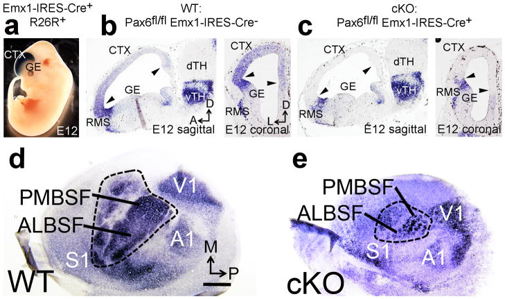
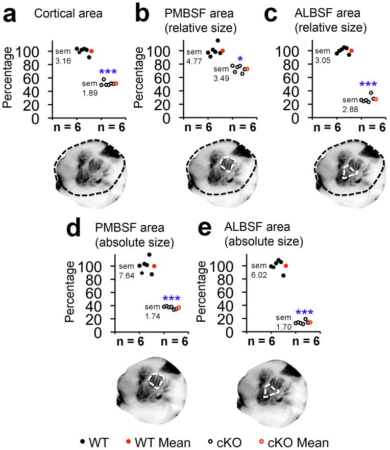
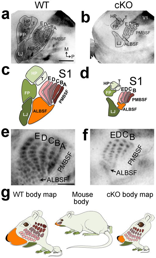
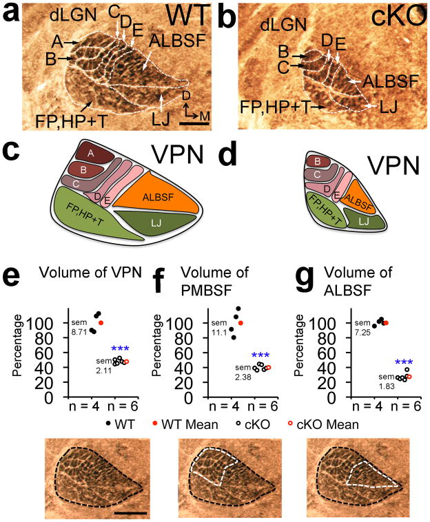
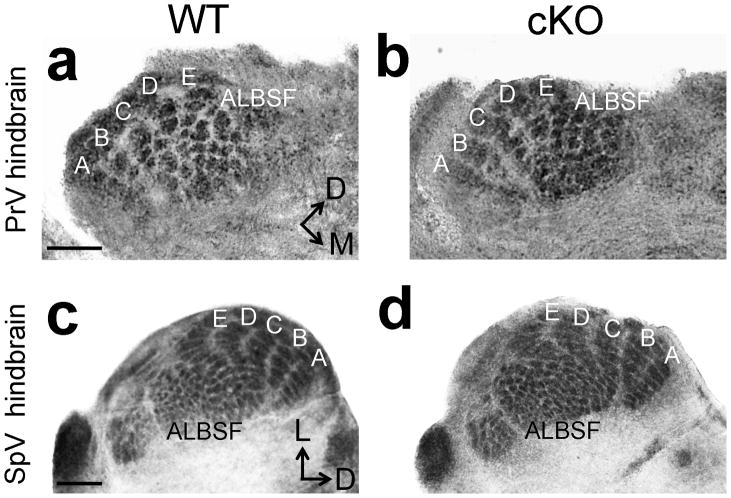
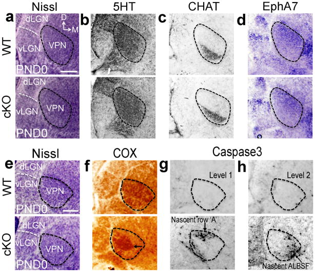
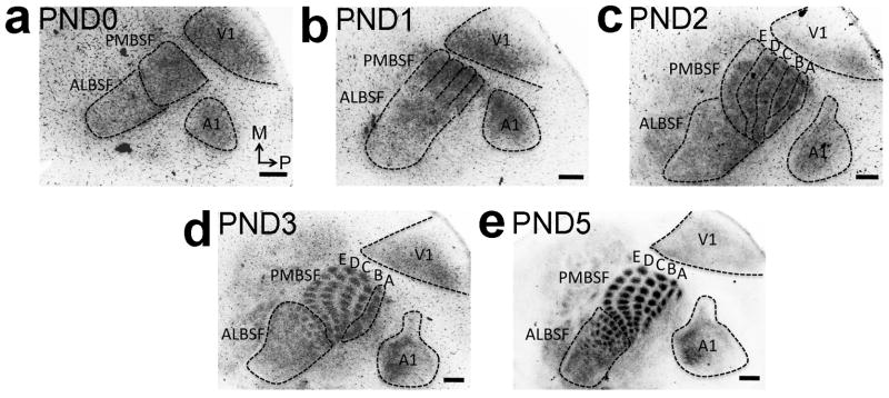
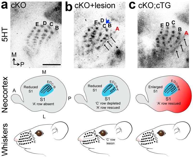
The primary somatosensory cortex (S1) contains a complete body map that mirrors the subcortical maps developed by peripheral sensory input projecting to the sensory hindbrain, the thalamus and then S1. Peripheral changes during development alter these maps through 'bottom-up' plasticity. Unknown is how S1 size influences map organization and whether an altered S1 map feeds back to affect subcortical maps. We show that the size of S1 in mice is significantly reduced by cortex-specific deletion of Pax6, resulting in a reduced body map and loss of body representations by an exclusion of later-differentiating sensory thalamocortical input. An initially normal sensory thalamus was repatterned to match the aberrant S1 map by apoptotic deletion of thalamic neurons representing body parts with axons excluded from S1. Deleted representations were rescued by altering competition between thalamocortical axons using sensory deprivation or increasing the size of S1. Thus, S1 size determined the resolution and completeness of body maps and engaged 'top-down' plasticity that repatterned the sensory thalamus to match S1.


Conflict of interest statement

The authors declare no competing financial interests.

Figures

Figure 1
Figure 1. Pax6 specifies S1 area identity in cortical progenitors and determines S1 size. a
Cre mediated recombination by Emx1-IRES-Cre crossed to the R26R reporter line revealed robust βgal staining restricted to dorsal telencephalon including neocortex at embryonic day (E) 12. b: In situ hybridization (ISH) for Pax6 mRNA in Pax6fl/fl Emx1-IRES-Cre “Wildtype” (WT) E12 embryos reveals graded expression of Pax6 in cortical progenitors (between arrowheads), and strong expression in the rostral migratory stream (RMS) bordering the ganglionic eminence (GE) and in ventral thalamus (vTH). c: In Pax6fl/fl Emx1-IRES-Cre+(cKO) E12 embryos Pax6 mRNA is selectively deleted from cortical progenitors (between arrowheads) but remains at normal levels in the RMS and vTH. d–e: ISH on tangential sections of flattened cortex at PND7 for the sensory area marker RORβ shows that deletion of Pax6 specifically from cortical progenitors using Emx1-IRES-Cre (cKO) results in a significant reduction of S1 size compared to wildtype. Abbreviations: M: medial, P: posterior, dTH: dorsal thalamus, S1: primary somatosensory area, V1: primary visual area, A1: primary auditory area, ALBSF: anterior lateral barrel subfield, PMBSF: posterior medial barrel sub field. Scale bar in a, 0.5mm.
Figure 2
Figure 2. Quantitation of absolute and relative sizes of S1 representations in cKO mice
Neocortical surface area and size of S1 barrel fields were quantified at PND7 using 5HT staining on tangential sections of flattened cortex. Overall cKO neocortex (a) was 51.6% +/− 1.89% the size of wildtype (p < 0.0001, t = 18.5, df = 33, n = 6). cKO PMBSF was 72.8% +/− 3.49% the relative size (b, p = 0.0015, t = 6.3, df = 5, n = 6) and 37.1% +/− 1.74% the absolute size (d, p < 0.0001, t = 14.1, df = 10, n = 6)of wildtype PMBSF. ALBSF was only 27.4% +/− 2.88% the relative size (c, p < 0.0001, t = 38.5, df = 5, n = 6) and 14% +/− 1.70% the absolute size (e, p < 0.0001, t = 24.1, df = 10, n = 6) of wildtype ALBSF. Abbreviations: df: degrees of freedom, sem: standard error of the mean, *: statistically significant, ***: highly statistically significant.
Figure 3
Figure 3. S1 size dictates resolution and completeness of body map
5HT staining on tangential sections of flattened cortex at PND7 reveals that all primary components of the S1 body map and barrels were miniaturized in cKO mice (outlines a–b, cartoons c–d). Most ALBSF barrels and the entire PMBSF barrel row ‘A’ were absent in S1 of cKO mice. Arrows in e and f mark ALBSF (a–b, higher magnification in e–f). g: Comparison of actual mouse body proportions to their representations in the S1 body map illustrates highly magnified representation of the face, including the nose/ALBSF and whiskers/PMBSF (wildtype body map), compared to the remaining body (limbs, trunk). The small reduced and incomplete body representations in S1 of cKO mice produce a distorted body map that illustrates abnormal representations of the sensory periphery (cKO body map) compared to wildtype. Abbreviations: FP: forepaw map, HP: hindpaw map, T: map of the trunk/rest of the body, LJ: lower jaw map. Scale bar in a, 0.5mm, e, 250μm.
Figure 4
Figure 4. VPN thalamus was re-patterned in cKO mice to match reduced size and in complete body map of S1
Cytochrome oxidase (COX) staining was performed on PND7 coronal sections to measure VPN volume and to reveal barreloid patterning. (a,b) Compared to wildtype, barreloid row ‘A’ and most ALBSF barreloids were absent in VPN of cKO mice. (c,d)Schematics of the VPN body maps as in a and b. Quantification of VPN body map: (e) VPN volume (47.8% +/− 2.11% of wildtype, p< 0.0001, t = 9.9, df = 8 n = 6), (f) PMBSF barreloid volume (40.08% +/− 2.38% of wildtype, p< 0.0001, t = 8.4, df = 8, n = 6) and (g) ALBSF barreloid volume (28.62% +/− 1.83% of wildtype, p< 0.0001, t = 15.6, df = 8, n = 6). Abbreviations: D: dorsal, VPN: ventroposterior nucleus, dLGN: dorsal lateral geniculate nucleus, A–E: barreloids of whisker rows ‘A’–‘E’, ALBSF: barreloids of anterior snout whiskers, FP, HP+T: barreloids of the paws and rest of the trunk/body, LJ: barreloids of the lower jaw map. Scale bar in a,e, 250μm.
Figure 5
Figure 5. Trigeminal hindbrain nuclei have normal somatotopic patterning in cKO mice
COX staining on coronal sections of PND7 hindbrain through the trigeminal nuclei reveals indistinguishable barrelette size, number and patterning between control (a,c) and cKO mice (b,d). Scale bar in a,c, 250μm.
Figure 6
Figure 6. Top-down plasticity re-patterns VPN through exaggerated apoptosis selective for VPN neurons representing body parts deleted from S1 in cKO
At PND0, prior to VPN re-patterning in cKO mice, Nissl-stained coronal sections reveal normal VPN size and architecture in cKO mice compared to wildtype (outlines a,e). Consecutive sections stained for the VPN markers, 5HT (b) and choline acetyltransferase (CHAT) (c) immunostaining, and EphA7 in situ hybridization (d), confirm that VPN in PND0 wildtype and cKO mice have similar size and internal subdivisions revealed by expression of molecular markers in PND0 cKO mice (a–e). In PND0 wildtype mice, adjacent to Nissl-stained sections (e), robust COX histochemical staining was evident throughout VPN (f) and only few apoptotic neurons positive for cleaved Caspase3 were scattered across VPN (g–h). In PND0 cKO mice, VPN resembles wildtype in size and cell density with Nissl staining (e), but has aberrant COX staining with a robust COX-positive central core (arrow in f) and a COX-negative surround that contains dense clusters of Caspase3-positive cells (g–h), that were coincident with representations absent at PND7, including row ‘A’ and much of ALBSF barreloids (arrows in g,h). Abbreviation: dLGN: dorsal lateral geniculate nucleus; vLGN: ventral lateral geniculate nucleus. Scale bar in a,e: 250μm.
Figure 7
Figure 7. Late differentiation of VPN TCA representations of PMBSF row ‘A’ and ALBSF in S1
Spatiotemporal progression of the differentiation of body representations formed by TCAs from VPN in the cortical plate of S1 was analyzed by GFP immunohistochemistry on tangential sections of flattened cortex from RORα-IRES-Cre; ROSA26-GAP43-eGFP mice (also see Supplementary Fig. 4). a: At PND0, more GFP-positive TCAs are evident in nascent PMBSF compared to the future ALBSF that is still sparsely innervated by GFP-positive TCAs. b: Although not segregated by septa yet, bands of nascent rostral barrel rows ‘B’, ‘C’, ‘D’ and ‘E’ are evident at PND1 (PMBSF outline b). c: TCAs forming the immature band for nascent caudal row ‘A’ differentiate within the S1 cortical plate significantly later and are first seen at PND2 (outlines of row ‘A’-‘E’ in c). d: By PND3 septa between individual barrels of row ‘B’ to ‘E’ and the later almost ALBSF barrels are formed and fully differentiated. Conversely row ‘A’, still appears as a band (d). e: Individually separated row ‘A’ barrels with differentiated septa are first identifiable later at PND5. Number (n) of brains analysed at each age: PND0: n= 8, PND1: n= 9, PND2: n= 8, PND3: n= 8, PND5: n=7. Scale bar in a–e, 250μm.
Figure 8
Figure 8. Altering competitive balance among VPN TCAs rescues representations deleted from S1 body map in cKO
(a) 5HT staining on tangential sections of flattened cortex at PND7 shows that only 4 barrel rows were evident in PMBSF in S1 in cKO mice, with row ‘A’ being absent. (b) Lesion of the ‘C’ row of large facial whisker follicles in cKO mice within a few hours of birth diminishes ‘C’ barrel row in S1 (blue arrowhead in b) and partially rescues VPN TCA input to row ‘A’ in S1 PMBSF (black arrows b), resulting in five rows in PMBSF as in wildtype compared to four PMBSF rows in un-lesioned cKO mice. (c)Increasing S1 size in Pax6 cKO mice by cortex-specific expression of a Pax6 transgene (cKO; cTG) also partially rescues the ‘A’ row in PMBSF of S1. Abbreviations: A: anterior, TCA: thalamocortical axons. Scale bar in a: 250μm.

References

    1. Kaas JH. The evolution of complex sensory systems in mammals. J Exp Biol. 1989;146:165–176. - PubMed
    1. Krubitzer L, Kahn DM. Nature versus nurture revisited: an old idea with a new twist. Prog Neurobiol. 2003;70:33–52. - PubMed
    1. Erzurumlu RS, Murakami Y, Rijli FM. Mapping the face in the somatosensory brainstem. Nat Rev Neurosci. 2010;11:252–263. - PMC - PubMed
    1. Erzurumlu RS, Gaspar P. Development and critical period plasticity of the barrel cortex. Eur J Neurosci. 2012;35:1540–1553. - PMC - PubMed
    1. Krubitzer LA, Seelke AM. Cortical evolution in mammals: the bane and beauty of phenotypic variability. Proc Natl Acad Sci U S A. 2012;109 (Suppl 1):10647–10654. - PMC - PubMed

Publication types

MeSH terms