. 2013 Jul 5;2(4):e000096.
doi: 10.1161/JAHA.113.000096.

Arginase-II induces vascular smooth muscle cell senescence and apoptosis through p66Shc and p53 independently of its l-arginine ureahydrolase activity: implications for atherosclerotic plaque vulnerability

Affiliations

Arginase-II induces vascular smooth muscle cell senescence and apoptosis through p66Shc and p53 independently of its l-arginine ureahydrolase activity: implications for atherosclerotic plaque vulnerability

Yuyan Xiong et al. J Am Heart Assoc. .

Abstract

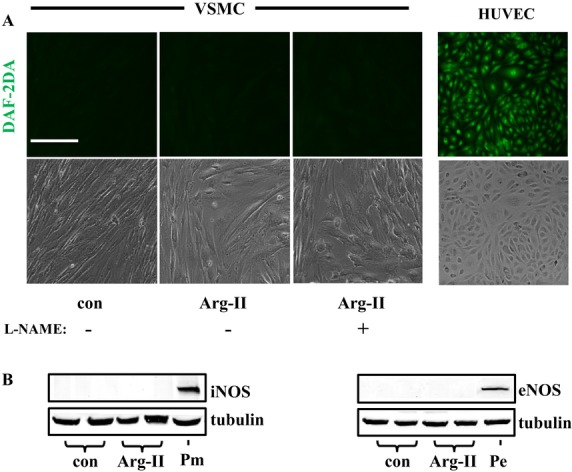
Background: Vascular smooth muscle cell (VSMC) senescence and apoptosis are involved in atherosclerotic plaque vulnerability. Arginase-II (Arg-II) has been shown to promote vascular dysfunction and plaque vulnerability phenotypes in mice through uncoupling of endothelial nitric oxide synthase and activation of macrophage inflammation. The function of Arg-II in VSMCs with respect to plaque vulnerability is unknown. This study investigated the functions of Arg-II in VSMCs linking to plaque vulnerability.

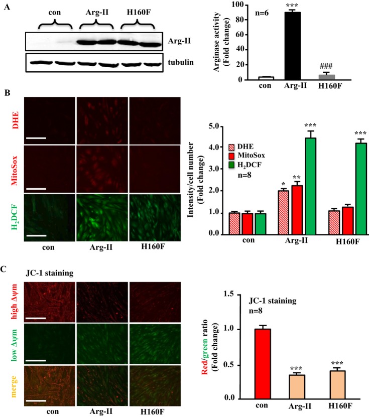
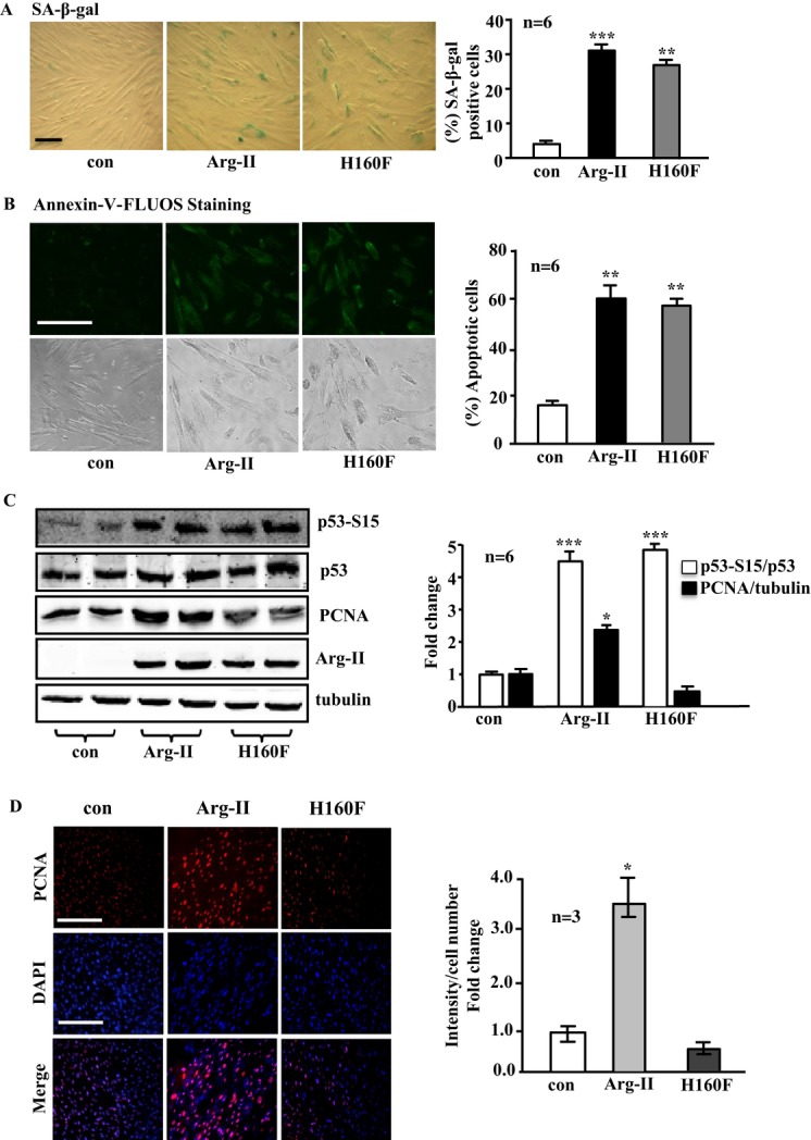
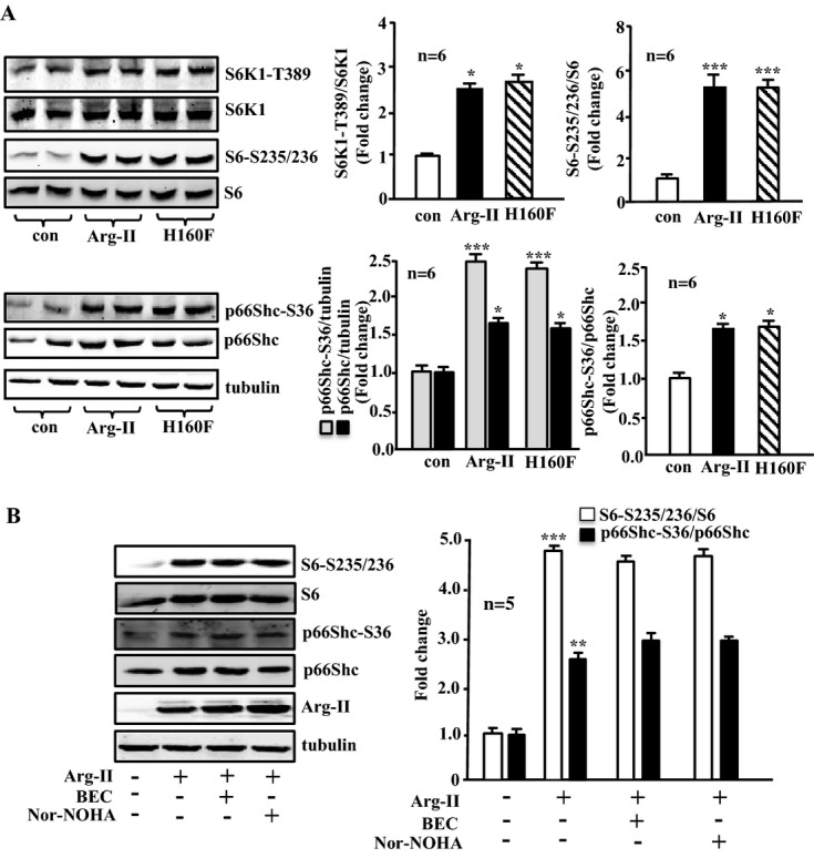
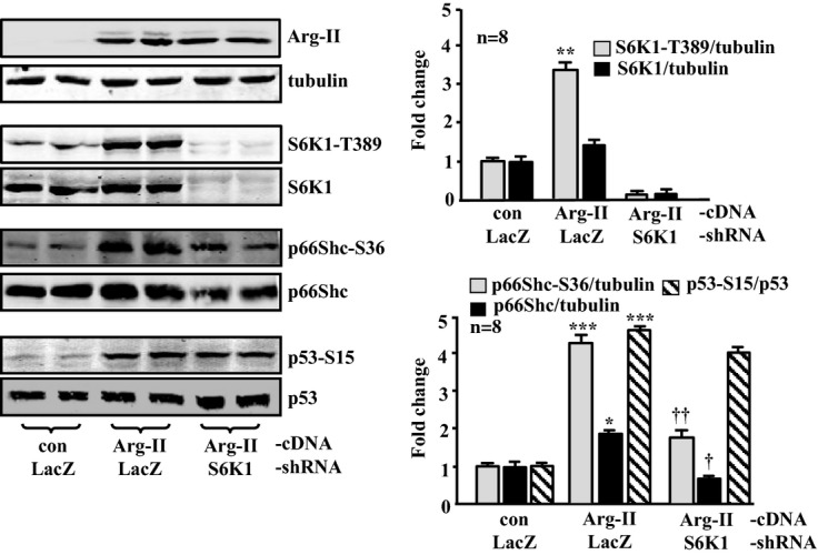
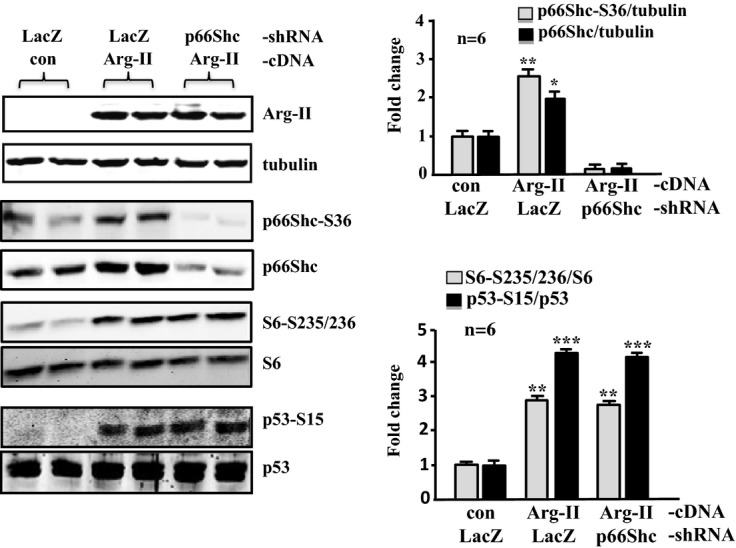
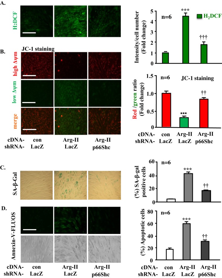
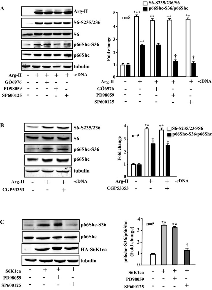
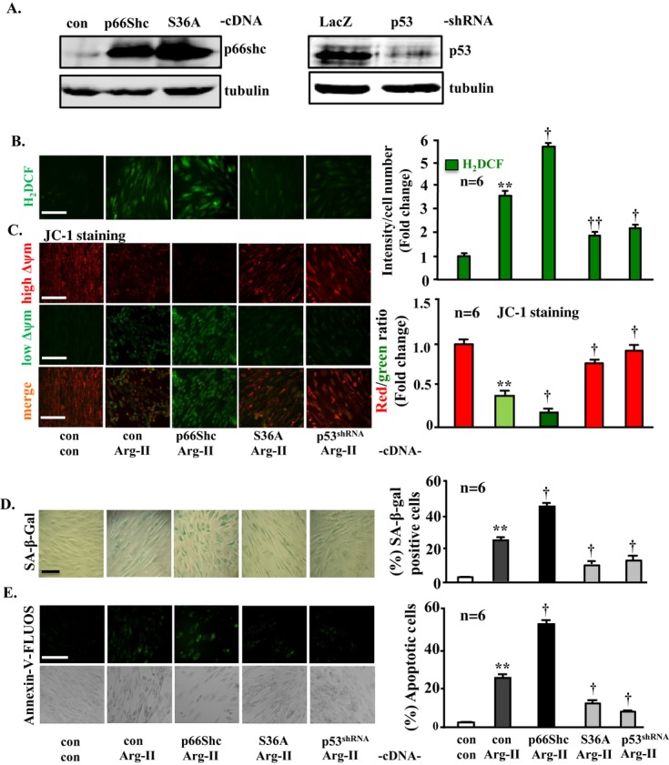
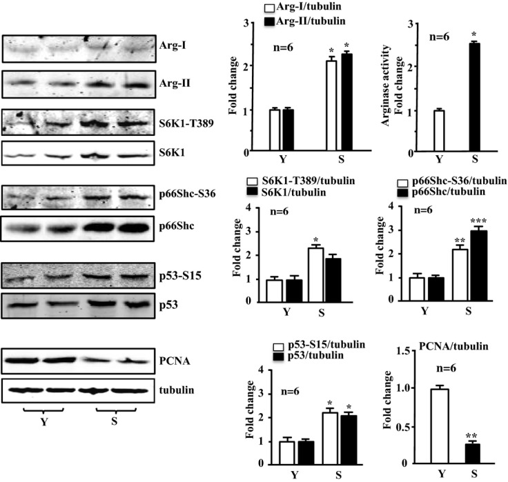
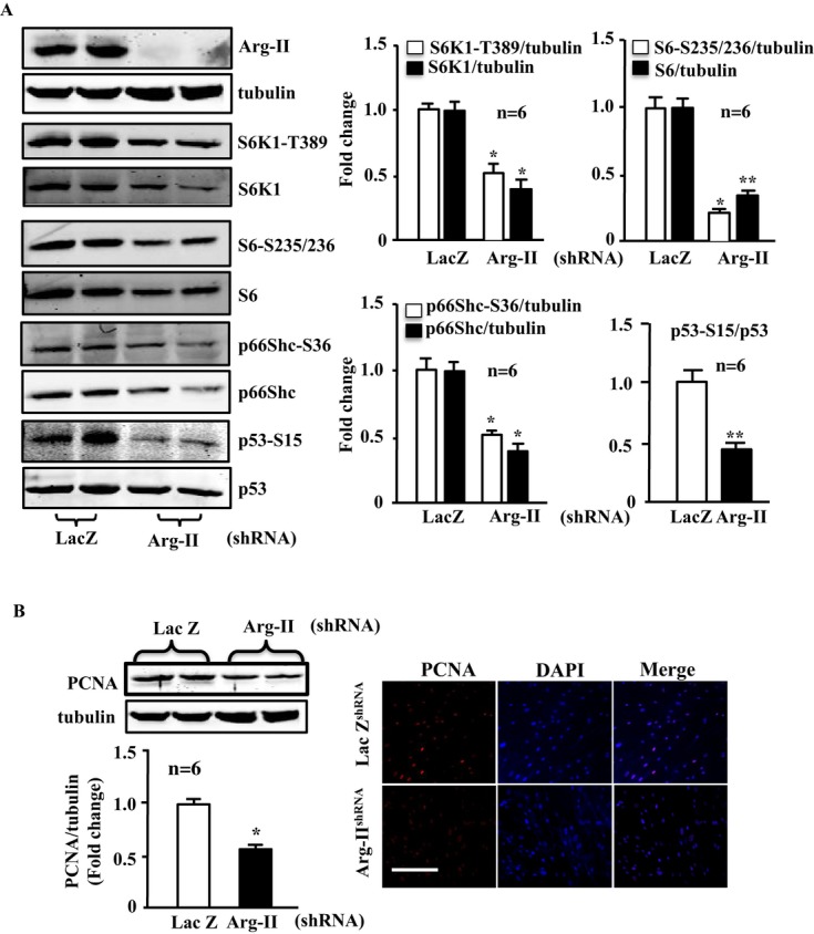
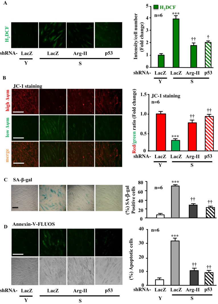
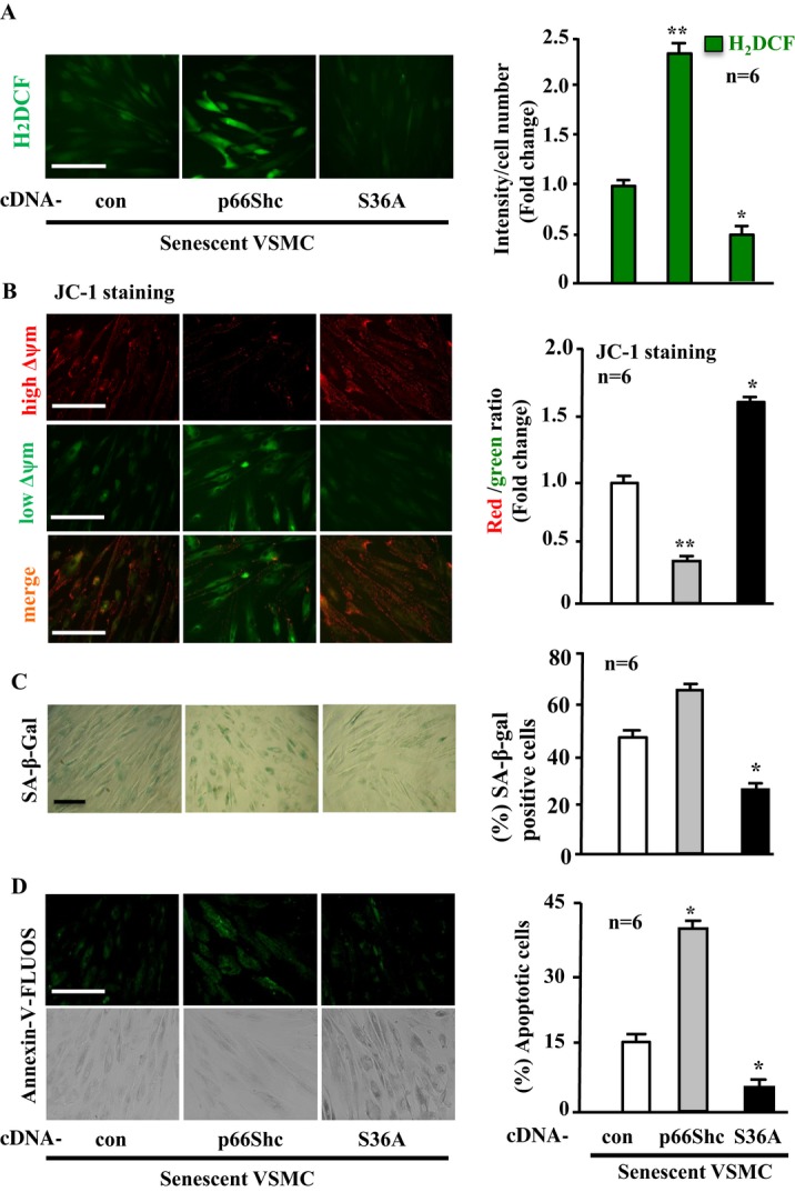
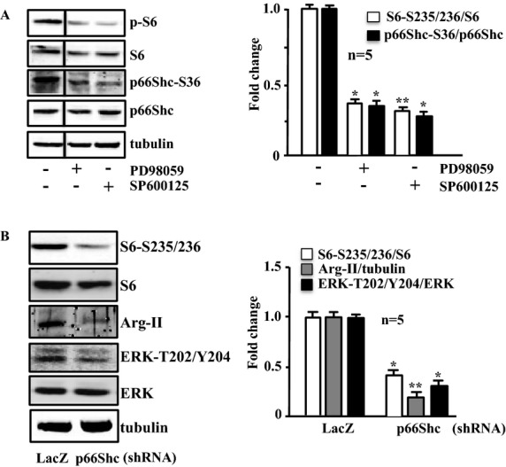
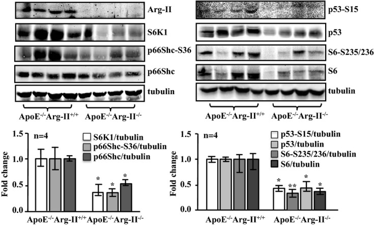
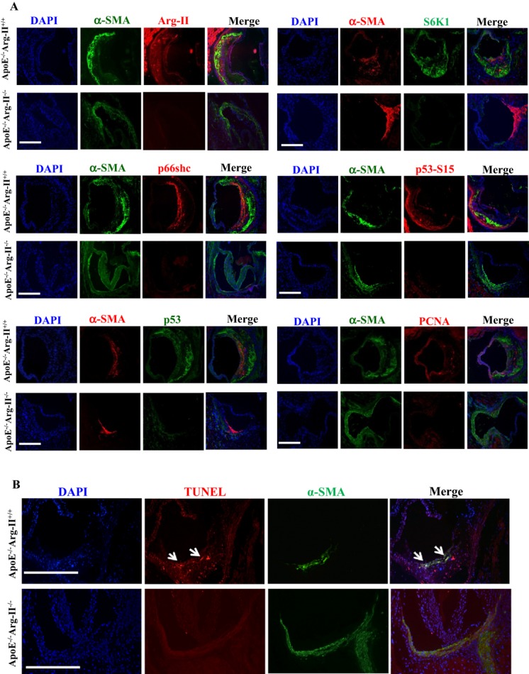
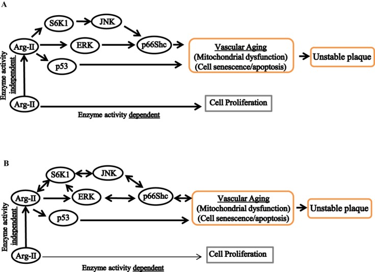
Methods and results: In vitro studies were performed on VSMCs isolated from human umbilical veins, whereas in vivo studies were performed on atherosclerosis-prone apolipoprotein E-deficient (ApoE(-/-)) mice. In nonsenescent VSMCs, overexpressing wild-type Arg-II or an l-arginine ureahydrolase inactive Arg-II mutant (H160F) caused similar effects on mitochondrial dysfunction, cell apoptosis, and senescence, which were abrogated by silencing p66Shc or p53. The activation of p66Shc but not p53 by Arg-II was dependent on extracellular signal-regulated kinases (ERKs) and sequential activation of 40S ribosomal protein S6 kinase 1 (S6K1)-c-Jun N-terminal kinases (JNKs). In senescent VSMCs, Arg-II and S6K1, ERK-p66Shc, and p53 signaling levels were increased. Silencing Arg-II reduced all these signalings and cell senescence/apoptosis. Conversely, silencing p66Shc reduced ERK and S6K1 signaling and Arg-II levels and cell senescence/apoptosis. Furthermore, genetic ablation of Arg-II in ApoE(-/-) mice reduced the aforementioned signaling and apoptotic VSMCs in the plaque of aortic roots.

Conclusions: Arg-II, independently of its l-arginine ureahydrolase activity, promotes mitochondrial dysfunction leading to VSMC senescence/apoptosis through complex positive crosstalk among S6K1-JNK, ERK, p66Shc, and p53, contributing to atherosclerotic vulnerability phenotypes in mice.

Keywords: apoptosis; arginase; p53; p66Shc; vascular smooth muscle cells.


Figures

Figure 1.
Figure 1.
Arginase‐II (Arg‐II) overexpression enhances cytoplasmic and mitochondrial O2•− generation in an l‐arginine ureahydrolase activity‐dependent manner, promotes production of H2O2, and decreases mitochondrial membrane potential (Δψm) independently of its enzymatic activity in young VSMCs. Young VSMCs were transduced with empty vector rAd/CMV as control (con), rAd/CMV‐Arg‐II (Arg‐II), or rAd/CMV‐Arg‐II‐H160F (H160F, an inactive Arg‐II mutant). Seventy‐two hours posttransduction, the cells were subjected to (A) immunoblotting analysis for detection of overexpressed Arg‐II taking tubulin as a loading control (left) and arginase activity assay (right). B, DHE and MitoSox staining for detection of cytoplasmic and mitochondrial O2•−, respectively, and H2DCF staining for detection of H2O2 production. C, Δψm assessment using a JC‐1 Mitochondrial Membrane Potential Detection Kit. Red fluorescence JC‐1 signal is indicative of healthy cells with high Δψm, whereas green fluorescence JC‐1 signal is indicative of unhealthy cells with low Δψm. Quantification of the signals is shown in the corresponding right‐sided panels (n=6 or 8 as indicated in the graphs). **P<0.01, ***P<0.001 vs control group, ###P<0.001 vs Arg‐II. Scale bar=0.2 mm. VSMC indicates vascular smooth muscle cell; rAd, recombinant adenovirus; DHE, dihydroethidium; H2DCF, 2′,7′‐dichlorofluorescein; CMV, cytomegalovirus.
Figure 2.
Figure 2.
NOS/NO is not involved in the actions of Arg‐II in VSMCs. VSMCs were transduced with empty vector rAd/CMV as control or rAd/CMV‐Arg‐II. Seventy‐two hours posttransduction, cells were treated with 5 mmol/L L‐NG‐Nitroarginine methyl ester (L‐NAME) for 1.5 hours and then subjected to (A) DAF‐2DA staining for nitric oxide (NO) production. Human umbilical veins endothelial cells (HUVECs) were used as a positive control for NO staining. B, Immunoblotting analysis of iNOS and eNOS (P, positive control). Macrophage (Pm) was used as a positive control for iNOS (left), whereas HUVECs (Pe) was used as a positive control for eNOS (right). Scale bar=0.2 mm. These experiments were repeated 3 times. NOS indicates nitric oxide synthase; VSMC, vascular smooth muscle cell; con, control; rAd, recombinant adenovirus; DAF‐2DA, 4,5‐diaminofluoresceine acetate; iNOS, inducible nitric oxide synthase; eNOS, endothelial nitric oxide synthase; Arg‐II, arginase‐II.
Figure 3.
Figure 3.
Overexpression of Arg‐II in young VSMCs causes cell senescence and apoptosis independently of its enzymatic activity, whereas it enhances cell proliferation in an enzymatic activity–dependent manner. The young VSMCs were as described in Figure 1. A, SA‐β‐gal staining for senescent cells. B, Annexin‐V‐FLUOS staining for detection of apoptotic cells. C, Immunoblotting analysis of p53‐S15, p53, PCNA, and Arg‐II levels. Tubulin served as a loading control. D, Representative images showing immunofluorescence staining of PCNA level (red) in VSMCs followed by counterstaining with DAPI (blue). The merged images are also shown. Bar graphs show quantifications of the corresponding signals (n=6 or 3). *P<0.05, **P<0.01, ***P<0.001 vs control. Scale bar=0.2 mm. Arg‐II indicates arginase‐II; VSMC, vascular smooth muscle cell; con, control; SA‐β‐gal, senescence‐associated β‐galactosidase; PCNA, proliferating cell nuclear antigen; DAPI, 4′6‐diamidino‐2‐phenyl‐indole, dihydrochloride.
Figure 4.
Figure 4.
Overexpression of Arg‐II in young VSMCs activates S6K1 and p66Shc independently of its enzymatic activity. A, Young VSMCs were transduced as in Figure 1. Shown is immunoblotting analysis of phosphorylated S6K1‐T389 and total S6K1, phosphorylated S6‐S235/236 and total S6, and phosphorylated p66Shc‐S36 and total p66Shc levels. Tubulin served as a loading control. B, Young VSMCs were transduced with empty vector rAd/CMV as control (Arg‐II: −) or rAd/CMV‐Arg‐II (Arg‐II: +). Cells were treated with or without the arginase inhibitor BEC (200 μmol/L) or nor‐NOHA (50 μmol/L) overnight during serum starvation before cell lysate preparation 72 hours posttransduction. Shown is the immunoblotting analysis of phosphorylated S6‐S235/236 and total S6, phosphorylated p66Shc‐S36, and total p66Shc levels and Arg‐II level. Tubulin served as a loading control. Bar graphs show quantifications of the signals (n=6 in A, n=5 in B). *P<0.05, **P<0.01, ***P<0.001 vs control. Arg‐II indicates arginase‐II; con, control; VSMC, vascular smooth muscle cell; rAd, recombinant adenovirus; BEC, S‐12‐bromoethyl‐l‐cystine; Nor‐NOHA, Nω‐hydroxy‐nor‐l‐arginine.
Figure 5.
Figure 5.
Silencing S6K1 in young VSMCs attenuates Arg‐II‐induced activation of p66Shc, but not that of p53. The young VSMCs were first transduced either with rAd/U6‐LacZshRNA as control or rAd/U6‐S6K1shRNA. Twenty‐four hours after the first transduction with rAd/U6‐shRNA, the cells were then transduced either with rAd/CMV as control (con) or with rAd/CMV‐Arg‐II to overexpress Arg‐II. Experiments were performed 72 hours after the second transduction (48 hours in 10% FCS‐DMEM plus overnight serum starvation in 0.2% BSA‐DMEM). Shown are immunoblotting analyses of Arg‐II levels and tubulin, S6K1‐T389 and total S6K1, p66Shc‐S36 and total p66Shc, and p53‐S15 and total p53. Tubulin served as a loading control. Quantification of the signals is shown in the bar graphs (n=8). *P<0.05, **P<0.01, ***P<0.001 vs control; †P<0.05, ††P<0.01 vs Arg‐II‐cDNA+LacZshRNA. Arg‐II indicates arginase‐II; VSMC, vascular smooth muscle cell; rAd, recombinant adenovirus.
Figure 6.
Figure 6.
Silencing p66Shc in young VSMCs does not affect Arg‐II‐induced activation of S6K1 or p53. The young VSMCs were first transduced either with rAd/U6‐LacZshRNA as control or rAd/U6‐p66ShcshRNA. Twenty‐four hours after the first transduction with rAd/U6‐shRNA, cells were then transduced either with rAd/CMV as control (con) or rAd/CMV‐Arg‐II. Experiments were carried out 72 hours after the second transduction. Immunoblotting analysis (left) reveals the overexpression of Arg‐II, the silencing efficiency of p66Shc, and the effects on S6‐S235/236 and S6, p53‐S15, and total p53. Tubulin served as a loading control. Quantification of the signals is shown in the bar graphs on the right (n=6). *P<0.05, **P<0.01, ***P<0.001 vs control. Arg‐II indicates arginase‐II; VSMC, vascular smooth muscle cell; rAd, recombinant adenovirus; SA‐β‐gal, senescence‐associated β‐galactosidase; H2DCF, 2′,7′‐dichlorofluorescein.
Figure 7.
Figure 7.
Silencing p66Shc in young VSMCs prevents Arg‐II‐induced mitochondrial dysfunction and cell senescence and apoptosis. The young VSMCs were transduced as described in Figure 6. A, H2DCF staining for detection of H2O2. B, Δψm Assessment by JC‐1 staining. C, SA‐β‐gal staining for senescent cells. D, Annexin‐V‐FLUOS staining for apoptotic cells. Quantification of the signals is shown in the corresponding bar graphs (n=6). ***P<0.001 vs control; ††P<0.01, †††P<0.001 vs Arg II. Scale bar=0.2 mm. Arg‐II indicates arginase‐II; VSMC, vascular smooth muscle cell; con, control; SA‐β‐gal, senescence‐associated β‐galactosidase; H2DCF, 2′,7′‐dichlorofluorescein; rAd, recombinant adenovirus.
Figure 8.
Figure 8.
Effects of signaling inhibitors on Arg‐II‐ or S6K1‐induced phosphorylation of p66Shc‐S36. Immunoblotting analysis of the signaling and protein levels as indicated. A and B, Young VSMCs were transduced with empty‐vector rAd/CMV as control (Arg‐II: −) or rAd/CMV‐Arg‐II (Arg‐II: +). Cells were treated with or without the signaling inhibitor overnight during serum starvation before cell lysate preparation 72 hours posttransduction. Cells were treated with Gö6976 (1 μmol/L), PD98059 (50 μmol/L), or SP600125 (20 μmol/L) or with CGP53353 (1 μmol/L) as indicated. C, Young VSMCs were transduced with empty‐vector rAd/CMV as control (S6K1ca: −) or rAd/CMV‐HA‐S6K1ca (an active mutant of S6K1; S6K1ca: +). Cells were treated with PD98059 or SP600125 as described in A and B. Quantification of the signals is shown in the corresponding bar graphs (n=5). *P<0.05, **P<0.01, ***P<0.001 vs control; †P<0.05 vs Arg‐II (in A) or vs S6K1ca (in C). Arg‐II indicates arginase‐II; VSMC, vascular smooth muscle cell; SA‐β‐gal, senescence‐associated β‐galactosidase; H2DCF, 2′,7′‐dichlorofluorescein; rAd, recombinant adenovirus.
Figure 9.
Figure 9.
Phosphorylation of p66Shc at S36 as well as p53 signaling is required for the actions of Arg‐II in young VSMCs. The young VSMCs were first transduced with either rAd/CMV empty vector as control (con) or rAd/CMV‐p66Shc, ‐p66Shc‐S36A; rAd/U6‐LacZshRNA, or rAd/U6‐p53shRNA. Twenty‐four hours after the first transduction, the cells were then transduced with either rAd/CMV as control (con) or rAd/CMV‐Arg‐II to overexpress Arg‐II as indicated. Experiments were performed 72 hours after the second transduction. A, Immunoblotting analysis showing the overexpression of p66Shc and p66Shc‐S36A (left) and the efficient silencing of p53 (right). B, H2DCF staining for detection of H2O2. C, Δψm assessment by JC‐1 staining. D, SA‐β‐gal staining for senescent cells. E, Annexin‐V‐FLUOS staining for apoptotic cells. Quantification of the signals is shown in the corresponding bar graphs (n=6). **P<0.01 vs control; †P<0.05, ††P<0.01 vs Arg II. Scale bar=0.2 mm. Of note, in B through E, the images of the experimental group with LacZshRNA+Arg‐II are not shown because of space limitation and because the data are similar to those of the experimental group with con+Arg‐II. Arg‐II indicates arginase‐II; VSMC, vascular smooth muscle cell; SA‐β‐gal, senescence‐associated β‐galactosidase; H2DCF, 2′,7′‐dichlorofluorescein; rAd, recombinant adenovirus.
Figure 10.
Figure 10.
Enhanced expression and activity of arginases, S6K1, p66Shc, and p53 and decreased PCNA level in senescent VSMCs. Immunoblotting analysis of Arg‐I, Arg‐II, S6K1‐T389 and total S6K1, p66Shc‐S36 and total p66Shc, p53‐S15 and total p53, and PCNA expression, as well as arginase activity in the senescent VSMCs (S) compared with the young cells (Y). Quantification of the signals is shown in the corresponding bar graphs (n=6). *P<0.05, **P<0.01, ***P<0.001 vs young cells (Y). Arg‐II indicates arginase‐II; VSMC, vascular smooth muscle cell; PCNA, proliferating cell nuclear antigen; S6K1, ribosomal protein S6 kinase 1.
Figure 11.
Figure 11.
Effects of silencing Arg‐II in senescent VSMCs on activation of S6K1‐p66Shc and p53 and on PCNA level. Senescent VSMCs were transduced with either rAd/U6‐LacZshRNA or rAd/U6‐Arg‐IIshRNA. Seventy‐two hours after transduction cell lysates were prepared and subjected to (A) immunoblotting analysis of Arg‐II, tubulin, S6K1‐T389 and total S6K1, S6‐S235/236 and total S6, p66Shc‐S36 and total p66Shc, p53‐S15, and total p53. Quantification of the signals is shown in the right bar graphs (n=6). *P<0.05, **P<0.01 vs young cell (Y). B, Immunoblotting analysis and immunofluorescence staining of PCNA. Quantification of the immunoblotting signals of PCNA is shown in the bar graphs underneath the immunoblotting. *P<0.05 vs LacZ‐shRNA. Arg‐II indicates arginase‐II; VSMC, vascular smooth muscle cell; PCNA, proliferating cell nuclear antigen; DAPI, 4′6‐diamidino‐2‐phenyl‐indole, dihydrochloride.
Figure 12.
Figure 12.
Silencing Arg‐II or p53 in senescent VSMCs prevents H2O2 production and decreased Δψm as well as senescence and apoptosis. Young (Y) and senescent (S) VSMCs were transduced with either rAd/U6‐LacZshRNA or rAd/U6‐Arg‐IIshRNA as indicated. Seventy‐two hours posttransduction, the cells were subjected to (A) H2DCF staining for detection of H2O2. B, JC‐1 staining for analysis of Δψm. C, SA‐β‐gal staining for senescent cells. D, Annexin‐V‐FLUOS staining for apoptotic cells. Quantification of the signals is shown in the corresponding bar graphs (n=6). ***P<0.001 vs Y+LacZshRNA; †P<0.05, ††P<0.01 vs S+LacZshRNA. Scale bar=0.2 mm. Arg‐II indicates arginase‐II; VSMC, vascular smooth muscle cell; H2DCF, 2′,7′‐dichlorofluorescein; SA‐β‐gal, senescence‐associated β‐galactosidase.
Figure 13.
Figure 13.
Enhanced phosphorylation of p66Shc‐S36 accounts for the phenotypic changes of senescent VSMCs. Senescent VSMCs were transduced with either rAd/CMV empty vector as control (con), rAd/CMV‐p66Shc or ‐p66Shc‐S36A (S36A) as indicated. Seventy‐two hours posttransduction, the cells were subjected to (A) H2DCF staining for detection of H2O2. B, JC‐1 staining for analysis of Δψm. C, SA‐β‐gal staining for senescent cells. D, Annexin‐V‐FLUOS staining for apoptotic cells. Quantification of the signals is shown in the corresponding bar graphs (n=6). *P<0.05, **P<0.01 vs con. Scale bar=0.2 mm. Arg‐II indicates arginase‐II; VSMC, vascular smooth muscle cell; H2DCF, 2′,7′‐dichlorofluorescein; SA‐β‐gal, senescence‐associated β‐galactosidase; rAd, recombinant adenovirus.
Figure 14.
Figure 14.
Interaction between S6K1, JNK, ERKs, and p66Shc in senescent VSMCs. Immunoblotting analysis of the parameters as indicated. A, Senescent VSMCs were either untreated (−) or treated with PD98059 (50 μmol/L) or SP600125 (20 μmol/L) overnight. The line between the first and second lanes indicates cutting of the same blots. B, Senescent VSMCs were transduced with either rAd/U6‐LacZshRNA or rAd/U6‐p66ShcshRNA as indicated. Experiments were performed 72 hours posttransduction. Quantification of the signals is shown in the corresponding bar graphs (n=5). *P<0.05, **P<0.01 vs untreated (in A) or LacZshRNA (in B). Arg‐II indicates arginase‐II; VSMC, vascular smooth muscle cell; JNK, c‐Jun N‐terminal kinases; ERK, extracellular signal‐regulated kinases; rAd, recombinant adenovirus.
Figure 15.
Figure 15.
Ablation of Arg‐II in atherosclerosis‐prone ApoE−/− mice reduces signaling of S6K1, p66Shc, and p53 in the aortas. ApoE−/−Arg‐II+/+ and ApoE−/−Arg‐II−/− mice were fed a high‐fat diet for 10 weeks. Aortas from mice of both genotypes were cleaned of perivascular tissues and subjected to immunoblotting analysis of Arg‐II, S6K1, p66Shc‐S36 and total p66Shc, p53‐S15 and total p53, and S6‐S235/236 and total S6. Tubulin served as a loading control. Quantification of the signals is shown in the corresponding lower panels (n=4). *P<0.05, **P<0.001 vs ApoE−/−Arg‐II+/+. Arg‐II indicates arginase‐II; ApoE, apolipoprotein E; S6K1, ribosomal protein S6 kinase 1.
Figure 16.
Figure 16.
Ablation of Arg‐II in atherosclerosis‐prone ApoE−/− mice reduces levels of Arg‐II, S6K1, p66Shc, p53‐S15, and p53, as well as PCNA in VSMCs along with decreased apoptotic VSMCs in atherosclerotic plaque. ApoE−/−Arg‐II+/+ and ApoE−/−Arg‐II−/− mice were fed a high‐fat diet for 10 weeks. A, AR‐cryosections (7 μm) were subjected to immunofluorescence costaining of Arg‐II, S6K1, p66Shc, p53‐S15 and p53, and PCNA with anti‐α‐smooth muscle actin antibody (α‐SMA) for VSMCs. Mouse anti‐α‐SMA (green) was used for costaining with rabbit antibodies against Arg‐II, p66Shc, p53‐S15, and PCNA (red), whereas rabbit anti‐α‐SMA (red) was used for costaining with mouse anti‐p53 and ‐S6K1 (green). Alexa Fluor 488–conjugated goat anti‐mouse IgG (green) and Alexa Fluor 594–conjugated goat anti‐rabbit F(ab)2 (red) were used as secondary antibodies. All sections were counterstained with DAPI. Representative images of individual staining and merged images are shown (n=4). B, Representative images showing apoptotic cells detected by TUNEL staining (red) in the plaques in aortic roots of ApoE−/−Arg‐II+/+ and ApoE−/−Arg‐II−/− mice. All sections were stained for VSMCs with anti‐α‐SMA (green) followed by counterstaining with DAPI for nuclei (blue). The merged images are also shown (n=4). The white arrows indicate apoptotic VSMCs. Scale bars=0.2 mm. Arg‐II indicates arginase‐II; ApoE, apolipoprotein E; S6K1, ribosomal protein S6 kinase 1; VSMC, vascular smooth muscle cell; PCNA, proliferating cell nuclear antigen; DAPI, 4′6‐diamidino‐2‐phenyl‐indole, dihydrochloride; TUNEL, terminal dUTP nick end‐labeling.
Figure 17.
Figure 17.
Schematic summary of the major findings of the study. A, Elevated Arg‐II leads to phosphorylation/activation of p66Shc through S6K1‐JNK and ERK in parallel. In addition, Arg‐II induces phosphorylation/activation of p53 independently of the S6K1‐JNK‐p66Shc pathway. Both S6K1‐JNK‐p66Shc and p53 are involved in Arg‐II‐induced H2O2 and mitochondrial dysfunction, which ultimately cause both VSMC senescence and apoptosis, contributing to plaque vulnerability. Arg‐II exerts all these effects independently of its l‐arginine ureahydrolase activity, whereas it is required for VSMC proliferation in an enzymatic activity–dependent manner. B, In senescent VSMCs, Arg‐II, S6K1, JNK, ERK, and p66Shc form a complex positive crosstalk network resulting in acceleration of VSMC aging. Arg‐II indicates arginase‐II; S6K1, ribosomal protein S6 kinase 1; VSMC, vascular smooth muscle cell; JNK, c‐Jun N‐terminal kinases; ERK, extracellular signal‐regulated kinases.

Comment in

References

    1. Libby P, Aikawa M. Stabilization of atherosclerotic plaques: new mechanisms and clinical targets. Nat Med. 2002; 8:1257-1262 - PubMed
    1. Finn AV, Nakano M, Narula J, Kolodgie FD, Virmani R. Concept of vulnerable/unstable plaque. Arterioscler Thromb Vasc Biol. 2010; 30:1282-1292 - PubMed
    1. Wang JC, Bennett M. Aging and atherosclerosis: mechanisms, functional consequences, and potential therapeutics for cellular senescence. Circ Res. 2012; 111:245-259 - PubMed
    1. Clarke MC, Figg N, Maguire JJ, Davenport AP, Goddard M, Littlewood TD, Bennett MR. Apoptosis of vascular smooth muscle cells induces features of plaque vulnerability in atherosclerosis. Nat Med. 2006; 12:1075-1080 - PubMed
    1. Matthews C, Gorenne I, Scott S, Figg N, Kirkpatrick P, Ritchie A, Goddard M, Bennett M. Vascular smooth muscle cells undergo telomere‐based senescence in human atherosclerosis: effects of telomerase and oxidative stress. Circ Res. 2006; 99:156-164 - PubMed

Publication types

MeSH terms

Substances