. 2013 Aug 1;191(3):1029-42.
doi: 10.4049/jimmunol.1300658. Epub 2013 Jul 5.

Butyrophilin 3A1 plays an essential role in prenyl pyrophosphate stimulation of human Vγ2Vδ2 T cells

Affiliations

Butyrophilin 3A1 plays an essential role in prenyl pyrophosphate stimulation of human Vγ2Vδ2 T cells

Hong Wang et al. J Immunol. .

Abstract

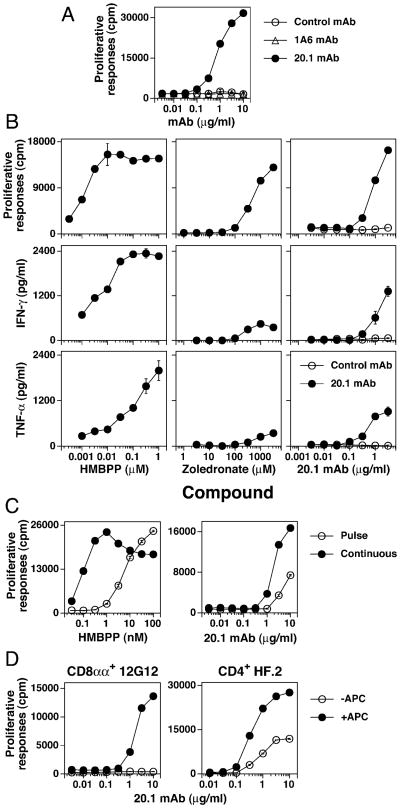
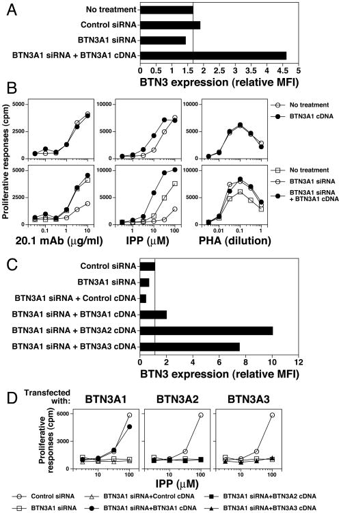
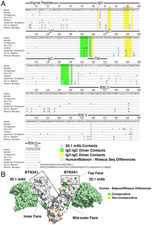
Most human γδ T cells express Vγ2Vδ2 TCRs and play important roles in microbial and tumor immunity. Vγ2Vδ2 T cells are stimulated by self- and foreign prenyl pyrophosphate intermediates in isoprenoid synthesis. However, little is known about the molecular basis for this stimulation. We find that a mAb specific for butyrophilin 3 (BTN3)/CD277 Ig superfamily proteins mimics prenyl pyrophosphates. The 20.1 mAb stimulated Vγ2Vδ2 T cell clones regardless of their functional phenotype or developmental origin and selectively expanded blood Vγ2Vδ2 T cells. The γδ TCR mediates 20.1 mAb stimulation because IL-2 is released by β(-) Jurkat cells transfected with Vγ2Vδ2 TCRs. 20.1 stimulation was not due to isopentenyl pyrophosphate (IPP) accumulation because 20.1 treatment of APC did not increase IPP levels. In addition, stimulation was not inhibited by statin treatment, which blocks IPP production. Importantly, small interfering RNA knockdown of BTN3A1 abolished stimulation by IPP that could be restored by re-expression of BTN3A1 but not by BTN3A2 or BTN3A3. Rhesus monkey and baboon APC presented HMBPP and 20.1 to human Vγ2Vδ2 T cells despite amino acid differences in BTN3A1 that localize to its outer surface. This suggests that the conserved inner and/or top surfaces of BTN3A1 interact with its counterreceptor. Although no binding site exists on the BTN3A1 extracellular domains, a model of the intracellular B30.2 domain predicts a basic pocket on its binding surface. However, BTN3A1 did not preferentially bind a photoaffinity prenyl pyrophosphate. Thus, BTN3A1 is required for stimulation by prenyl pyrophosphates but does not bind the intermediates with high affinity.


Figures

Figure 1
Figure 1
Anti-BTN3 mAb, 20.1, stimulates Vγ2Vδ2 T cells to proliferate and secrete cytokines. (A) Vγ2Vδ2 T cells proliferate in response to the 20.1 but not the 1A6 mAb, although both are specific for BTN3. The 12G12 Vγ2Vδ2 T cell clone was cultured with control IgG1 mAb, 1A6 mAb, or 20.1 mAb in the presence of mitomycin C-treated Va2 cells. The cells were pulsed with [3H]-thymidine after 24 h and harvested 18 h later. (B) 20.1 mAb stimulates both proliferation and cytokine production by Vγ2Vδ2 T cells. 12G12 T cells were cultured continuously with the 20.1 mAb or the P3 control mAb in the presence of mitomycin C-treated Va2 cells. For HMBPP and zoledronate, mitomycin C-treated Va2 cells were pulsed with the compounds for 1 h, washed, and then cultured with the 12G12 T cell clone. Proliferative responses were measured as in (A). Supernatants were collected at 16 h for the measurement of TNF-α and IFN-γ. (C) APC pulsed with the 20.1 mAb stimulate 12G12 T cells. Mitomycin C-treated Va2 cells were pulsed for 1 h with HMBPP or the 20.1 mAb. The Va2 cells were then washed and cultured with the 12G12 Vγ2Vδ2 T cell clone. Proliferative responses were measured as in (A). (D) CD8αα+ and CD4+ Vγ2Vδ2 T cell clones exhibit different APC dependency for stimulation by the 20.1 mAb. The CD8 αα+ 12G12 clone and the CD4+ HF.2 clone were cultured with the 20.1 mAb in the presence or absence of mitomycin C-treated Va2 (for 12G12) or CP.EBV (for HF.2). Cell proliferations were measured as in (A). Note that the HF.2 clone proliferates in response to the 20.1 mAb in the absence of APC whereas the 12G12 clone does not.
Figure 2
Figure 2
20.1 mAb specifically stimulates diverse Vγ2Vδ2 T cells in a Vγ2Vδ2 TCR-dependent manner. (A) 20.1 mAb specifically stimulates the expansion of Vγ2Vδ2 T cells from blood mononuclear cells. PBMC from five donors were cultured with media, HMBPP (100 nM), a control mAb (10 μg/ml), or the 20.1 mAb (10 μg/ml). On day 3, IL-2 was added to 1 nM. After 9 d, cells were stained with the indicated mAbs and analyzed by flow cytometry. (B) Specific stimulation of Vγ2Vδ2 T cell clones by the 20.1 mAb. CD8αα+/CD4- CD8- (solid bars) and CD4+ (open bars) T cell clones expressing different V genes were stimulated with the 20.1 mAb (1 μg/ml) or PHA-P (1:1000) with Va2 APC and [3H]-thymidine incorporation measured at day 2. Stimulation index was calculated as the ratio between the 20.1 and control mAb response. PHA stimulation indices were >2.5 for all clones and averaged 12.6-fold. (C) 20.1 mAb stimulation is mediated by the Vγ2Vδ2 TCR. The DBS43 Vγ2Vδ2 TCR transfectant and the parent β J.RT3-T3.5 cell line line were incubated with mitomycin C-treated Va2 APC and either the 20.1 mAb or HMBPP for 24 h. The supernatants were then harvested and used to stimulate the IL-2-dependent proliferation of HT-2 T cells.
Figure 3
Figure 3
Stimulation by the 20.1 mAb is not due to accumulation of IPP in APC. (A) Stimulation of Vγ2Vδ2 T cells by the 20.1 mAb or by HMBPP is relatively resistant to statin inhibition. Statin inhibition of Vγ2Vδ2 T cell proliferation to HMBPP (0.1 μM), risedronate (1 mM), or the 20.1 mAb (3.16 μg/ml) was tested using mitomycin C-treated Va2 cells that were pulsed with HMBPP or risedronate in the presence of either mevastatin or pravastatin for 1 h, washed, and cultured with 12G12 T cells in the presence of the statins. The 20.1 mAb was continuously present in the culture. After 24 h, the cells were pulsed with [3H]-thymidine and harvested 18 h later. Results are shown as the percentage of the control proliferative response without the statins. (B) 20.1 mAb treatment does not increase IPP or ApppI levels. MCF-7 cells were untreated or incubated for 24 h with a control mAb (5 μg/ml), the 20.1 mAb (5 μg/ml), or zoledronate (25 μM). Cells were then harvested, washed, and lysed with acetonitrile for determination of IPP and ApppI levels by liquid chromatography/mass spectrometry.
Figure 4
Figure 4
siRNA inhibition of BTN3A1 in APC abolishes IPP stimulation of Vγ2Vδ2 T cells. (A) siRNA inhibition of BTN3A1, BTN3A2, and BTN3A3 expression. HeLa cells were transfected with either control siRNA or with siRNAs specific for each of the members of the BTN3 family. After 72 h, transfected HeLa cells were stained with the 20.1 mAb and BTN3 surface expression determined by flow cytometry. (B) siRNA inhibition of BTN3A1 greatly reduces IPP stimulation of Vγ2Vδ2 T cells. siRNA transfected HeLa cells were used as APC for 20.1 mAb, IPP, and PHA stimulation of the 12G12 Vγ2Vδ2 T cell clone. Note that for each of the siRNA shown, there was at least one additional siRNA with a similar effect (i.e. oligo A for BTN3A1, oligo C for BTN3A2, and oligo A and C for BTN3A3).
Figure 5
Figure 5
Re-expression of BTN3A1 but not BTN3A2 or BTN3A3 in siRNA-treated APC restores IPP stimulation of Vγ2Vδ2 T cells. (A) Transfection of BTN3A1 cDNA restores siRNA-inhibited BTN3 expression on HeLa cells. HeLa cells were transfected with the indicated BTN3 siRNA or cotransfected with BTN3 siRNA and cDNA. After 72 h, the transfectants were stained with either PE-conjugated isotype control mAb or the 20.1 mAb and analyzed by flow cytometry. The relative mean fluorescence intensity (MFI) was calculated as 20.1 mAb MFI minus isotype control mAb MFI. (B) Transfection of BTN3A1 cDNA in APC restores BTN3 siRNA-inhibited IPP stimulation of Vγ2Vδ2 T cells. HeLa cells were transfected with the indicated siRNA or co-transfected with the indicated siRNA and cDNA. After 72 h, the transfectants were harvested, washed, and treated for 1 h with mitomycin C. The transfectants were then cultured with half-log dilutions of the 20.1 mAb, IPP, or PHA followed by addition of 12G12 T cells. Proliferative responses were assessed as in Fig. 1A. (C) Transfection of BTN3A1, BTN3A2, or BTN3A3 cDNA restores BTN3 expression on HeLa cells treated with BTN3 siRNA. HeLa cells were transfected with BTN3A1 siRNA oligo B or cotransfected with BTN3A1 siRNA oligo B and the indicated BTN3 cDNA as detailed in (A). After 72 h, BTN3 expression was assessed by flow cytometry. (D) Transfection of BTN3A1 but not BTN3A2 or BTN3A3 cDNA in BTN3 siRNA-treated APC restores IPP stimulation of Vγ2Vδ2 T cells. HeLa cells were transfected with BTN3A1 siRNA oligo B or co-transfected with BTN3A1 siRNA oligo B and the indicated BTN3 cDNA. After 72 h, the transfectants were harvested, washed, and treated for 1 h with mitomycin C. The transfectants were then cultured with half-log diluted IPP followed by addition of 12G12 T cells. Proliferative responses were assessed as in Fig. 1A.
Figure 6
Figure 6
BTN3A1 dimers have a basic pocket on the binding face of the intracellular B30.2 domain but do not bind with high affinity to a photoaffinity prenyl pyrophosphate. (A) The surface potential of the extracellular BTN3A1 dimer. The surface potential was calculated in PyMOL using the APBS plugin and is colored from red (-10 kT) to blue (+10 kT). (B) Basic pocket on the binding face of the BTN3A1 B30.2 domain. Left panels, Model of BTN3A1 B30.2 domain (gray color) superimposed on the crystal structure of the TRIM21 B30.2 domain (blue color) with variable regions 1-4 labeled. Middle, right panels, Surface potentials of the B30.2 domains of BTN3A1 and TRIM21 from top (upper panels) and side views (bottom panels) were calculated as in A. The IgG Fc domain binds to the top of TRIM21 via the v1-v4 regions. (C) The extracellular domain of recombinant BTN3A1 predominantly exists as a dimer. The extracellular domain of BTN3A1 was expressed in E. coli, refolded, and size separated by Superdex 200 gel filtration. The position of the BTN3A1 dimer and monomer are indicated. (D) Biotin-BZ-C-C5-pyrophosphate (diphosphate) does not preferentially label BTN3A1 or BTN3A2. Recombinant BTN3A1, recombinant BTN3A2, or ovalbumin (OVA) were incubated with a biotin-benzophenone-C-C5-pyrophosphate label (structure is shown) and exposed to 350 nm UV light for 90 min on ice. Two hundred fifty nanograms of each protein were then separated on a 10-20% SDS-PAGE gel. Proteins were blotted and biotinylated proteins visualized with streptavidin-HRP.
Figure 7
Figure 7
BTN3-expressing baboon and rhesus monkey APC present prenyl pyrophosphates and the 20.1 mAb to stimulate human Vγ2Vδ2 T cells. (A) Baboon herpesvirus-transformed B cells express BTN3. Baboon B, human B, and mouse T cells were stained with PE-conjugated-isotype-control or PE-20.1 mAbs and analyzed by flow cytometry. (B) Rhesus monkey monocytes and lymphocytes express BTN3. Rhesus and human PBMC were isolated and stained as in (A). The histogram plots of cells gated for monocytes and lymphocytes are shown. (C) Baboon B cells can present nonpeptide Ags to human Vγ2Vδ2 T cells. Human CP.EBV B cells and two monkey B cell lines were treated with mitomycin C for 1 h and then either pulsed for 1 h with HMBPP or risedronate, or continuously cultured with the 20.1 mAb or PHA-P. Human 12G12 Vγ2Vδ2 T cells were then added and proliferative responses assessed as in Fig. 1A. (D) Baboon B cells present prenyl pyrophosphates and the 20.1 mAb to human HF.2 Vγ2Vδ2 T cells. Human HF.2 Vγ2Vδ2 T cells were cultured with HMBPP, the 20.1 mAb, or PHA-P in the presence or absence of mitomycin-treated CP.EBV human B cells or GAB-LCL baboon B cells and proliferative responses assessed as in Fig. 1A. (E) Rhesus and human PBMC present prenyl pyrophosphates and the 20.1 mAb to human HF.2 Vγ2Vδ2 T cells (as in D).
Figure 8
Figure 8
Amino acid differences between human and monkey BTN3A1 localize to the outer face of the BTN3A1 dimer. (A) Amino acid alignment for primate BTN3A1. The amino acid sequences for various primate BTN3A1 proteins were aligned using the Clustal W method. Locations of IgV, IgC, and B30.2 structural domains are indicated. Sequences shaded yellow highlight the IgV to IgC contacts whereas sequences shaded green highlight the IgC to IgC contacts. Red asterisks indicate residues making up the 20.1 mAb epitope. (B) Conservative and nonconservative amino acid differences between humans and baboons/rhesus monkeys localize to the outer face of the BTN3A1 dimer. The BTN3A1 extracellular dimer in complex with the 20.1 mAb is shown with conservative (shaded green) and nonconservative (shaded orange) differences located on the structure. Conserved areas on the top, inner face, and mid-outer face are boxed.
Figure 9
Figure 9
Models for BTN3A1 involvement in prenyl pyrophosphate stimulation. In model 1, BTN3A1 binds prenyl pyrophosphates extracellularly with or without the contribution of a second protein with direct contract of the prenyl pyrophosphate to the Vγ2Vδ2 TCR. In model 2, the BTN3A1 B30.2 domain associates with a protein that either binds prenyl pyrophosphates directly or stabilized the binding of the prenyl pyrophosphates to the B30.2 domain intracellularly causing a change in BTN3A1 conformation or distribution leading to Vγ2Vδ2 T cell stimulation. In model 3, BTN3A1 acts as a ligand binding to a costimulatory or inhibitory receptor on Vγ2Vδ2 T cells. In model 4, BTN3A1 functions as an APC signaling receptor.

Similar articles

Cited by

References

    1. Morita CT, Jin C, Sarikonda G, Wang H. Nonpeptide antigens, presentation mechanisms, and immunological memory of human Vγ2Vδ2 T cells: discriminating friend from foe through the recognition of prenyl pyrophosphate antigens. Immunol Rev. 2007;215:59–76. - PubMed
    1. Hintz M, Reichenberg A, Altincicek B, Bahr U, Gschwind RM, Kollas AK, Beck E, Wiesner J, Eberl M, Jomaa H. Identification of (E)-4-hydroxy-3-methyl-but-2-enyl pyrophosphate as a major activator for human γδ T cells in Escherichia coli. FEBS Lett. 2001;509:317–322. - PubMed
    1. Puan KJ, Jin C, Wang H, Sarikonda G, Raker AM, Lee HK, Samuelson MI, Märker-Hermann E, Pasa-Tolic L, Nieves E, Giner JL, Kuzuyama T, Morita CT. Preferential recognition of a microbial metabolite by human Vγ2Vδ2 T cells. Int Immunol. 2007;19:657–673. - PubMed
    1. Kunzmann V, Bauer E, Wilhelm M. γ/δ T-cell stimulation by pamidronate. N Engl J Med. 1999;340:737–738. - PubMed
    1. Gober HJ, Kistowska M, Angman L, Jenö P, Mori L, De Libero G. Human T cell receptor γδ cells recognize endogenous mevalonate metabolites in tumor cells. J Exp Med. 2003;197:163–168. - PMC - PubMed

Publication types

MeSH terms