

Interaction of 11C-tariquidar and 11C-elacridar with P-glycoprotein and breast cancer resistance protein at the human blood-brain barrier
- PMID: 23833270
- PMCID: PMC3882137
- DOI: 10.2967/jnumed.112.118232
Interaction of 11C-tariquidar and 11C-elacridar with P-glycoprotein and breast cancer resistance protein at the human blood-brain barrier
Abstract
The adenosine triphosphate-binding cassette transporters P-glycoprotein (Pgp) and breast cancer resistance protein (BCRP) are 2 major gatekeepers at the blood-brain barrier (BBB) that restrict brain distribution of several clinically used drugs. In this study, we investigated the suitability of the radiolabeled Pgp/BCRP inhibitors (11)C-tariquidar and (11)C-elacridar to assess Pgp density in the human brain with PET.
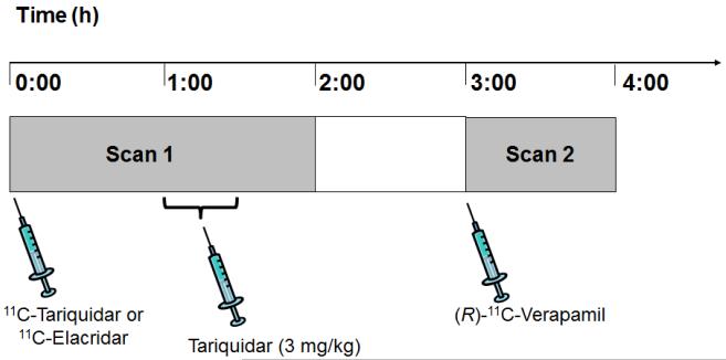
Methods: Healthy subjects underwent a first PET scan of 120-min duration with either (11)C-tariquidar (n = 6) or (11)C-elacridar (n = 5) followed by a second PET scan of 60-min duration with (R)-(11)C-verapamil. During scan 1 (at 60 min after radiotracer injection), unlabeled tariquidar (3 mg/kg) was intravenously administered. Data were analyzed using 1-tissue 2-rate-constant (1T2K) and 2-tissue 4-rate-constant (2T4K) compartment models and either metabolite-corrected or uncorrected arterial input functions.
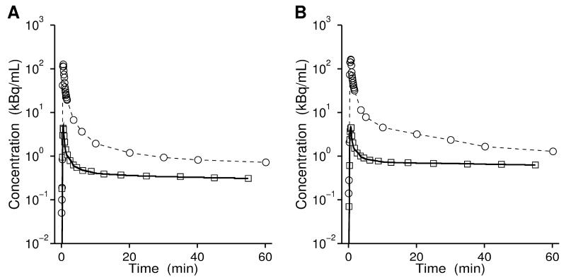
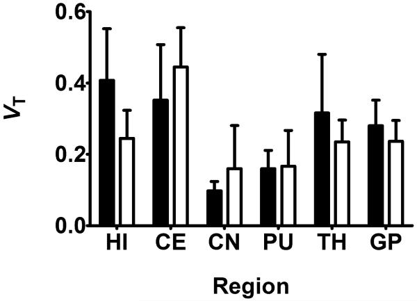
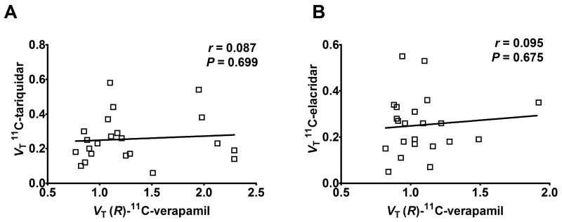
Results: After injection of (11)C-tariquidar or (11)C-elacridar, the brain PET signal corrected for radioactivity in the vasculature was low (~0.1 standardized uptake value), with slow washout. In response to tariquidar injection, a moderate but statistically significant rise in brain PET signal was observed for (11)C-tariquidar (+27% ± 15%, P = 0.014, paired t test) and (11)C-elacridar (+21% ± 15%, P = 0.014) without changes in plasma activity concentrations. Low levels of radiolabeled metabolites (<25%) were detected in plasma up to 60 min after injection of (11)C-tariquidar or (11)C-elacridar. The 2T4K model provided better data fits than the 1T2K model. Model outcome parameters were similar when metabolite-corrected or uncorrected input functions were used. There was no significant correlation between distribution volumes of (11)C-tariquidar or (11)C-elacridar and distribution volumes of (R)-(11)C-verapamil in different brain regions.
Conclusion: The in vivo behavior of (11)C-tariquidar and (11)C-elacridar was consistent with that of dual Pgp/BCRP substrates. Both tracers were unable to visualize cerebral Pgp density, most likely because of insufficiently high binding affinities in relation to the low density of Pgp in human brain (∼1.3 nM). Despite their inability to visualize Pgp density, (11)C-tariquidar and (11)C-elacridar may find use as a new class of radiotracers to study the interplay of Pgp and BCRP at the human BBB in limiting brain uptake of dual substrates.
Keywords: 11C-elacridar; 11C-tariquidar; P-glycoprotein; blood-brain barrier; breast cancer resistance protein.
Figures


References
-
- Kodaira H, Kusuhara H, Ushiki J, Fuse E, Sugiyama Y. Kinetic analysis of the cooperation of P-glycoprotein (P-gp/Abcb1) and breast cancer resistance protein (Bcrp/Abcg2) in limiting the brain and testis penetration of erlotinib, flavopiridol, and mitoxantrone. J Pharmacol Exp Ther. 2010;333:788–796. - PubMed
-
- Szakács G, Paterson JK, Ludwig JA, Booth-Genthe C, Gottesman MM. Targeting multidrug resistance in cancer. Nat Rev Drug Discov. 2006;5:219–234. - PubMed
Publication types
MeSH terms
Substances
Grants and funding
LinkOut - more resources
Full Text Sources
Other Literature Sources
Miscellaneous