Review
. 2013 Jul 5;113(2):223-37.
doi: 10.1161/CIRCRESAHA.111.300084.

Current management of calcific aortic stenosis

Affiliations
Review

Current management of calcific aortic stenosis

Brian R Lindman et al. Circ Res. .

Abstract

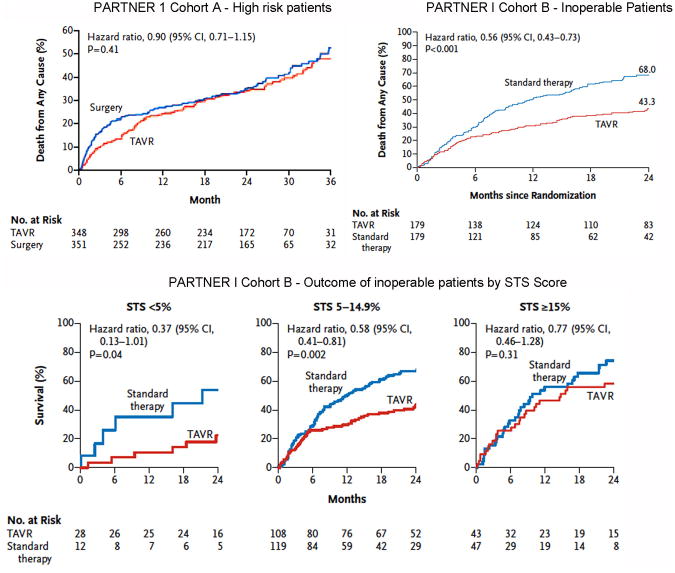
Calcific aortic stenosis is a progressive disease with no effective medical therapy that ultimately requires aortic valve replacement (AVR) for severe valve obstruction. Echocardiography is the primary diagnostic approach to define valve anatomy, measure aortic stenosis severity, and evaluate the left ventricular response to chronic pressure overload. In asymptomatic patients, markers of disease progression include the degree of leaflet calcification, hemodynamic severity of stenosis, adverse left ventricular remodeling, reduced left ventricular longitudinal strain, myocardial fibrosis, and pulmonary hypertension. The onset of symptoms portends a predictably high mortality rate unless AVR is performed. In symptomatic patients, AVR improves symptoms, improves survival, and, in patients with left ventricular dysfunction, improves systolic function. Poor outcomes after AVR are associated with low-flow low-gradient aortic stenosis, severe ventricular fibrosis, oxygen-dependent lung disease, frailty, advanced renal dysfunction, and a high comorbidity score. However, in most patients with severe symptoms, AVR is lifesaving. Bioprosthetic valves are recommended for patients aged >65 years. Transcatheter AVR is now available for patients with severe comorbidities, is recommended in patients who are deemed inoperable, and is a reasonable alternative to surgical AVR in high-risk patients.

Keywords: aortic valve stenosis; heart failure; heart valve diseases; heart valve prosthesis; hypertrophy, left ventricular.


Figures

Figure 1
Figure 1. Diagnostic Imaging of Aortic Stenosis
Transvalvular mean and peak gradients and the aortic valve VTI are obtained by continuous wave Doppler across the aortic valve (a). The LV outflow tract VTI is obtained from pulse wave Doppler in the LV outflow tract (b) and used along with the aortic valve VTI to calculate the AVA by the continuity equation. The valve morphology, opening, and calcification can be seen clearly on transesophageal echocardiographic imaging (c) and the amount of calcification in the valve leaflets quantified by CT (d).
Figure 2
Figure 2. Left Ventricular Response to Pressure Overload from Aortic Stenosis
All images are from patients with AVA <1 cm2 and preserved ejection fraction (EF >50%). Hypertrophic remodeling of the LV frequently leads to marked concentric LVH (arrow) (a). Diastolic dysfunction commonly develops as shown by mitral valve inflow with shortened deceleration time of 130 msec indicating restrictive diastolic filling (b) and reduced tissue Doppler e′ (arrow) at the septal annulus of 3 cm/sec (c). Although EF may be preserved, systolic dysfunction is demonstrated by reduced LV longitudinal systolic strain shown as color coded speckle tracking in a long axis view (d) or in a map of the LV with the apex in the center and the base at the circumference of a circle (e) (normal LV longitudinal systolic strain is closer to - 20%). Myocardial fibrosis contributes to systolic and diastolic dysfunction, shown by picrosirius red staining of LV tissue from a patient with AS undergoing valve replacement (f).
Figure 3
Figure 3. Clinical Decision Making on Timing of AVR
Decisions about whether and when to recommend aortic valve replacement (AVR) are based upon integrating these factors.
Figure 4
Figure 4. Outcomes for Patients with Aortic Stenosis by Jet Velocity
(A) Kaplan–Meier plot for survival free of symptoms of aortic stenosis by peak aortic velocity <3.50 m/s, 3.50–4.00 cm/s, and >4.00 cm/s (log rank P < 0.0001) in a study of 183 initially asymptomatic adults with moderate to severe aortic stenosis and normal left ventricular systolic function. From Stewart RA, et al. Reprinted with permission. (B) Kaplan–Meier event-free survival curves according to maximum aortic velocity in 163 initially asymptomatic aortic stenosis patients with a normal LV ejection fraction and an indexed AVA of 0.6 cm2/m2 or less. The mean±SD survival rates at two and 4 years are indicated. From Lancellotti et al. Reprinted with permission.
Figure 5
Figure 5. Approach to the Diagnosis of Aortic Stenosis
2D, two-dimensional; AS, aortic stenosis; AVA, aortic valve area; AR, aortic regurgitation; AVR, aortic valve replacement; DSE, dobutamine stress echocardiography; ETT, exercise treadmill testing; LV, left ventricular; LVEF, left ventricular ejection fraction; PA pulmonary artery. * A subset of patients presents with low flow, low gradient severe AS with preserved EF, characterized by a stroke volume index <35 ml/m2 and usually accompanied by LVH, a very calcified valve, small LV chamber, and reduced longitudinal systolic strain. See text for details. Surgical AVR is appropriate in most patients. Transcatheter AVR is recommended in inoperable patients and may be reasonable in patients with high surgical risk.
Figure 6
Figure 6. Outcomes in the PARTNER I Trial
From Kodali et al. and Makkar et al. Reprinted with permission.

Similar articles

Cited by

References

    1. Stewart BF, Siscovick D, Lind BK, Gardin JM, Gottdiener JS, Smith VE, Kitzman DW, Otto CM. Clinical factors associated with calcific aortic valve disease. Cardiovascular Health Study. J Am Coll Cardiol. 1997;29:630–634. - PubMed
    1. Otto CM, Lind BK, Kitzman DW, Gersh BJ, Siscovick DS. Association of aortic-valve sclerosis with cardiovascular mortality and morbidity in the elderly. N Engl J Med. 1999;341:142–147. - PubMed
    1. Nkomo VT, Gardin JM, Skelton TN, Gottdiener JS, Scott CG, Enriquez-Sarano M. Burden of valvular heart diseases: a population-based study. Lancet. 2006;368:1005–1011. - PubMed
    1. Owens DS, Katz R, Takasu J, Kronmal R, Budoff MJ, O'Brien KD. Incidence and progression of aortic valve calcium in the Multi-ethnic Study of Atherosclerosis (MESA) Am J Cardiol. 2010;105:701–708. - PMC - PubMed
    1. Novaro GM, Katz R, Aviles RJ, Gottdiener JS, Cushman M, Psaty BM, Otto CM, Griffin BP. Clinical factors, but not C-reactive protein, predict progression of calcific aortic-valve disease: the Cardiovascular Health Study. J Am Coll Cardiol. 2007;50:1992–1998. - PubMed

Publication types

LinkOut - more resources