

Selective doxorubicin drug eluting beads chemoembolization of hypovascular hepatocellular carcinoma using cone beam computed tomography
- PMID: 23833414
- PMCID: PMC3698885
- DOI: 10.4103/0971-3026.111472
Selective doxorubicin drug eluting beads chemoembolization of hypovascular hepatocellular carcinoma using cone beam computed tomography
Abstract
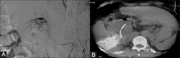
Hepatocellular carcinoma (HCC) of the liver is the third most common cause of cancer-related deaths in the world. Only one-third of patients with HCC are suitable candidates for hepatic resection. Transarterial chemoembolization (TACE) is performed in unresectable HCC. Drug-eluting beads (DEB) TACE is a modification of TACE, in which doxorubicin beads are used as embolizing material. These beads deliver the drug and embolize the vessels; however, it carries the risk of non-target embolization and it is difficult in cases with absent arterial blush on digital subtraction angiography (DSA). This is resolved using C-arm cone-beam computed tomography in the DSA suite. It identifies the tumor-feeding vessels, their area of supply, and differentiates between tumor and normal liver parenchyma. In addition, it is very useful in the embolization of hypovascular HCC. It helps and guides the radiologist in performing TACE effectively and also prevents non-target embolization of normal liver parenchyma.
Keywords: Cone-beam computed tomography; hepatocellular carcinoma; transarterial chemoembolization.
Conflict of interest statement
Figures


Similar articles
-
Cone-Beam Computed Tomography-Based Spatial Prediction of Drug Dose After Transarterial Chemoembolization Using Radiopaque Drug-Eluting Beads in Woodchuck Hepatocellular Carcinoma.Invest Radiol. 2022 Aug 1;57(8):495-501. doi: 10.1097/RLI.0000000000000864. Epub 2022 Mar 4. Invest Radiol. 2022. PMID: 35239613 Free PMC article.
-
Quantification of perfusion reduction by using 2D-perfusion angiography following transarterial chemoembolization with drug-eluting beads.Abdom Radiol (NY). 2018 May;43(5):1245-1253. doi: 10.1007/s00261-017-1296-z. Abdom Radiol (NY). 2018. PMID: 28840307
-
Intraprocedural C-arm dual-phase cone-beam CT: can it be used to predict short-term response to TACE with drug-eluting beads in patients with hepatocellular carcinoma?Radiology. 2013 Feb;266(2):636-48. doi: 10.1148/radiol.12112316. Epub 2012 Nov 9. Radiology. 2013. PMID: 23143027 Free PMC article.
-
Conventional vs drug-eluting beads transarterial chemoembolization for hepatocellular carcinoma.World J Hepatol. 2017 Jun 28;9(18):808-814. doi: 10.4254/wjh.v9.i18.808. World J Hepatol. 2017. PMID: 28706579 Free PMC article. Review.
-
Transarterial chemoembolization using drug eluting beads for the treatment of hepatocellular carcinoma: Now and future.Clin Mol Hepatol. 2015 Dec;21(4):344-8. doi: 10.3350/cmh.2015.21.4.344. Epub 2015 Dec 24. Clin Mol Hepatol. 2015. PMID: 26770921 Free PMC article. Review.
Cited by
-
Detection of hepatocellular carcinoma feeding vessels: MDCT angiography with 3D reconstruction versus digital subtraction angiography.BMC Med Imaging. 2024 Sep 18;24(1):250. doi: 10.1186/s12880-024-01408-z. BMC Med Imaging. 2024. PMID: 39294600 Free PMC article.
-
Agitated saline sonography: a simple technique for intraprocedural feeder identification during transcatheter arterial chemoembolization of hepatocellular carcinoma.Diagn Interv Radiol. 2016 May-Jun;22(3):269-72. doi: 10.5152/dir.2015.15356. Diagn Interv Radiol. 2016. PMID: 27015444 Free PMC article.
References
-
- Llovet JM, Burroughs A, Bruix J. Hepatocellular carcinoma. Lancet. 2003;362:1907–17. - PubMed
-
- Itsubo M, Koike K, Tsuno S, Osada M, Komuro O, Shimada N, et al. Subsegmental transcatheter arterial embolization for small hepatocellular carcinoma. Hepatogastroenterology. 2002;49:735–9. - PubMed
-
- Del PP, Maddeo A, Zabbialini G, Piti A. Chemoembolization of hepatocellular carcinoma with drug eluting beads. J Hepatol. 2007;47:157–8. - PubMed
-
- Iwazawa J, Ohue S, Mitani T, Abe H, Hashimoto N, Hamuro M, et al. Identifying feeding arteries during TACE of hepatic tumors: Comparison of C-arm CT and digital subtraction angiography. AJR Am J Roentgenol. 2009;192:1057–63. - PubMed
LinkOut - more resources
Full Text Sources
Miscellaneous
