Laser in dentistry: An innovative tool in modern dental practice
- PMID: 23833485
- PMCID: PMC3700144
- DOI: 10.4103/0975-5950.111342
Laser in dentistry: An innovative tool in modern dental practice
Abstract
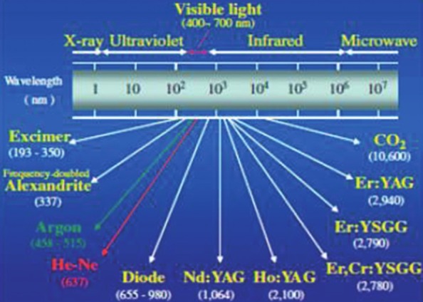
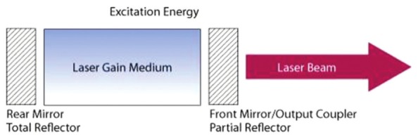
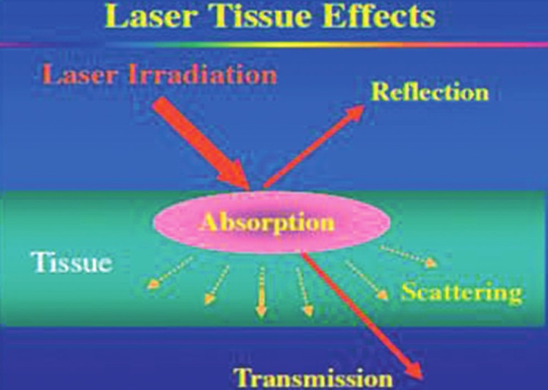
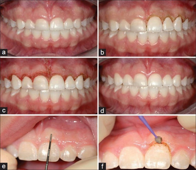
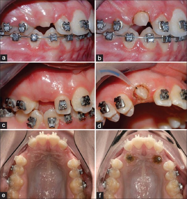
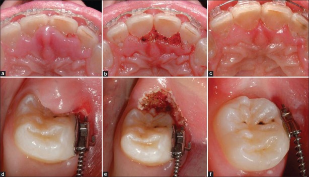
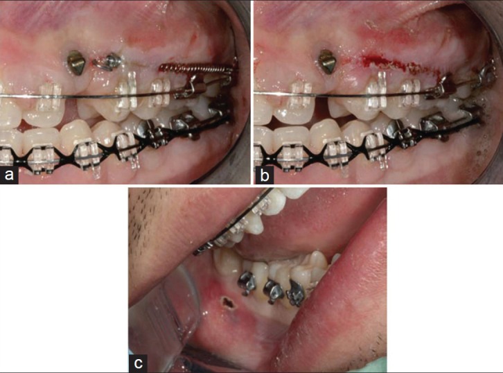
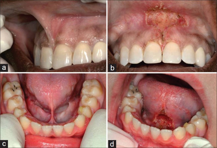
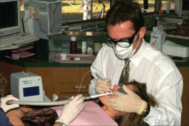
The term LASER is an acronym for 'Light Amplification by the Stimulated Emission of Radiation'. As its first application in dentistry by Miaman, in 1960, the laser has seen various hard and soft tissue applications. In the last two decades, there has been an explosion of research studies in laser application. In hard tissue application, the laser is used for caries prevention, bleaching, restorative removal and curing, cavity preparation, dentinal hypersensitivity, growth modulation and for diagnostic purposes, whereas soft tissue application includes wound healing, removal of hyperplastic tissue to uncovering of impacted or partially erupted tooth, photodynamic therapy for malignancies, photostimulation of herpetic lesion. Use of the laser proved to be an effective tool to increase efficiency, specificity, ease, and cost and comfort of the dental treatment.
Keywords: Dental application; lasers; photostimulation.
Conflict of interest statement
Figures
References
-
- Maiman TH. Stimulated optical radiation in ruby lasers. Nature. 1960;187:493.
-
- Walsh LJ. Dental lasers: Some basic principles. Postgrad Dent. 1994;4:26–9.
-
- Pick RM, Miserendino LJ. Chicago: Quintessence; 1995. Lasers in dentistry; pp. 17–25.
-
- Goldman L, Goldman B, Van-Lieu N. Current laser dentistry. Lasers Surg Med. 1987;6:559–62. - PubMed
-
- Frentzen M, Koort HJ. Lasers in dentistry: New possibilities with advancing laser technology. Int Dent J. 1990;40:323–32. - PubMed