. 2013 Aug;13(8):1963-75.
doi: 10.1111/ajt.12333. Epub 2013 Jul 8.

Transplant tolerance to pancreatic islets is initiated in the graft and sustained in the spleen

Affiliations

Transplant tolerance to pancreatic islets is initiated in the graft and sustained in the spleen

N Gagliani et al. Am J Transplant. 2013 Aug.

Abstract

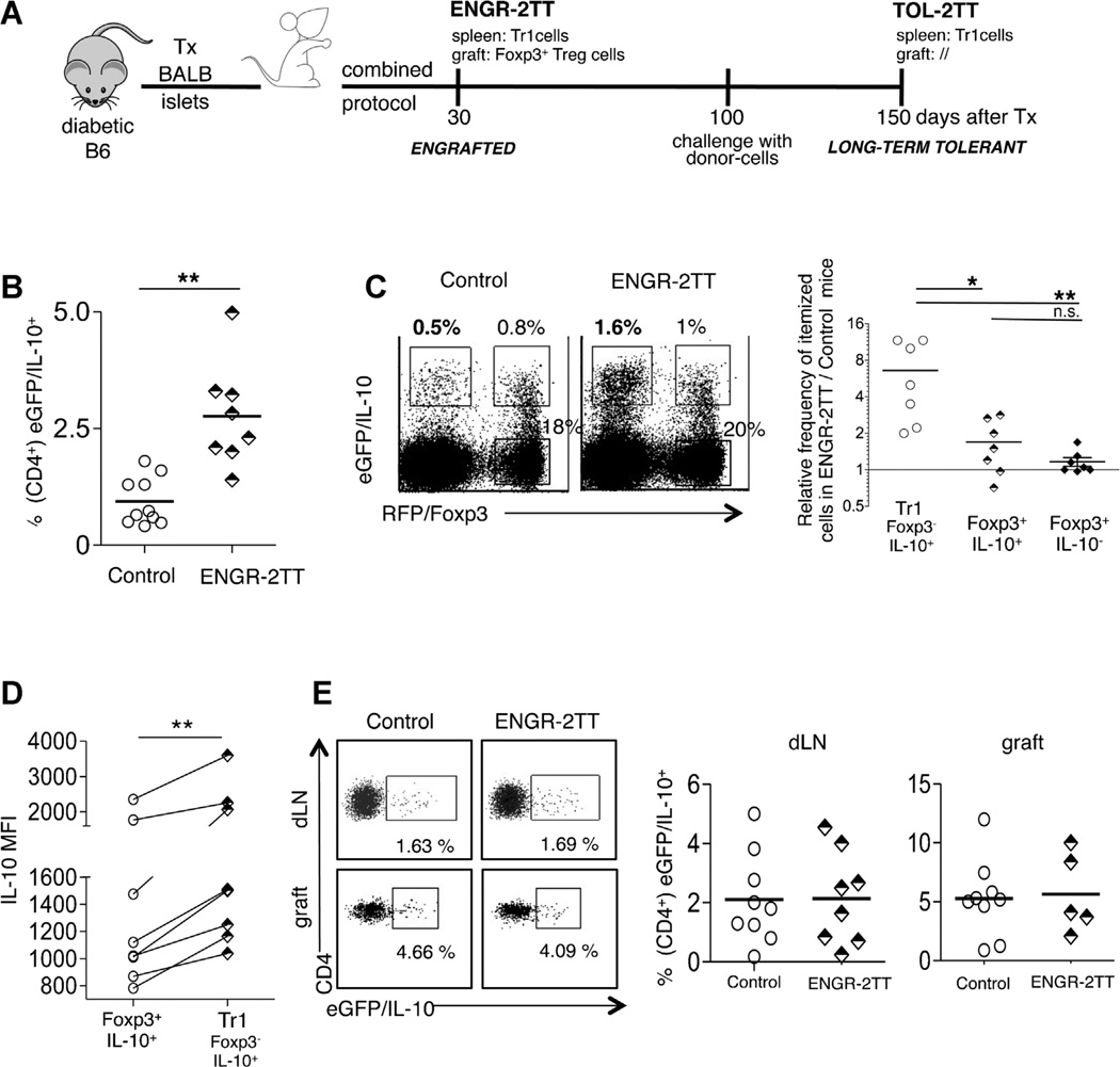
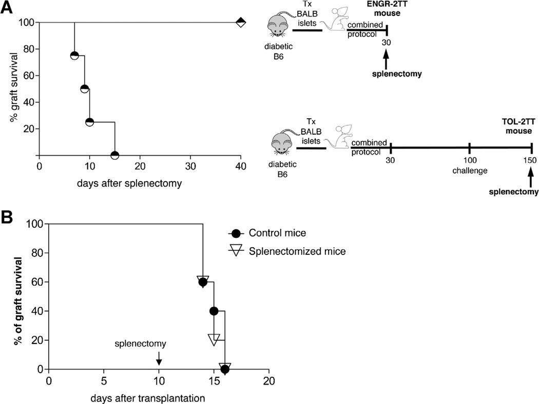
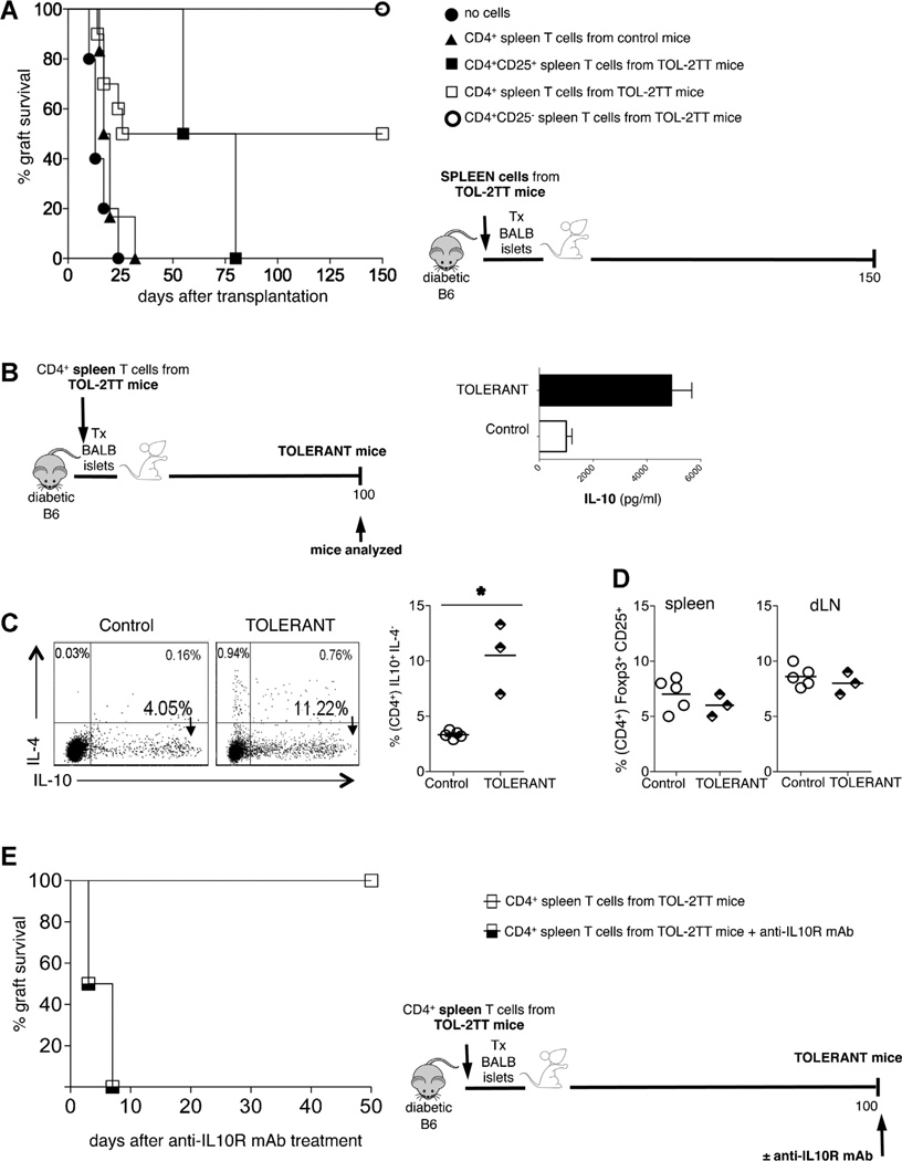
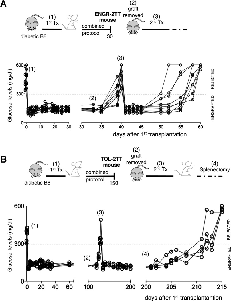
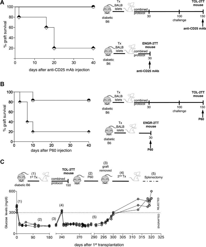
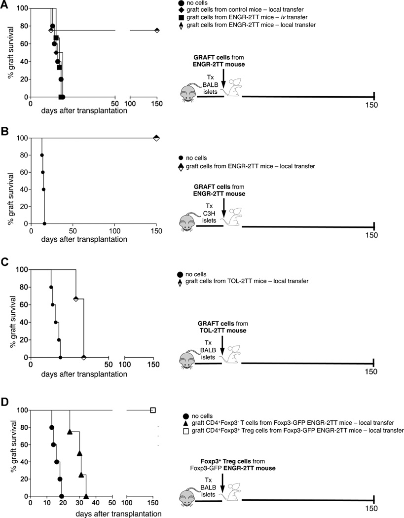
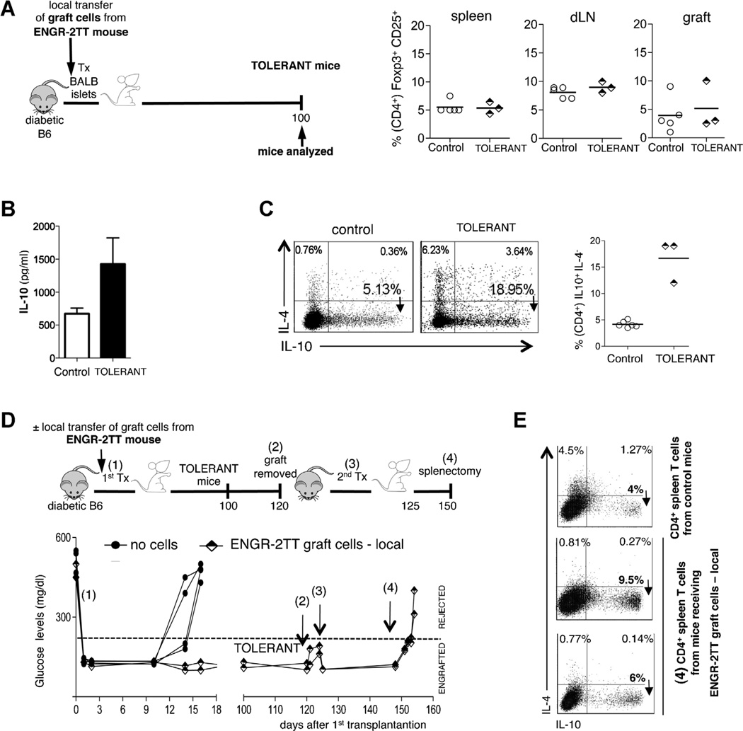
The immune system is comprised of several CD4(+) T regulatory (Treg) cell types, of which two, the Foxp3(+) Treg and T regulatory type 1 (Tr1) cells, have frequently been associated with transplant tolerance. However, whether and how these two Treg-cell types synergize to promote allograft tolerance remains unknown. We previously developed a mouse model of allogeneic transplantation in which a specific immunomodulatory treatment leads to transplant tolerance through both Foxp3(+) Treg and Tr1 cells. Here, we show that Foxp3(+) Treg cells exert their regulatory function within the allograft and initiate engraftment locally and in a non-antigen (Ag) specific manner. Whereas CD4(+) CD25(-) T cells, which contain Tr1 cells, act from the spleen and are key to the maintenance of long-term tolerance. Importantly, the role of Foxp3(+) Treg and Tr1 cells is not redundant once they are simultaneously expanded/induced in the same host. Moreover, our data show that long-term tolerance induced by Foxp3(+) Treg-cell transfer is sustained by splenic Tr1 cells and functionally moves from the allograft to the spleen.

Keywords: Graft; T regulatory cells; spleen; transplant tolerance.


Conflict of interest statement

Disclosure

The authors of this manuscript have no conflicts of interest to disclose as described by the American Journal of Transplantation.

Figures

Figure 1
Figure 1. The combined protocol induces Tr1 cells selectively in the spleen of 2TT-mice
(A) A schematic depicting of the 2TT mouse model where diabetic B6 mice are transplanted with islets from BALB mice and are treated with the combined protocol for 30 days. (B) Diabetic double reporter B6 Fir/Tiger mice were transplanted with BALB islets and were either left untreated and analyzed once they rejected the graft (Control) or treated with the combined protocol and analyzed 30 days after transplantation (ENGR-2TT). The frequency of CD4+ eGFP/IL-10+ cells in the spleen of all control and ENGR-2TT mice is shown. (C) One representative dot plot of splenic cells isolated from one control and one ENGR-2TT mouse gated on CD4+ T cells is shown (left panel). The frequencies of CD4+ Foxp3IL-10+ (Tr1), Foxp3+IL-10+and Foxp3IL-10 in the spleen of ENGR-2TT mice were averaged and divided by the average of the same cell types found in the spleen of control untreated mice (right panel). (D) The IL-10MFI of Foxp3+IL-10+andof CD4+ Foxp3IL-10+ (Tr1), gated on CD4+T cells residing in the spleen of all control and ENGR-2TTmice, is plotted. Lines link analyses performed in the same mouse. (E) One representative dot plot of cells isolated from the dLN and from the graft of one control and one ENGR-2TT mouse is shown (left panel). The frequencies of CD4+ eGFP/IL-10+ in the dLN and in the graft of all control and ENGR-2TT mice are plotted (right panel). Lines represent mean values (*p < 0.05; **p < 0.005).
Figure 2
Figure 2. Transplant tolerance is maintained in the spleen
(A) Graft survival in ENGR-2TT mice splenectomized 35 days (diamonds, n = 7) or 150 days (circles, n = 4) after transplantation is shown. The cartoon depicting the specific experiment is displayed in proximity. (B) Diabetic B6 mice were transplanted with BALB islets (n = 10). Mice were left untreated and 10 days after transplantation a group of mice were splenectomyzed (n = 5) while others were not (n = 5). Percentage of graft survival is shown.
Figure 3
Figure 3. Splenic CD4+CD25T cells contain Tr1 cells and have a strong regulatory capacity
(A) Diabetic B6 mice were transplanted with BALB islets and left untreated (filled circles n = 5), or the day before transplant were injected with CD4+ spleen T cells isolated from transplanted and untreated control mice (filled triangles, n = 6), or with CD4+CD25+ spleen T cells from TOL-2TT mice (filled squares, n = 2), or with CD4+spleen T cells isolated from TOL-2TT mice (empty squares, n = 10), or with CD4+CD25 spleen T cells from TOL-2TT mice (empty circles, n = 4). The graft survival curves are shown on the left while the cartoon depicting the experiment is shown on the right. (B) Mice that became tolerant upon the mere transfer of CD4+ spleen T cells isolated from TOL-2TT mice were analyzed 100 days after cell transfer (TOLERANT) and B6 mice transplanted and untreated were analyzed as soon as they rejected the graft (control). The cartoon depicting the experiment is shown on the left while the amount of IL-10 released from CD4+ T cells isolated from the spleen of tolerant (black bars, n = 3) and control (white bars, n = 5) mice is shown on the right (mean ± SEM). (C) Representative dot plots of IL-10 and IL-4 producing CD4+ T cells from one control and one tolerant mouse are shown on the left. The results collected from all mice analyzed are shown on the right. Bars represent mean values (*p < 0.05). (D) Frequency of Foxp3+CD25+cells (gated on CD4+T cells) in the spleen and dLN of control and tolerant mice. (E) Mice that became tolerant upon the mere transfer of CD4+ spleen T cells isolated from TOL-2TT mice were either injected with the anti-IL-10R mAb (half-black squares, n = 2) or with PBS (empty squares, n = 3) 100 days after transplantation. The percentage of graft survival is shown on the left while the cartoon depicting the specific experiment is shown on the right.
Figure 4
Figure 4. Transplant tolerance initiates within the graft
(A) Diabetic B6 mice were transplanted with BALB islets (1) and were treated with the combined protocol. Thirty days after transplantation the graft was removed from ENGR-2TT mice (2). A second islet transplant was performed in the controlateral kidney of mice returned diabetic (3). The cartoon depicting the specific experiment and time points is shown on top, while the blood glucose levels of each of the animal tested (n = 10) are shown below. (B) Diabetic B6 mice were transplanted with BALB islets (1) and were treated with the combined protocol. One hundred fifty days after transplantation the graft was removed from TOL-2TT mice (2). A second islet transplant was performed in the controlateral kidney of mice returned diabetic (3) and upon normalization of the glucose levels mice were splenectomized (4). The cartoon depicting the specific experiment and time points is shown on top, while the blood glucose levels of each of the animal tested (n = 4) are shown below.
Figure 5
Figure 5. Foxp3+ Treg cells play a key role in tolerance induction
(A) Graft survival in ENGR-2TT (diamonds, n = 5) or in TOL-2TT (circles, n = 4) mice injected with anti-CD25 mAb is shown. The cartoon depicting the specific experiment is displayed in proximity. (B) Graft survival in ENGR-2TT (diamonds, n = 5) or in TOL-2TT (circles, n = 6) injected with P60 is shown. The cartoon depicting the specific experiment is displayed in proximity. (C) Diabetic B6 mice were transplanted with BALB islets (1) and were treated with the combined protocol. One hundred fifty days after transplantation the TOL-2TT mice were treated with P60 (2). Two hundred days after transplant the graft was removed from TOL-2TT mice (3) and a second islet transplant was performed in the controlateral kidney of mice returned diabetic (4). Upon normalization of the glucose levels, all mice were splenectomized (5). The cartoon depicting the specific experiment and time points is shown on top, while the blood glucose levels of each of the animal tested (n = 3) are shown below.
Figure 6
Figure 6. The graft of 2TT mice is rich in Foxp3+ T cells with regulatory capacity
(A) Diabetic B6 mice were transplanted with BALB islets and left untreated (filled circles n = 5), or the day before the transplant were injected locally with the cells isolated from the graft of transplanted and untreated control mice (filled diamonds, n = 3), or intravenously (filled squares, n = 3) or locally (half-black diamonds, n = 4) with cells isolated from the graft of ENGR-2TT mice. The graft survival curves are shown on the left while the cartoon depicting the experiment is shown on the right. (B) Diabetic B6 mice were transplanted with BALB islets and left untreated (filled circles n = 5), or the day before transplant of third party islets were injected locally with the cells isolated from the graft of ENGR-2TT mice (half-black diamonds, n = 3). The graft survival curves are shown on the left while the cartoon depicting the experiment is shown on the right. (C) Diabetic B6 mice were transplanted with BALB islets and left untreated (filled circles n = 5), or the day before the transplant were injected locally with the cells isolated from the graft of TOL-2TT mice (half-black diamonds, n = 3). The graft survival curves are shown on the left while the cartoon depicting the experiment is shown on the right. (D) Diabetic B6 mice were transplanted with BALB islets and left untreated (filled circles n = 5), or the day before the transplant were injected locally with CD4+ Foxp3∼ T cells isolated from the graft of Foxp3-GFP ENGR-2TTmice (filled triangles, n = 4) or with CD4+ Foxp3+T cells isolated from the graft of Foxp3-GFP ENGR-2TTmice (empty squares, n = 3). The graft survival curves are shown on the left while the cartoon depicting the experiment is shown on the right.
Figure 7
Figure 7. Graft infiltrating cells from ENGR-2TT mice transfer long-term tolerance, which is sustained by the spleen
(A) Mice that became tolerant upon the mere transfer of cells isolated from the graft of ENGR-2TT mice were analyzed 100 days after cell transfer (TOLERANT, n = 3). Age matched B6 mice transplanted and untreated were analyzed as soon as they rejected the graft (control n = 5). The cartoon depicting the experiment is shown on the left, while the percentages of CD25+Foxp3+ (among CD4+T cells) in the spleen, dLN and in the graft of all mice analyzed are reported on the right (bars represent mean values). (B) The amount of IL-10 released from CD4+ T cells isolated from the spleen of control (white bars, n = 5) and tolerant (black bars, n = 3) mice after in vitro polyclonal stimulation are shown (mean ± SEM). (C) Representative dot plots of IL-10/IL-4 producing CD4+ T cells from one control and one tolerant mouse are shown on the left. The results collected from all mice analyzed are shown on the right (bars represent mean values). (D) Diabetic B6 mice were transplanted with allogeneic islets and the day before transplant were left untreated (filled squares, n = 4) or were injected locally with graft-cells from ENGR-2TTmice (half-black diamonds, n = 2) (1). One hundred twenty days after transplantation, the graft was removed from the tolerant mice which had received the local transfer of graft-cells (2). Five days later, a second islet transplant was performed with islets of the original donor (3). Twenty-five days after the second transplant, mice were splenectomized (4). The cartoon depicting the specific experiment and time points is shown on top, while the blood glucose levels of each of the animal tested are shown below. (E) CD4+ T cells were isolated from the spleens collected at the time point (4) and the frequency of IL-10/IL-4 producing CD4+ T cells was tested by intracellular staining. As control, splenic CD4+ T cells from age-matched transplanted and untreated mice were used.

Comment in

References

    1. Workman CJ, Szymczak-Workman AL, Collison LW, et al. The development and function of regulatory T cells. Cell Mol Life Sci. 2009;66(16):2603–2622. - PMC - PubMed
    1. Belkaid Y, Chen W. Regulatory ripples. Nat Immunol. 2010;11(12):1077–1078. - PMC - PubMed
    1. Littman DR, Rudensky AY. Th17 and regulatory T cells in mediating and restraining inflammation. Cell. 2010;140(6):845–858. - PubMed
    1. Li XC, Turka LA. An update on regulatory T cells in transplant tolerance and rejection. Nat Rev Nephrol. 2010;6(10):577–583. - PubMed
    1. Feuerer M, Hill JA, Mathis D, et al. Foxp3+ regulatory T cells: Differentiation, specification, subphenotypes. Nat Immunol. 2009;10(7):689–695. - PubMed

Publication types

MeSH terms